Symptoms Diagnosis Treatment Recommendations and prevention Isosporosis (cystoisosporosis) of cats is a parasitic disease caused by protozoa (single-celled parasites) of the genus Cystoisospora (Isospora, Cystoisospora). The parasites are species-specific, that is, cat isospores are not infectious to dogs or humans, but are transmitted from cat to cat. In adult animals, infestation (infection) with isospores most often does not lead to the appearance of clinical symptoms. Isospora belongs to the family Eimeridae and is primarily an intracellular parasite of the intestinal epithelium. With minor invasion, isospores have virtually no effect on the cat’s condition; usually, immunity develops after invasion. At a high level of invasion, isospores provoke the death of the intestinal epithelium, which leads to softening of the stool, and the stool becomes unformed.
Infection occurs through ingestion of oocysts; in some cases, rodents may be reservoirs of species-specific isospores.
Cystoisosporosis in cats
Cystoisosporosis
is a protozoal disease of cats
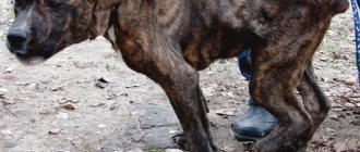
, dogs and fur-bearing animals, caused by various species of coccidia from the genus Cystoisospora. Mostly young animals are affected. Infestation is accompanied by diarrhea, emaciation, and sometimes death of animals.
Pathogens
cystoisosporosis of cats belong to the subkingdom Protozoa, phylum Apicomplexa, class Sporozoa, order Coccidiida, family Eimeriidae, genus Cystoisospora.
The following types of cystoisospores parasitize cats: Cystoisospora fe-lis, Cystoisospora rivelta, Cystoisospora cati. Cats infected with cystoisosporosis excrete immature (without spores) oocysts of the parasite into the external environment along with feces. In the external environment, under favorable conditions (temperature 20-25°C, sufficient humidity and free access of air), the process of sporulation begins in the isolated oocysts. Initially, two sporoblasts are formed in each oocyst by fission, and each sporoblast, in turn, is divided into four sporozoites. Thus, a mature (sporulated) oocyst contains eight sporozoites. The process of sporulation of oocysts is completed within 3-4 days. Mature oocysts, along with food and water, enter the gastrointestinal tract of cats, where sporozoites are released from the oocyst membranes and invade the epithelial cells of the small intestinal mucosa, causing cystoisosporosis.
In the epithelial cells of the intestine, a process of multiple asexual reproduction occurs (schizogony stage), as a result of which many new individuals are formed - merozoites. Merozoites, destroying epithelial cells, leave them and invade others, where they again multiply in a schizogonal way. This process is repeated 2-3 times, after which schizogony is replaced by gametogony (stage of sexual reproduction). In epithelial cells, not merozoites are formed, but micro- and macrogametocytes, they enter the intestinal lumen, where microgametes fertilize macrogametes and the resulting zygote is covered with a thick three-layer membrane, becoming an oocyst. Such immature oocysts are released into the external environment with the feces of infested animals.
A feature of cystoisospores (compared to Eimeria) is the ability of the oocysts of these parasites to infect the internal organs of mouse-like rodents and cats, where, resembling in its ultrastructure a sporozoite located in a parasitiform vacuole, after one or two divisions it is preserved without further development in the organs and tissues of these animals. Cystoisospora develops further in the dog's body. Mice and cats are considered by different authors as intermediate or reservoir hosts of cytoisospores. The prepotent period of development of the parasite in the body of the definitive host (cat) is shortened after eating infested reservoir hosts compared to conventional infection through oocysts. Thus, in C. felis this period is 7-8 days.
Etiology.
Cats can be infested with several types of cystoisospora. In the external environment, cystoisospore oocysts remain viable for months. The source of infection is sick and recovered animals that excrete cytoisospore oocysts in their feces. Cytoisospore oocysts can be found in cat feces at any time of the year, but are more often recorded in the summer.
Features of oocysts
In appearance, oocysts are bodies up to 33x19 microns in size, oval in shape, at the end of which there are two ends, slightly narrowed. Oocysts have a pale, smooth shell. Such isosporosis pathogens can even live in the soil for a long time before they enter the human or animal body.
But isospore in the body does not always cause illness and leads to disruption of the gastrointestinal tract. Often people, cats, and dogs are simply carriers of such a pathogen for a long time. If a cat has a strong immune system, its physical condition will not change. But the situation is completely different with pets that are weakened by long-term illnesses or have a weak immune system from birth.
In this case, the oocysts begin to actively influence the body. What happens to a kitten or weak adult animal? His digestion process is significantly disrupted, and also, which is no less dangerous, nutrients are not absorbed correctly.
Symptoms and treatment of lice in cats
Lice eaters are one of the most common types of ectoparasites that infect representatives of the cat family. Many cat owners confuse these insects with ticks and fleas. However, this is an independent type of parasite, so the methods for identifying and combating them have a number of features. Lice eaters pose a serious threat to a cat's health. In this connection, every owner should be able to identify the symptoms of trichodectosis in their pet and promptly begin to eliminate and treat them.
Lice eaters are one of the most common types of ectoparasites that infect representatives of the cat family. Many cat owners confuse these insects with ticks and fleas. However, this is an independent type of parasite, so the methods for identifying and combating them have a number of features. Lice eaters pose a serious threat to a cat's health. In this connection, every owner should be able to identify the symptoms of trichodectosis in their pet and promptly begin to eliminate and treat them.
How does the disease manifest itself?
Within a week or two after the oocysts enter the cat’s body, dangerous symptoms of isosporosis can be observed. The process moves even faster when small kittens are infected. An attentive owner can immediately notice changes in the physical condition of the pet. The main symptoms of the disease caused by isospores:
- There is an immediate decrease in appetite.
- The cat is quickly losing weight.
- Weakness, apathy, and indifference to games and communication appear.
- The animal vomits and feels sick.
- Watery diarrhea is also present.
- Due to the symptoms listed above, dehydration occurs.
- You may notice blood in the animal's stool.
- The coat loses its shine and may fall out.
The most important thing in this situation is quick diagnosis and treatment. The deterioration of the condition occurs very quickly, every hour matters. To save an animal, small or adult, you must immediately, when the first symptoms appear, take it to a veterinary clinic, to a good specialist.
Symptoms similar to isosporiasis are also observed with cryptosporidiosis - diarrhea, vomiting, weakness, dehydration. An accurate diagnosis is established only after laboratory tests. But in any case, a quick reaction from the owner and contacting the clinic on the first day is important.
Isosporosis in cats
The simplest single-celled microorganism, namely feline isospora, provokes a protozoal disease - isosporosis in cats. The parasite settles in the intestines and, under favorable conditions, leads an active life, causing diarrhea, loss of appetite, and exhaustion. Adult pets most often do not show symptoms; cats are only carriers of the pathogen. The risk group includes kittens, animals in shelters and nurseries, old and weakened individuals. With timely treatment, the disease has a favorable prognosis, otherwise the animal may die from dehydration.
Recommendations
To prevent isosporosis, it is necessary to maintain cleanliness in the room where the cat lives. To treat a room from isospores, chlorine-containing detergents are usually used; hot steam treatment also works well.
To prevent epidemics, nurseries and shelters must be kept clean, wet cleaning must be carried out daily, and animals must have their own bowls and individual toilets.
If your cat has had isosporosis, it is important to know that for some time it will be a source of infection, so do not introduce it to other animals and practice only indoor keeping.
Isosporosis in cats
(c) Veterinary center for the treatment and rehabilitation of animals “Zoostatus”.
Varshavskoe highway, 125 building 1.
Isosporosis (cystoisosporosis) of cats is a parasitic disease caused by protozoa (single-celled parasites) of the genus Cystoisospora (Isospora, Cystoisospora). The parasites are species-specific, that is, cat isospores are not infectious to dogs or humans, but are transmitted from cat to cat. In adult animals, infestation (infection) with isospores most often does not lead to the appearance of clinical symptoms. Isospora belongs to the family Eimeridae and is primarily an intracellular parasite of the intestinal epithelium. With minor invasion, isospores have virtually no effect on the cat’s condition; usually, immunity develops after invasion. At a high level of invasion, isospores provoke the death of the intestinal epithelium, which leads to softening of the stool, and the stool becomes unformed.
Infection occurs through ingestion of oocysts; in some cases, rodents may be reservoirs of species-specific isospores.
(c) Veterinary center for the treatment and rehabilitation of animals “Zoostatus”.
Methods of infection
Isosporosis (synonyms for the disease - coccidiosis, isosporosis in cats) is transmitted when oocysts of the parasite enter the animal's body. Then they begin to actively mature in the intestines.
The main routes of transmission of isospores:
- through contaminated water;
- through food containing oocysts;
- when eating intermediate hosts of parasites (small rodents);
- from mother to baby.
The active development of single-celled organisms leads to the fact that they begin to leak through the intestinal lining into the blood vessels, and then into the liver, spleen and lymph nodes.
Isosporosis of dogs and cats
Isosporosis of dogs and cats is a parasitic disease caused by several species of protozoa of the genus Isospora. The disease in dogs and cats is accompanied by diarrhea, polyuria, dehydration, exhaustion and intoxication of the animal's body. With a severe degree of infection, the death of animals occurs. In dogs, the causative agents of infection are Isospora bigemina and I. Canis, in cats – Isospora bigemina, I. Felis, I. Rivolta.
Development cycle . Dogs and cats with isosporosis are the final (definitive) hosts. The initial stage of reproduction in them takes place in the small intestine. As a result, oocysts are formed, which are released into the external environment with feces, where sporogony occurs. In this case, two sporocysts are formed in the oocyst and each of them contains four sporozoites. When a mature oocyst enters the body of an intermediate host (many animal species), sporozoites emerge from it and invade the internal organs of the animal, where they transform into trophozoites - Toxoplasma gondii. Gametogony and schizogony occur in the small intestine of dogs and cats. Along with the intestinal stage of parasitism, isospores can also be endogenous, when they penetrate the internal organs - liver, spleen, lungs, lymph nodes, brain.
Epizootological data . The disease is widespread. The disease affects many species of animals and birds of various age groups. In puppies and kittens the disease is more severe than in adult cats and dogs. Carnivores become infected with isospores mainly by consuming food and water infested with oocysts. Newborn puppies become infected from their mother by licking their nipples and the hair around them. Infection is possible through intermediate hosts - mice, rats, waterfowl. Unsanitary conditions, poor living conditions, the presence of helminthic infestations, and poor feeding of animals contribute to infection.
Oocysts can survive in the external environment for up to two years. They are very resistant to chemicals. It is believed that domestic animals are natural carriers and reservoirs of the pathogen. More often, stray and hunting dogs become ill with isosporosis.
Pathogenesis. The development of the disease depends on the species, breed, age and especially the dose of infection. The development of the sexual stages of pathogens usually causes hemorrhagic inflammation of the small intestine, and less often the large intestine. They multiply in epithelial (enterocytes) and subepithelial cells of the intestinal villi. Towards the end of the development of the pathogen, desquamation of the epithelium occurs, which leads to impaired digestion, especially membrane digestion, starvation and intoxication of the body. Absorption of products of inflammation and metabolism of isospores, poorly digested food and toxins of putrefactive microflora leads to depression of the nervous system, damage to the liver, kidneys and general intoxication.
Immunity in cats and dogs with isosporosis lasts 2-6 months, after which they can become infected again.
Symptoms and course of the disease . In dogs and cats, the clinical manifestation of the disease occurs 5-7 days after infection. The disease is accompanied by frequent bowel movements with the release of liquid stool with large amounts of mucus and blood. Stool is often red or orange in color. When examining the visible mucous membranes, their pallor is noted; they are often jaundiced. Appetite is very variable, but often reduced. In severe cases of isosporosis, dogs and cats completely refuse to eat food. The general condition is depressed. Animals are inactive and prefer to lie down more. Polyuria increases. In this case, the urine becomes dark with a sharp putrid odor. Exhaustion and emaciation develop. The abdominal wall is tense and painful upon palpation. The skin loses its elasticity. The coat loses its shine and becomes disheveled. With improper treatment and a poor feeding ration, the disease becomes more severe and ends in the death of the animal. Body temperature, as a rule, rises 0.5-2 degrees above normal.
The chronic course of isosporosis is characterized by: periodic vomiting (vomiting in a dog); dysbiosis (dysbacteriosis in cats, dysbiosis in dogs); periodic diarrhea alternating with constipation (constipation in a cat, constipation in a dog); depressed state; liver dysfunction (liver disease in dogs, liver disease in cats).
With a weak degree of infection, clinical signs of the disease are not typical or may be absent. Diarrhea is periodically recorded (diarrhea in dogs, diarrhea in cats), animals are stunted in growth, and rickets often develop in young animals (Hypovitaminosis D in dogs (rickets).
Pathological data . When autopsying the corpses of dead animals, changes are found mainly in the small intestine. The intestinal mucosa shows signs of catarrhal or hemorrhagic inflammation, with multiple pinpoint and streak hemorrhages. The serous membrane has a bluish-red tint.
Diagnosis . The diagnosis of isosporosis is made comprehensively, taking into account the medical history and characteristic clinical signs. Intravital diagnosis is confirmed by the detection of oocysts in fecal matter using the Fulleborn or Darling method. Posthumously - based on the results of an autopsy and laboratory examination of scrapings from the mucous membrane of the small intestine for the detection of oocysts. In laboratory conditions, their genus and species are determined, for which feces or scrapings are moistened with a 2% solution of potassium dichromate and kept in a thermostat at a temperature of 25-30 degrees for 2-5 days for sporulation of oocysts.
Differential diagnosis . When carrying out differential diagnosis, isosporosis is distinguished from parvoviral enteritis of dogs, panleukopenia of cats, pasteurellosis by bacteriological and virological studies to detect the pathogen. Based on the collected medical history and clinical signs, it is necessary to distinguish from food and drug poisoning.
Treatment . Treatment of the disease should be comprehensive and begin with diet therapy. Sick dogs and cats are first given a fasting diet and then a bland diet. Diet therapy is described in more detail in the article - gastroenteritis in dogs. Feed should be easily digestible, have mucous properties and not irritate the intestinal mucosa.
Chemical coccides are considered to be the best therapeutic drugs. It is prescribed to sick dogs and cats for 3 days. Administered orally with food at the rate of 0.024 g per 1 kg of body weight.
In the absence of this drug, sulfonamide drugs can be prescribed: norsulfazole with food or norsulfazole sodium with water at a dose of 0.03-0.04 g per 1 kg of animal weight, sulfadimezin (in combination with chloridine) or separately at the rate of 0.1-0.4 g for cats per 1 kg of body weight every 6-8 hours, etc. Sulfonamides are usually used for 7 days and under the supervision of a veterinarian. Their effect can be enhanced by the simultaneous administration of antibiotics: chloramphenicol, tetracycline, baytril, bicillin, etc.
Cause of the disease
A dangerous disease is caused by a microorganism that penetrates directly into the gastrointestinal tract. There are two types of parasites that cause isosporosis - felis and isspora rivolta. In the gastrointestinal tract, dangerous microorganisms undergo two stages of reproduction - asexual and sexual (schizogony and gametogony).
Once ingested by an adult in the form of an oocyst, they cause only minor symptoms of infection. This may be weakness, vomiting. Sometimes the symptoms are not expressed at all. But for a kitten this parasite is a real danger. Not only does the small animal suffer from severe symptoms, but it usually eventually dies.
Who is most susceptible to isosporosis disease? These are kittens under the age of six months, as well as adults, but they have very weak immunity. Particular attention should be paid to representatives of those breeds that initially do not have a strong immune system and resistance to disease.
When diagnosing the disease, instead of “isosporosis” the synonyms “istoisosporosis” and “coccidiosis” can be used.
What is coccidia?
Isospora Felis
Therapeutic measures
Isosporosis requires symptomatic and specific treatment not only in humans, but also in animals. Most often, a protozoal disease of this type is detected already at the chronic stages of development, which significantly complicates therapy.
Among the most effective drugs against isosporosis are:
- nitrofurans;
- sulfonamides;
- coccidiostatics.
When carrying out etiotropic treatment, there is a need to prescribe symptomatic drugs. To exclude the addition of infectious diseases, the temperature of animals is measured, and a blood test is taken from people for laboratory testing. Meals during therapy should be light and small. It is important to ensure that there is no overeating.
source
Collapse
Therapeutic techniques
How is cystoisosporosis treated in cats? For this, regular biseptol . For every kilogram of live weight there should be 15 mg of the drug. Frequency of application – twice a day. But here it must be emphasized that experienced specialists warn against the use of “human” medicines. work just as well .
As we have already emphasized, one of the most important tasks in the treatment of this disease is the rapid relief of dehydration. For this purpose, Ringer's solutions, normal saline and similar mixtures are administered intravenously. They not only eliminate dehydration processes, but also help relieve intoxication in the cat’s body. Since the veins in kittens collapse almost instantly, in their case the solutions have to be injected subcutaneously, or even simply drunk orally.
Treatment can be stopped only when at least three negative stool test results for oocysts are obtained. Moreover, it is very desirable that these checks be carried out at intervals of two to three days.