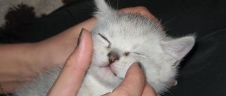
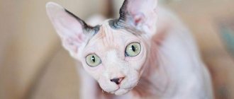
Ear diseases in cats can be non-contagious or contagious in origin.
The most common non-contagious ear diseases in cats include:
- Inflammation of the middle and inner ear - otitis media.
- Inflammation of the external ear is an inflammation of the skin of the auricle and external auditory canal.
- Hematoma is an accumulation of blood under the skin of the ear.
- Lymphoextravasate is an accumulation of lymph under the skin of the auricle.
- Auricular necrosis is the death of the ear cartilage.
- Foreign bodies in the ear canal.
- Neoplasms.
Ear hematoma in cats
Hematoma in cats occurs as a result of mechanical damage to the auricle - blows, bites from other cats, insects, scratching.
With a hematoma, blood flows from the blood vessels of the auricle into the tissue under significant pressure, pushing these tissues apart and forming a cavity. The size of the hematoma depends on the strength of blood pressure in the damaged vessel, as well as on the degree of compliance of the tissues located near it.
A hematoma occurs quickly and its volume increases until the pressure from the stretched tissue becomes equal to the pressure in the damaged blood vessel. After this, the spilled blood clots, and a blood clot forms in the blood vessel.
Most often, hematomas in cats occur on the inner surface of the ear and much less often on the outside. The damaged ear increases in size, hangs down, and the swelling is painful and hot to the touch. If the hematoma is not treated, the pain only increases, and the hematoma itself can become infected with secondary microflora, which can ultimately lead to necrosis of the ear cartilage.
During a clinical examination of such a cat, a veterinarian notes the following symptoms:
- We observe anxiety and nervousness in the cat.
- The cat almost constantly shakes its head from side to side.
- He constantly scratches his damaged ear with his paws.
- When trying to stroke the cat's head, it becomes aggressive.
Treatment. Treatment of auricular hematoma is not very difficult. If no more than 48 hours have passed since the formation of the ear hematoma, then the cat owner fixes the ears with a bandage on the back of the head and applies cold. In the future, in order to resolve the hematoma, it is necessary to use heat and apply locally irritating ointments.
If the hematoma cannot be cured at home, the owner needs to contact the nearest veterinary clinic. In a veterinary clinic, the veterinarian will open the resulting hematoma, remove blood clots from it, wash the resulting cavity with a solution of novocaine with antibiotics and give recommendations to the owner so that the hematoma resolves safely.
Lymphoextravasate in dogs
Classification of lymphatic extravasates. Lymphatic extravasation can be superficial, in which lymph is poured into the subcutaneous tissue, and deep, when lymph accumulates between the muscles, under deep fascia and aponeuroses. When there is a significant admixture of blood in the lymph, they speak of hemolymph extravasation.
Pathogenesis. Violation of the integrity of the lymphatic vessels is accompanied by the outpouring of lymph, first into the area of injury, and then its penetration into the surrounding tissues with extensive separation and the formation of many pockets. This is facilitated by poor coagulation of lymph, maceration of connective tissue fibers under the influence of lymph, which has collected and is mechanically pushed through loose fiber when the muscles are contracted. The pathogenesis of reactive inflammatory processes with lymphatic extravasation is similar to the pathogenesis with bruises.
Lymphoextravasate in dogs
Clinical signs. Lymphatic extravasates can be localized in various parts of the body. In cattle, they most often occur in the area of the hips, perineum, abdominal and chest wall, and in horses in the area of the withers, poll and dewlap. At this time, a slight, slightly painful inflammatory swelling with a slight increase in local temperature occurs at the site of injury. After resorption of the edema, a sharply separated swelling clearly protrudes, when pressure is applied to it, wavy movements of fluid from one part of the cavity to another are felt - undulation.
Lymphoextravasates are characterized by slow and prolonged development of swelling. It reaches its greatest size several days and even weeks after the injury. Despite the significant accumulation of lymph, the skin in the affected area is not tense. It seems that the volume of the cavity that has formed is much larger than the volume of lymph that has accumulated in it.
It is also characteristic of lymphatic extravasates that the inflammatory reaction and pain with them are mild, there is no local increase in temperature and there is no general reaction of the body.
When the swelling is punctured, a clear or slightly lemon-yellow liquid is obtained - lymph, sometimes mixed with fibrin. The presence of blood in the punctuation indicates hemolymph extravasation.
Forecast. With lymphatic extravasation, the prognosis is favorable in most cases, since the animal usually recovers even when it has extensive lymphatic extravasation. In case of complications of lymphatic extravasation by the development of microbes in them, the prognosis depends on the type of microorganisms that caused the difficulty.
Lymphatic extravasation of the auricle in cats
Lymphatic extravasation is an accumulation of lymph in a cavity formed as a result of tissue dissection and rupture of lymphatic vessels.
Lymphatic extravasation of the auricle in a cat occurs for the same reasons as a hematoma.
It develops slowly in a cat and is characterized by the development of a contouring swelling in the ear area; there is no local increase in temperature.
The diagnosis is made by clinical signs. To clarify the diagnosis, a puncture of the resulting swelling is performed.
Treatment. With this disease, unlike a hematoma, it is strictly forbidden to use cold or heat. In case of this disease, the owner must contact a veterinary clinic. Where the drained lymph will be aspirated using a syringe. If this procedure does not lead to the desired results, then the veterinarian resorts to surgery, which boils down to making a skin incision and more thoroughly removing the contents of the cavity and applying small sutures.
Clinical picture
How does this pathology manifest itself in practice? If we are talking about the external variety, then a noticeable swelling forms on the surface of the organ affected by the blow, which can gradually increase. It is soft to the touch; when palpated, a slight “crackling” is sometimes felt.
We suggest you read: Treatment of sheep with pasteurellosis
A distinctive feature of “pure” lymph extravasate is that the translucent contents are clean, transparent, reddish or yellowish in color. This pathology differs from hematomas. In addition, the palpable area maintains normal local temperature. What is even more important is that the liquid in the cavity seems to “flow”; there is a feeling of the presence of “extra” space there. Hematomas, recall, have a tight, tense surface.
Identifying internal “lymphatic leaks” can be much more difficult. A more or less reliable sign is the presence of traces of an impact (scratches, abrasions) on the affected area. In combination with a large and painless swelling, this can serve as sufficient grounds for making the correct diagnosis.
Necrosis of the auricle
Necrosis of the auricle in a cat can occur as a result of:
- Transfer of purulent processes from surrounding tissues to the auricle.
- With prolonged squeezing of the auricle.
- Infection of hematomas, lymphatic extravasates with pathogenic microflora and with an abscess in the ear area.
When a purulent process develops in the area of the auricle and in the absence of proper and necessary treatment, the resulting abscess opens, forming areas of skin necrosis (necrosis), resulting in ulcers appearing on the auricle.
During a clinical examination, the ear cartilage begins to be visible through the areas of damage, and its blood circulation is disrupted. The cartilage itself becomes black in color and emits an unpleasant putrid odor. With necrosis, the cartilage tissue rots, and the ear becomes deformed.
Treatment. Treatment of auricular necrosis should be carried out in a veterinary clinic. A veterinary specialist performs either a complete amputation of the auricle or a necrotic part of it, followed by a course of treatment with antibiotics.
Foreign body in the external auditory canal
Foreign bodies can get parts of plants, insect larvae, sand, lice into the ear canal, wax plugs and other objects can form.
Sometimes the presence of a foreign body in a cat’s ear does not cause any concern and may go unnoticed by the animal’s owners. Most often, a foreign body causes irritation and inflammation in the external auditory canal.
Treatment. Treatment should be aimed at removing the foreign body from the ear. After removing it, the ear canal is washed with a solution of soda or a 3% solution of hydrogen peroxide. In order to reduce the cat's pain reaction, it is necessary to drop a few drops of camphor oil into the ear canal.
Dermatitis and eczema of the ears
With dermatitis, the cat's ear turns red and a rash appears on the skin. A sick cat begins to scratch its ears due to severe itching, increasing the symptoms of dermatitis. With dermatitis, hair begins to fall out from the damaged area of the skin. Food allergies cause dermatitis in cats. Streptococcosis (Streptococcosis of dogs and cats) can lead to dermatitis.
Parasitic otitis.
Parasitic otitis in cats occurs with otodectosis and notoedrosis (Otodectosis in cats, notoedrosis (pruritic scabies).
Surgery
Trying to get rid of severe itching, the cat often hits the sore ears with its paw, which leads to the rupture of blood vessels, the release of blood into the interstitial space and the formation of hematomas. When a lymphatic vessel is injured, lymphatic extravasation is formed.
In this case, only surgical removal of the formation, performed under local anesthesia, helps. Despite the fact that the cartilage remains intact, the shape of the auricle is disrupted, and a small scar remains.
Conservative methods of treating these complications do not give a positive result.
Otitis in cats
Otitis in cats can be due to inflammation of the outer, middle and inner ear.
Causes of otitis. The cause of otitis in cats can be food allergies, the presence of parasites (ticks, fleas), ear injuries, foreign bodies, etc. Otitis of the middle and inner ear can be the result of complications of inflammation of the outer ear.
Signs of otitis media . During a clinical examination of a sick cat, the skin of the sore ear is reddened, the cat rubs the sore ear with its paw, and tries to keep the sore ear folded and pressed to its head. If the pain in the ear is acute, “shooting,” the cat suddenly jumps up, looks around in fear, and screams. With constant pain, the cat does not allow the sore ear to be touched, avoids stroking the head, and presses the sore ear to its bedding.
With purulent otitis media, inflammatory exudate is released from the cat's ear; when touched, the ear gurgles and squelches. An unpleasant odor emanates from the diseased ear.
In advanced cases, in the absence of proper treatment, the cat’s body temperature rises, the cat becomes depressed, and there is no appetite. If timely treatment measures are not taken, the eardrum may be perforated, and the inflammatory process may spread to the brain.
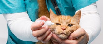
Treatment. Treatment of otitis media depends on the severity and severity of the inflammatory process in the ear. In the initial stage, the cat is prescribed special drops (Bars), Stop-Itching spray, antibacterial drugs (tylosin, etc.)
Prevention of otitis. Prevention of otitis in cats should be based on compliance with the rules of care and maintenance of cats. To help keep your pet healthy:
- Timely cleansing of the ear canal from accumulated wax. To do this, the pharmacy chain has recently recommended using a special lotion - “Dewdrop for the ears”, which is used to remove wax and inflammatory products from the auricle and external auditory canal.
- When bathing your cat, be careful not to let water get into its ears.
- Avoid keeping your cat in damp and cool areas to prevent hypothermia.
- In order to prevent infection with ear mites, try to avoid contact with stray cats.
Periodically disinfect and decontaminate the cat's place of residence and cat care items.
Ear diseases in cats are quite common. However, they can be either contagious or non-contagious. Cat ears have a rather complex anatomy; very often the source of the problem can be hidden deep inside.
Therefore, in order to make an accurate diagnosis and prescribe adequate treatment, it is best to contact a qualified veterinarian.
Causes
Ear diseases in cats can occur for various reasons:
- as a result of various injuries;
- penetration of infection;
- parasites.
Sometimes the culprit may be a lack of proper care and unsanitary conditions. The likelihood of diseases increases significantly with improper feeding, which contributes to problems with the immune system. Sometimes ear diseases develop as a secondary infection, being a complication of more serious diseases.
Also find out what other reasons a cat has hot ears>>>.
Symptoms and diagnosis
Ear diseases in cats are quite varied. The most common of them are discussed below.
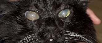
Ear scabies
The second name for this disease is otodectosis. You should be suspicious if your cat's ears itch, she shakes her head, and bald spots form around her ears.
The cause of this disease is a microscopic ear mite. In the process of its life, it makes passages in the skin layer, thereby causing unbearable itching.
Later, irritation joins it, and the affected area from the ears spreads to the entire head. This is one of the most common reasons why a cat scratches its ears.
Notoedrosis
Another fairly common parasitic disease. Its cause is a microscopic mite that cannot be seen with the naked eye.
With notoedrosis, a cat's ears, areas in the nose and brow ridges become bald. The animal experiences unbearable itching. In the absence of appropriate treatment, a characteristic bald spot also forms in the chest and back area.
Then the affected areas become crusty and begin to crack, causing unbearable pain to the cat.
Find out more about the symptoms and treatment of ear mites in cats>>>.
And a little about secrets.
The story of one of our readers, Irina Volodina:
I was especially distressed by my eyes, which were surrounded by large wrinkles, plus dark circles and puffiness. How to completely remove wrinkles and bags under the eyes? How to deal with swelling and redness? But nothing ages or rejuvenates a person more than his eyes.
But how to rejuvenate them? Plastic surgery? I found out - no less than 5 thousand dollars. Hardware procedures - photorejuvenation, gas-liquid peeling, radio lifting, laser facelift? A little more affordable - the course costs 1.5-2 thousand dollars. And when will you find time for all this? And it's still expensive. Especially now. Therefore, I chose a different method for myself.
Treatment of ear mites in cats requires patience from owners, as treatment must be carried out 2 times a day for a month. The difficulty is that the disease is often accompanied by fungal and bacterial microflora. Therefore, if symptoms are detected, you should contact a veterinarian who will make an accurate diagnosis and develop a comprehensive treatment plan aimed at eliminating the clinical signs and the causative agent of the disease.
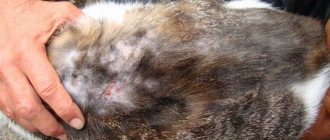
Dermatitis and eczema
Dermatitis and eczema in cats are quite common. They are especially common in animals kept in apartment conditions.
The main reason for their occurrence is various metabolic disorders, resulting in severe allergies and, as a result, dermatitis or eczema. Stress and bad heredity can also cause them.
Red ears that are hot to the touch should immediately alert the owner. This is usually a symptom of an incipient allergy. Later, sores appear on the ears, which itch unbearably. The cat begins to scratch them vigorously, thereby aggravating the situation.
The main difference between eczema and dermatitis is the nature of the skin rashes. With eczema, ulcers appear on the ears, which subsequently form a continuous weeping surface. With dermatitis, the lesions are focal and more like severe scratching.
Treatment
It is not recommended to treat ear diseases in cats at home. You can simply try to alleviate your pet's condition before going to the veterinary clinic. At the veterinary clinic, medications will be selected for the animal and adequate therapy will be prescribed.
If a cat has a leaky ear, and there is no opportunity to go to a veterinary clinic in the near future, you can carry out the initial treatment yourself.
For this you will need hydrogen peroxide or furatsilin solution. They use a cotton or gauze swab to thoroughly wash the inner surface of the auricle. In this case, dried dirt and crusts should be soaked and should not be pulled off with force. After this, special ear drops are placed in the ear.
Treatment for ear scabies at home should also begin with preliminary cleaning of the ears. After this, special drops are placed in the ear. Which can be purchased at any veterinary pharmacy. After 7-10 days, the treatment must be repeated.
Lymphextravasate
Signs of lymphatic extravasation development are similar to hematoma.
The reasons for the development of lymphatic extravasation are in many ways similar to the reasons for the occurrence of hematoma.
Clinical signs are also almost the same. The only difference is the treatment. The use of any thermal procedures - cold or heat - is strictly prohibited .
Therapy should only be carried out by a veterinarian.
Therapy should only be carried out by a veterinarian. In a hospital setting, fluid is pumped out using a syringe. In more complex cases, surgical opening of the skin and removal of exudate, followed by the use of antiseptics and suturing, is recommended.
The symptoms of otitis are in many ways similar to many ear diseases.
With otitis media, ear discharge and redness are observed.
The pet's behavior changes, the cat becomes restless and aggressive, ear discharge, redness and swelling of the diseased organ are observed. There may be an unpleasant odor coming from the sink. In more complex cases, appetite disappears and the temperature rises.
At the beginning of treatment, clean the affected area with a cotton swab.
Treatment is carried out in a complex: local treatment and drug therapy.
- First of all, clean the affected area with a cotton swab dipped in a solution of boric acid, hydrogen peroxide or furatsilin.
- Dry with a cloth.
- Use ear drops.
- It is possible to relieve pain with novocaine blockade.
- A course of antibiotic therapy and medications that support the general condition of the animal are prescribed - immunostimulants, vitamins.
- At the same time, they provide the cat with a complete diet with vitamin and mineral supplements.
Preventive measures
The main prevention of ear diseases in cats is to regularly clean them using special lotions, drops and gels. The condition of your pet's ears should be monitored constantly. Especially if your pet is prone to producing large amounts of wax.
After bathing, your cat's ears should be checked for any remaining water. If this is not done, otitis may develop. It is also necessary to carry out preventive treatment against parasites once a month. Since they are the ones who in most cases become the source of diseases.
Do you want to know more about the article or something? Call +79774692712, we will advise you.
The thinnest blood and lymphatic vessels located on the inside of the auricle in cats and dogs are often quite fragile, especially in older animals susceptible to chronic inflammatory processes or suffering from a lack of vitamins and microelements. In some cases, even a slight mechanical impact can lead to rupture of blood vessels, bleeding under the skin, and sometimes the formation of pockets filled with lymph or blood between the skin and the cartilage of the auricle - lymphoextrovases or hemolymphoextrovases.
Ear hematomas are most often seen in dogs with floppy ears, but they can also occur in dogs with any other type of ear. The same is true for cats.
What it is?
The lymphatic system is largely “guided” by the same laws as the circulatory system. Moreover, lymphatic vessels run parallel to blood vessels. Accordingly, any injuries fraught with loss of integrity of the latter are also dangerous for the lymphatic system. In general, lymphatic extravasation is a pathology in which the contents of the lymphatic vessels leak into the surrounding tissues.
The phenomenon is often confused with edema of various etiologies, which significantly complicates their treatment, as well as identifying the causes that led to the development of the disease. What are the reasons?
As already noted, any injuries that can lead to bleeding can lead to the release of lymph from the vessels: cuts, lacerations, biting wounds, puncture wounds, etc. But the phenomenon we describe occurs only in cases of blows with blunt objects and severe bruises: closed damage to tissues and blood vessels occurs, lymph leaks into the tissue. Under the pressure of the liquid, a cavity is formed, which, in fact, is called “extravasate”.
Attention! In many cases, this term refers to lymphatic extravasation of the dog's ear. The tissues of the auricle are thin and delicate, and therefore, when the vessels are damaged, they easily expand under the pressure of the lymph leaving the vessels.
Deep extravasates are formed much less frequently. The fluid, escaping into the dense tissues, forms a swelling, which, based on external signs, can easily be confused with ordinary edema. If the affected area is small, even an experienced veterinarian will mistake it for the consequences of an ordinary bruise. In principle, there is nothing wrong with this, since over time everything will resolve on its own. Of course, this does not happen in all cases, and sometimes the consequences of lymphatic extravasation have to be eliminated by resorting to surgery.
Causes of lymphoextrovasation in cats and dogs
In most animals, auricular hematoma develops as a result of excessive head shaking or chronic, persistent scratching and damage to the ear. This can occur immediately after bathing the animal, due to chronic otitis media or an allergic reaction, which causes severe itching and inflammation. The presence of parasites (for example, demodex mites) or a foreign object in the auricle can also indirectly affect this pathological process.
Shaking your head can cause these tiny blood or lymph vessels to rupture, bursting and spilling into the surrounding area, gradually peeling the skin away from the cartilage tissue. The accumulated exudate causes excessive irritation, and the animal begins to shake its head even more, thereby aggravating the process. If the situation is not resolved, then blood or lymph continues to accumulate under the skin in greater and greater quantities, increasingly peeling the skin away from the cartilage and forming a large pocket, which in some cases can block the ear canal.
In some cases, constant shaking of the head can eventually lead to spontaneous rupture of the hematoma and splashing of its contents outward. However, a responsible pet owner should never allow such a development to occur.
General information
Lymph nodes and vessels play an integral role in the functioning of the immune system, acting as blood filters and transport arteries for lymphocytes. There are many lymphatic vessels in the body, there are large ducts and “reservoirs” for the accumulation of lymph. So here it is. The word “lymphoextravasate” consists of three parts: “lymph”, “extra” and “vazat”. Well, everything is clear with lymph, “extra” means above, from outside, and the word “vazat” means vessel.
Simply put, this pathology involves the release of lymph into the tissues surrounding the lymphatic vessels. As is easy to understand, this becomes possible only as a result of some kind of injury, strong blows, “March sprees,” etc.
More often we are talking about hemolymphoextravasates. This is the name of “mixed” pathology, in which not only lymph, but also blood leaks into the surrounding tissues. This disease should not be confused with inflammatory phenomena in the lymphatic system. That is, in cases where your cat suffers from some kind of infectious disease, and he has swollen, hot lymph nodes that clearly protrude even through the skin, there is no question of extravasation in principle.
Attention! The most harmless phenomenon of this type is lymphatic extravasation of the auricle. Again, in 90% of cases there is no talk of true (!) damage to the lymphatic system! Most likely, this term almost always refers to a “banal” hematoma of the auricle, which can develop due to blows, with strong and constant scratching (allergies, ear mites, etc.).
Diagnosis and treatment of auricular hematomas
Usually, this problem is quite clearly visible visually and a veterinarian, upon examination, can easily diagnose lymphatic extrovase in a cat or dog. If the swelling is soft in consistency and warm, this indicates a small hematoma, but if it is tense and hot, then most likely this hematoma fills a large area of the auricle.
Treatment of hemolymphoextrovasation of the auricle in cats and dogs includes not only a solution to relieve swelling, but also determining the true cause of its occurrence and resolving this causative factor. There are several different options aimed at reducing the volume of the hematoma, and the first of them is pumping out the exudate using a syringe and needle. The surface of the hematoma is treated with an antiseptic antibacterial solution that prevents the spread of infection, and a syringe is used to puncture the skin on the inside of the ear at an angle of 90° or 45° and, if possible, completely pump out the contents of the pocket. Quite often, without removing the needle, a second syringe is connected to it, with a special solution of antibiotic, hormonal and anti-inflammatory drugs that promote the development of adhesive inflammation; and is introduced in a small volume into the cavity of the pocket. This is the simplest and most inexpensive way to solve the problem, but it has many disadvantages.
Diagnosis and treatment
In general, the entire diagnosis of lymphatic extravasation is based on the principles described above: absence of pain, presence of traces of mechanical trauma. The veterinarian will take samples of your pet's blood, bladder secretions or swelling, and review his medical history.
The main task is to identify those diseases that could lead to the development of similar symptoms. If the veterinarian discovers them, measures should be taken to eliminate them as soon as possible. How is lymphoextravasate treated in cats?
The easiest way is with extravasates on the ears. As a rule, the operation does not even require anesthesia (with the exception of particularly large, aggressive cats). The surface of the bladder is carefully dissected, the secretion is pumped out, and the cavity is washed with an antiseptic solution. The resulting wound is sutured, preventing complete “fusion” of the edges of the cut (so that ichor and other fluids can flow in). If you follow the simplest measures to care for the animal after surgery, postoperative damage will heal within a few days.
With internal “lymph flows” it is more difficult. The cat is provided with absolute rest; nervous and excitable animals are prescribed sedatives. Using an aspiration needle, the contents of the cavities are removed. Do not apply cold compresses under any circumstances! The surface of the skin is lubricated with alcohol tincture of iodine. But in cases where the affected area is large, you still have to resort to surgical intervention.
In this case, the animal is put into a state of general anesthesia, the skin is cut, the contents of the resulting cavity are removed, it is washed, exfoliated, crushed tissue is excised, and the postoperative wound is sutured. Note that there is no need to wash the incision after the operation: it is covered with a sterile bandage, which is changed regularly. If a specialist suspects that the wound may fester, drainage with an antiseptic is left in it.
Source
Surgical methods for the treatment of lymphoextrovase in cats and dogs
There are several surgical techniques for solving the problem of lymphoextrovase and hemolymphoextrovase in cats and dogs. All of them involve evacuating the hematoma exudate and placing one or more sutures to create an adhesion - a cohesive fusion between the skin of the ear and the cartilage of the auricle. To do this, through stitches are made in the damaged part of the auricle, the purpose of which is, first of all, to ligate the damaged lymphatic or blood vessels and prevent further leakage. The sutures are left in place for approximately three weeks to create favorable conditions conducive to the development of scar tissue, which exerts additional pressure and compresses long-term bleeding vessels, preventing the hematoma cavity from filling with lymph or blood.
All surgical methods for solving this problem, unfortunately, can lead to some degree of scarring of the auricle. In this case, scarring is not only inevitable, but also a desirable process, since the main cartilage of the animal’s auricle is damaged and thinned, and the resulting collagen scars, although not able to completely restore the functions of the cartilage, can still partially support it. The more scars are formed, the more irregular, corrugated in shape and hard to the touch the auricle after healing. If the owner of the animal does not seek veterinary help from specialists at all, then within several weeks or even months, provided that the hematoma does not rupture earlier, its contents gradually resolve, and the surrounding tissues grow into powerful dense scar tissue. This is an inevitable process. Not only will the animal experience severe discomfort and pain throughout this period, but complete drying out and severe deformation of the auricle as a whole are possible.
Diagnostic procedures
When diagnosing, all the same signs that we described above are taken into account. First, the veterinarian needs to look for marks on the dog's skin that indicate impacts. The owner is asked whether the dog was hit by a bicycle/car, whether it fought with other animals, whether it fell from obstacles during training...
Secondly, measures are being taken to identify other diseases, which, in some cases, can give similar symptoms:
- All diseases of the cardiovascular system.
- Pathologies of the urinary system.
- Extreme degree of exhaustion, in which hunger and cachectic edema are possible.
- Parasitic diseases, poisoning, in which the liquid part of the blood can escape through the walls of blood vessels.
- Generalized forms of allergic reactions. Histamine, abundantly released into the blood, also increases the porosity of blood vessels, the walls of which begin to resemble a colander.
Causes of auricular hematoma in cats and dogs
Since excessive shaking of the head and excessive trauma to the auricle with the front and rear paws is the main root cause of the occurrence and development of lymphatic extravasation in cats and dogs, it is necessary, first of all, to understand the cause of itching and discomfort in this area. This is important both for diagnosis and for treatment and prevention.
Often the cause of soreness is an obvious injury, bruise or wound to the ear, but more often it is an infection or a severe allergic reaction. The ear of a cat or dog is carefully examined using an otoscope; The contents of the auricle are cleaned out and a small amount is taken for microscopic examination for the presence of bacteria, yeast or mites. Any of these reasons should be considered for the diagnosis of lymphatic extrovase, since any of them causes irritation of the delicate skin of the ear canal.
If a specific microflora is detected, it is necessary to immediately determine its sensitivity to antibiotics and antifungal drugs; if a tick infestation is detected, then it is necessary to develop a scheme for acaricidal treatment of the animal, causing the least irritation of the ear canal.
If the roots of this problem are of allergic origin, then, if possible, it is advisable to establish what exactly it could develop into. If its cause is discovered, it is necessary to exclude this allergen, be it an environmental factor or a nutrition-related factor.
Particular attention should be paid to dogs with long ears. It is important to regularly inspect and clean them to keep your ears and ear canal clean.
Diagnostic methods
If the ears are swollen, you should consult a specialist who will diagnose the disease.
Lymphatic extravasation in tissues other than the auricle is not so easy to detect. In this case, swelling also occurs in the affected area, traces of bruises, bites, and scratches are visible, which is the cause of the disease. To exclude other causes of edema, it is necessary to show the animal to a veterinarian. The specialist will analyze the contents of the auricle for the presence of bacteria, yeast, mites, and inflammation. If swelling has developed in another part of the body, palpation and necessary follow-up tests are performed.