All malignant tumors are called cancer, and they received this name because of their irregular shape with ragged edges, reminiscent of tentacles. Cancerous tumors do not have clear boundaries and are accompanied by severe gnawing pain. Intestinal cancer (lymphoma) is rarely diagnosed in cats, but this type of tumor metastasizes widely and causes intestinal blockage.
The danger of intestinal cancer in cats, as in other animals, is that the stage of its development when the tumor can be completely removed is usually asymptomatic. But in any case, the earlier the disease is detected, the more favorable the treatment prognosis, so it is very important to consult a veterinarian at the first signs.
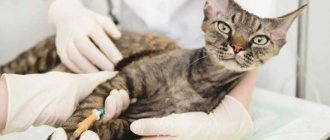
He will order a thorough examination, including a biochemical blood test to assess electrolyte balance and determine the amount of protein in the serum, a biopsy followed by cytology to determine the type of tumor, and x-rays to determine its size and location.
Etiology of cancer
The causes of the disease are not fully understood. It is possible that when feeding cats fried, fatty or salty foods, the risk of intestinal tumors increases, as in humans. But there is no exact research data. It has been established that Siamese cats have a breed predisposition to intestinal cancer.
Mostly animals over 11 years of age are affected. Lymphoma can be a consequence of feline viral leukemia , but in many animals with such cancer the pathogen has not been isolated, meaning the virus is only one of the factors.
The causes of adenocarcinoma and mastocytoma in cats remain unclear.
Intestinal cancer in a cat - how to detect and treat the disease in time?
Oncological diseases in domestic animals have not been sufficiently studied. Intestinal cancer is rare in cats, but often has an unfavorable outcome. The course of treatment and further prognosis depend on the owner and how early it is to take the animal to the veterinarian. Timely consultation with a doctor significantly increases the animal’s chances of starting a healthy, happy life.
Histological types
Various cancer cells can be found in the intestines. The most common types found in cats are:
- Alimentary lymphoma. In this case, cancer cells are formed from lymph. Since the disease is systemic, it often affects many organs and most parts of the intestine. It usually causes diarrhea, alternating with constipation. At the same time, the abdomen increases in size and the animal loses its appetite.
- Intestinal adenocarcinoma. With this pathology, cancer develops from the glandular epithelium. The prognosis depends on the presence or absence of metastases. The first sign of the disease is constipation and intestinal blockage. The pathology progresses rapidly, often giving metastases to the liver, kidneys, lungs and other organs.
- Intestinal mastocytoma. Originates from mast cells. Since these cells secrete heparin, histamine and other hormones that are responsible for inflammatory reactions, one of the striking symptoms is acute intestinal inflammation with severe diarrhea. Metastases are also observed frequently and are usually found in the lymph nodes.
The most common type of tumor in cats is lymphoma.
Intestinal adenocarcinoma in a pet
Intestinal adenocarcinoma is a fairly common malignant tumor in cats. The factors causing this disease have not been fully identified, but experiments have shown that nitrosamines may play some role. Intestinal adenocarcinomas can be nodular or ring-shaped.
At the time of diagnosis, metastases are often present, most often to the mesenteric lymph nodes. Other sites of metastasis include the liver, kidneys, peritoneum, lungs, and omentum.
Intestinal adenocarcinomas show aggressive local growth, and tumors often recur after removal.
Clinical signs
Intestinal adenocarcinomas have been observed in animals aged 3 to 13 years, but most cases occur in older cats. The average age for developing this disease is 10 to 12 years. Males are more susceptible to the disease than cats. No breed dependence was observed.
Tumors of the small intestine are accompanied by vomiting, weight loss, tarry stools, flatulence and rumbling in the abdomen.
Colon tumors are characterized by the presence of blood and mucus in the stool and a painful urge to defecate.
Other signs that appear with any form of the disease include loss of appetite, diarrhea and signs consistent with acute intestinal obstruction or perforation and peritonitis.
Diagnostics
Small intestinal adenocarcinomas are palpated transabdominally in the middle of the abdominal cavity. Distended loops of small intestine can also be detected by palpation, and a rectal examination may reveal tarry stools and bright scarlet blood.
Various paraneoplastic syndromes, including skin disorders and blood clot syndrome, are also associated with intestinal adenocarcinoma.
In addition to intestinal adenocarcinoma, the following conditions may cause similar symptoms: intestinal foreign body, irritable bowel syndrome, nutritional lymphoma, parasites, leiomyoma or leiomyosarcoma, and pancreatitis.
- Blood tests often show microcytic, hypochromic anemia due to chronic bleeding in the gastrointestinal tract. Due to exhaustion due to intestinal bleeding and dehydration, blood urea nitrogen may be elevated. A stool occult blood test may be positive.
- A plain X-ray may reveal a tumor in the abdomen or swollen loops of the small intestine. Contrast radiography is used to localize filling defects in intraluminal tumors, especially if gas accumulation makes ultrasound difficult to use. Chest X-ray is also recommended during diagnosis to identify possible metastases in the lungs.
- Abdominal ultrasound is more accurate than x-rays and can assess the degree of involvement of surrounding organs. In addition, ultrasound allows you to assess the condition of the intestinal walls. Intestinal adenocarcinomas in cats tend to have mixed echogenicity and are often asymmetrical.
- Endoscopy is used both to provide visualization of the damaged area and for biopsy. Ultrasound-guided fine-needle aspiration biopsy can detect epithelial tumor cells, thereby ruling out lymphoma.
- Because tumors are often located deep beneath the surface of the mucosa, endoscopic biopsy often does not provide sufficient information to make a diagnosis.
- Therefore, exploratory laparotomy and surgical biopsy are necessary to make a definitive diagnosis.
The treatment is surgery
Clinical picture
Depending on the location of the tumor (small or large intestine), symptoms vary slightly.
Small intestine
There are many nonspecific symptoms that may suggest cancer:
- loss of appetite;
- weight loss;
- vomiting or nausea;
- stomach ache;
- pallor of the mucous membranes.
Remember! All these signs develop gradually, which is why they often go unnoticed in the early stages.
characteristic symptoms may appear :
- When the intestines are blocked, severe abdominal pain appears, the animal constantly vomits, and bloody diarrhea without feces may be observed.
- When the wall is perforated, anemia quickly develops, the animal lies without strength, tachycardia is observed, the main symptom is ascites - abdominal dropsy.
- With lymphoma, diarrhea alternates with constipation, but the main diagnostic sign is low levels of protein in the blood.
- In the case of a mast cell tumor, particularly severe, incessant diarrhea is observed, which occurs due to the action of histamine.
- With adenocarcinoma, complete blockage of the intestine quickly develops.
Colon
Most often, the tumor is localized in the last third of the large intestine; rectal cancer is usually diagnosed. Regardless of the histological type of tumor, the pathology is always accompanied by severe constipation, followed by bloody diarrhea (without feces). The animal constantly tries to go to the toilet and experiences severe pain.
Treatment of peptic ulcer
Treatment for gastric ulcers in cats is symptomatic: water and electrolyte balance is restored, drugs with analgesic and antiemetic effects are prescribed, and a dietary regimen is prescribed.
If there are signs of dehydration, infusion therapy is performed. A dropper is installed, the basis of which is a 0.9% solution of sodium chloride with glucose or other components (as prescribed by the treating veterinarian).
If the development of a stomach ulcer is provoked by pathogenic microorganisms Helicobacter Pylori, antibacterial therapy is carried out, and drugs are prescribed to increase blood clotting. In severe cases, when the bleeding cannot be stopped, a gastrotomy is performed.
The next stage of treatment is the restoration of the cat’s body. In order to strengthen internal organs and systems, vitamins are prescribed:
- A (retinol),
- E (tocopherol),
- C (ascorbic acid),
- B6 (pyridoxine).
The key to successful treatment is the elimination of the factor that provoked the development of gastric ulcer. Upon completion of therapy, we recommend regularly visiting the veterinarian to eliminate the possibility of relapses.
Tumor diagnosis
It differs depending on which department is affected.
Small intestine
It is impossible to make a diagnosis during the initial examination and medical history, but palpation of the abdomen will clearly indicate the presence of a foreign object in the intestine. After this, the veterinarian will send the animal for an x-ray, where you can clearly see the organic origin of the “foreign object,” which will be the reason for the diagnosis.
You can suspect cancer on an X-ray based on the following signs:
- violation of the progress of the contrast agent;
- thickening of the intestinal wall;
- intestinal ulcers.
But to accurately determine the condition of the tumor and intestines, a number of procedures are necessary:
- A biochemical blood test will show changes in electrolyte balance, which is a reflection of the severity of the process.
- When the tumor penetrates the entire thickness of the intestine, a decrease in the amount of proteins in the serum can be seen.
- With metastases in the liver and other organs, corresponding changes will be found in the blood.
However, all this does not give an accurate indication of the histological composition of the tumor. This information can be obtained during abdominal surgery, when a biopsy can be taken and the tumor can be visualized.
Colon
Since the tumor is usually localized in the rectum, the simplest and most reliable way is a rectal examination . It is often done using a special instrument - an open proctoscope, which stretches the intestine, which makes it possible to see the tumor.
Simultaneously with proctoscopy, a biopsy sample is taken to determine the cytological structure of the tumor. In some cases, an X-ray is taken with a barium enema (for contrast), this is necessary to determine the spread of cancer along the intestine, as well as the presence of metastases.
Establishing diagnosis
If the listed symptoms appear, the doctor will prescribe a diagnosis; the presence of oncology can only be confirmed by a basic examination. This will help rule out similar pathologies of an infectious or autoimmune nature.
The cat will be assigned:
- Ultrasound of the intestine - to detect a tumor, determine its size and location;
- laboratory tests - to confirm the clinical diagnosis;
- biopsy - a referral is issued in doubtful cases to clarify the nature of the origin of the compaction.
After instrumental and laboratory diagnostics, the doctor issues a final conclusion. Based on the data obtained, therapy can be adjusted and the most effective program can be prescribed.
We will help you cure your pet. Call 33-94-45!
Stages of development and prognosis
There are several stages of cancer development:
- The tumor does not grow into the serosa (the outer lining of the intestine), often the lymph nodes are not involved in the process, and there are no metastases. If resection is performed correctly, the prognosis is favorable; in the presence of adenocarcinoma, the prognosis is cautious.
- The tumor grows throughout the entire depth of the intestine , including the serous membrane. In this case, the lymph nodes will already be involved. In some cases there are metastases, in others there are not; depending on their presence or absence, the prognosis can be cautious or favorable.
- The tumor grows into neighboring tissues and organs. In this situation, neoplasms will be found in many distant lymph nodes, as well as in the liver, kidneys, spleen and other organs. The prognosis is unfavorable.
Inflammatory bowel disease in cats
Idiopathic inflammation is a group of diseases of the digestive system with persistent symptoms but an unclear cause.
Cats of any gender, age and breed can get sick, but, as a rule, inflammation begins at the age of 7 years and older. Symptoms may come and go.
Symptoms of inflammatory bowel disease in cats
- Changes in appetite
- Weight fluctuations
- Diarrhea
- Nausea.
Inflammation is difficult to diagnose, as similar symptoms can indicate many other diseases.
Treatment of inflammatory bowel disease in cats
The goal of treatment is to eliminate diarrhea in the cat, and, consequently, increase weight and reduce the inflammatory process. If the cause is identified (diet disorders, drug reactions, increased bacteria or parasites), it must be eliminated. Sometimes a change in diet helps, sometimes it helps treatment and makes it possible to reduce the number of medications or completely eliminate them.
The veterinarian sometimes recommends the use of hypoallergenic or eliminated food. As long as your pet is on this diet (at least 4-6 weeks), he should not take medications without the approval of a veterinarian.
Inflammatory bowel disease can often be controlled through a combination of medications and diet, but a complete cure is rarely achieved and relapses are possible.
Prevention
Lymphosarcoma in cats is diagnosed quite often. The pathology can affect all organs of the animal, accompanied by the proliferation of tissues of the lymphatic system. More than half of patients diagnosed with abdominal sarcoma feel much better after undergoing chemotherapy, the symptoms of the disease decrease and they are able to live more than 2 years.
The main measures to prevent the disease are to prevent your pet from coming into contact with animals infected with the leukemia virus and immunodeficiency virus. In addition, it is important to follow the vaccination schedule and immunize against the feline leukemia virus.
Given the nature of the disease, it is necessary to monitor the state of the pet’s immune system. This means that it is important to organize the correct conditions for keeping the animal, diet and exercise. Cats with strong immunity are less likely to develop not only lymphomas, but also other systemic pathologies.
Source of the article: https://zen.yandex.ru/media/ivethelp/limfoma-kishechnika-u-koshek-v-chem-opasnost-i-vozmojno-li-lechenie-5d3825b5b5e99200c48b1cbe