Diaphragmatic hernia in a cat. Surgical treatment (June 2020).
Having your veterinarian tell you that they think your cat has a diaphragmatic hernia can be very scary and upsetting—with good reason, since this condition requires surgery and in some cases may be too much for your pet to handle. This article explains what a diaphragmatic hernia is and how it is treated.
So what is a diaphragmatic hernia?
The diaphragm itself is a large muscle that acts as a barrier between the chest space (which contains the heart and lungs) and the abdomen, which holds the stomach, liver, spleen and intestines, among other things. Although the diaphragm is a thin muscle, it is supposed to provide an airtight barrier, so if it is damaged, big problems can arise. Its function is to allow the chest to have negative pressure, which means the lungs can expand outward rather than collapse inward. The diaphragm moves down the body with each breath and the lungs with it, causing them to expand even further as your pet (or us) inhales. Because this muscle, when you exhale, it rises again, helping the lungs contract and expel air. This way you can see if the debris has any damage, it won't stop the cat (or any animal/human) from breathing, but they won't be able to breathe very well.
The problem is that some diaphragmatic hernias are emergencies and require immediate medical attention, while some may go undetected for several days - it all depends on how damaged the diaphragm is and the size of the hole in it. The problem also means that the organ filling the hole can cut off the blood supply, causing further worries and complications. As owners, most of the time they will be the first to realize something is not quite right.
How to act at home
If a pathology is suspected, it is necessary to take the cat to the veterinary clinic as quickly as possible. Most clinical cases of hernias are well treated.
You should not try to repair the hernia yourself or give any medications. Inexperienced actions can cause irreparable harm to your pet's health.
The owner must follow the veterinarian's instructions, provide the cat with the most comfortable living conditions, good care and a balanced diet. For several days after surgery, it is advisable to feed the cat low-fat beef broth. Then you can gradually introduce purees and other easily digestible foods.
Causes and symptoms of diaphragmatic hernia.
In cats, the main cause of a diaphragmatic hernia is trauma, usually from a car accident in which a car hits the cat in such a way as to cause a rupture. Some cats, especially younger ones, can also cause a diaphragmatic hernia by simply underestimating jumping between two surfaces and landing awkwardly or on the edge of one of the surfaces. Naturally, since their diaphragms are not as developed, they are thinner and can be damaged more easily.
This is why it is important, if you think your cat is involved in a traffic accident, to seek veterinary advice for them. They may have suffered other injuries that may even mask a diaphragmatic hernia.
The symptoms of a diaphragmatic hernia are quite simple -
- Rapid breathing - because there is no proper expansion of the lung due to a hernia.
- Decreased appetite is not always the case, but again depending on how many injuries there were and if the cat is sick.
- Lethargy and reluctance to move - You may see that your cat is calmer than usual, this could also mean pain, but they may also have discomfort when breathing. This is more common in kittens, who always seem to be on the go and also seem to have boundless energy!
Looking for free pet advice? Click here to join the UKs favorite pet community - PetForums.co.uk
Causes
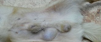
Most often we are talking about a lump on a cat’s stomach – a pathology of the abdominal cavity. Mechanically, bulging occurs when pressure from the inside exceeds the ability of the muscles to resist it from the outside. The same happens with inguinal, perineal, and diaphragmatic pathologies.
Causes of hernia in cats:
- Obesity, thinness
- Inactivity
- Pregnancy, childbirth
- Excessive loads
- Diseases of internal organs
- Age-related muscle thinning
- Injuries, consequences of surgery
- Congenital structural pathologies
- Constipation, urinary retention (pet straining)
Due to chronic diseases of the heart and respiratory system, a cough develops, which increases intra-abdominal pressure. The causes of intervertebral hernia in cats are poorly understood, but inheritance has been confirmed. Cranial diseases develop in utero, pulmonary diseases are a consequence of closed trauma or surgery.
The cause of umbilical hernia in kittens was previously thought to be the carelessness of the cat or the breeder. But it has been proven that the tension of the umbilical cord does not lead to expansion of the umbilical ring or protrusion of organs. Of course, this does not mean that you can break the rules of obstetrics.
Jacobs ulcer
Stomatitis
Anemia
Contrary to the myth, castrated and fertile cats suffer from inguinal hernias equally often. Changes occur with age or in animals suffering from constipation, inflammation of the anal glands - forced to push. An inguinoscrotal cyst can form if a gross mistake was made during castration, but this is a rare case for modern veterinary medicine.
Diagnosis of diaphragmatic hernia
If a veterinarian examines a cat with a suspected diaphragmatic hernia, they will still undergo a full examination. In one of the main areas they will look and listen, there is a chest. They will see if there are visible bruises around the chest area that indicate injury, and they will of course listen to the chest with a stethoscope.
Listening to the chest will give a very good indication along with the breathing rate (which will seem very high), the sounds in the chest will allow the veterinarian to know if the cat is having trouble getting enough air into the lungs.
They may take the cat for an x-ray, usually using only light sedation as they still want the cat to breathe for itself without full general anesthesia. A chest x-ray will provide a definitive diagnosis of a diaphragmatic hernia and a very good indication of how bad the hernia is.
Treatment of diaphragmatic hernia
Treatment for diaphragmatic hernia is surgery. The cat will be opened up so the surgeon can see inside and assess the extent of the damage. They will then repair the injury by carefully removing the organ causing the hernia, putting it back where it should be, and stitching up the hole. While the surgery is underway, the veterinary nurse will keep the cat stable under anesthesia and use a refrigeration bag to skillfully hold the cat while artificially breathing until the hole is repaired.
Before the cat is stitched up, the veterinarian will check that all other organs are in order and appear to have a good blood supply. In most cases, a needle is also inserted into the chest space before the cat wakes up and extra air is added so that there is negative pressure. By doing this, the cats' breathing will immediately improve.
Treatment, prognosis
The preferred treatment for hernias is surgery. It is quite simple, removal is possible already on the eighth day. The rehabilitation period in most cases passes without complications, the animal quickly returns to its usual way of life.
It is advisable to put a blanket on the cat so that he does not have the opportunity to lick the seams. To prevent infection, the operated area should be promptly treated with a disinfectant solution.
If discharge or inflammation of the sutures appears, you should inform the veterinarian who operated on the pet.
Small, ungrounded and non-hazardous formations, as a rule, are not operated on. These are mainly inguinal hernias. Alternative treatment is prescribed. The veterinarian repairs the hernia and then applies a fixing blanket, which the cat will have to wear for several months (from 1 to 3, based on the individual characteristics of the cat’s health).
If the reduction procedure is successfully performed, the hernia heals. However, it should be noted that this method is not entirely convenient. The owner will have to constantly ensure that the cat does not tear off the blanket and, if necessary, adjust the fixing device.
Diaphragmatic hernia requires a different approach to treatment. Compared to other types of neoplasms, the operation is more complex, involving opening the chest and returning all incorrectly located organs to their place.
The veterinarian sutures the diaphragm, then performs its plastic surgery, applying a special mesh or his own tissue. If organs are injured, they are resected. At the initial stage of an intervertebral hernia, drug therapy with anti-inflammatory and painkillers cannot be ruled out.
The animal's physical activity should be limited. If surgery cannot be avoided, the veterinarian removes part of the vertebra along with the prolapsed disc and in the area of the tumor. After surgery, the cat will need complete rest. For large-scale spinal cord injuries, euthanasia is indicated.
The prognosis is favorable in most cases. The main thing is to start the treatment process in a timely manner.
Forecast
Once the surgery is over, most cats will be absolutely fine. It will take some time for the injuries to heal, and during this time the cat must be kept calm by the owner - and definitely not allowed to jump anywhere! If the injury was caused by a car accident, another injury may also take several weeks to heal—especially in the case of broken bones.
Unfortunately, sometimes the injuries are too severe, and even with surgery, some cats are too poor to survive general anesthesia, especially if they have a suspected diaphragmatic hernia, which complicates anesthesia.
If you suspect your cat has been in an accident or is having trouble breathing, contact your veterinarian as an emergency.
Good day! Cat Balu was adopted from the street when she was 3 months old, 2 years old, fed 2 grades of raw beef, 2% curdled milk, rarely cottage cheese, chicken heart or raw stomachs, fed wet Whiskas for a week before illness (I live in the village, I couldn’t find anything else) 12/3/2017 in the evening I ate a piece of boiled mackerel, after a couple of hours the cat became ill, vomited all night with loose stools, a terrible smell from the mouth and butt, stopped eating and drinking, on December 5th I took him to the regional veterinary hospital, the doctor suggested poisoning (there are no tests or diagnostics there) I injected it to relieve intoxication and stop loose stools. On December 6, there was again loose yellow stool mixed with blood and the smells were even worse, again at the veterinary hospital they gave the same injections, the condition did not change. On December 7, I took her to a regional private veterinary clinic, everything further written from the words of the doctor: An ultrasound showed a diaphragmatic hernia, volvulus, urgent surgery is needed, you will take her home in 3 days. I left the cat at the clinic (daily communication with the doctor by phone, the clinic is 130 km from home). On December 8, he was operated on; he recovered well from the anesthesia, responded to stimuli, had a normal temperature, and had loose stools. On December 9, the same condition, but the cat turned out to have a calicivirus infection, they administered serum (it’s not clear when), if they knew, they wouldn’t take him to the clinic, he will infect others, and during the operation the liver was scratched in 3 places, but it’s not scary. On the 10th without changes, on the 11th I asked for blood biochemistry, the answer is, you have nowhere to spend the money, I insist on an analysis, the answer is I don’t see the point in this, because no matter what the analysis shows, the treatment regimen will not be changed, to the question about calicivirus how it could be without an increase in temperature and the rest, the answer is that it can be asymptomatic. On December 12, the same condition; on December 13, he ate a little wet food and was gnawing on dry food; To the question why is it dry in this condition, the answer is, what do you have against dry food? On December 17, I took the terribly frightened cat home, hoping that everything would be explained and told, but the doctor ran back and forth. Upon discharge, the liver is enlarged, about this, the answer to all questions is that in a couple of days it will return to normal; I asked about the intestinal volvulus, the answer was no volvulus; questions about nutrition - feed with dry hosts, I insist on natural food, feed well with natural food and give dry food and the cat will be healthy, come back for vaccinations in 2 weeks. A doctor from the regional veterinary hospital called and asked about the cat, after everything that was said she had a lot of questions and misunderstandings , from practice with calicivirus and surgical operations, fatalities, where are the symptoms, how calicivirus was determined, what does it mean that the liver was scratched and why it is enlarged, the extract contains only the disease code and no more information. On December 18, I called the doctor and said that the cat was all the way home I sneezed at home too, prescribed Xylin drops for children, asked for something for the liver and a speedy recovery, prescribed 1|4 tablets of Corsil, Thymalin IM 0.5 ml once a day, and on December 18 and 19 I was injected with Refkin 2.5% 0.3 ml IM was prescribed upon discharge, I am following the recommendations. Today I don’t like his condition, temperature 38, limping on 2 hind legs, constant rumbling in the stomach, enlarged liver, gases with an odor, there was a slight, or rather deep smell from the mouth after discharge, now it has intensified, he pooped once a day, today 3, in the evening the feces are viscous with liquid, the fur sticks out and constantly asks for food, on day 1 I fed Poyal canin for stomach problems dry, 3 times 50 ml of chicken broth without salt, day 2 dry and wet for the stomach, beef broth and a little boiled beef; today the same dry, a little broth and in the evening wet Purina for digestion, since there is no food in 2 nearby cities. I cried every day while the cat was in the hospital, I really want to help him, tell me what to do, how to cure the liver and how to properly switch to natural food.
Hello! kittens usually have a complex of diseases, including parasites and infections; kittens are unlikely to have other liver diseases of a non-infectious nature. Stool analysis will allow you to exclude parasites, worms and isosporosis, and you need to start with antiparasitic treatment. It seems that the operation should have been postponed until the infection problems were resolved.
- Vox audita latet, littera scripta manet - Despite all the desire to help you, there is no way to treat in absentia.
Good day ! Thank you for your answer, let's go get vaccinated and get tested!
IrenaPolezh writes: Good day! Thank you for your answer, let's go get vaccinated and get tested!
Vaccinations cannot be given.
- Vox audita latet, littera scripta manet - Despite all the desire to help you, there is no way to treat in absentia.
*Add. expenses are paid. materials: yes
Add. Ambulatory fee is paid. reception: yes
When purchasing a puppy, all veterinarians recommend paying attention to the presence or absence of a hernia. So what are hernias, what are they, where do they come from, what to do with them? These are the questions that potential owners and breeders ask themselves if they are faced with hernias in one way or another. A hernia is a temporary or permanent prolapse of internal organs through a natural or pathological opening with protrusion of the membrane lining the anatomical cavity. A hernia has its own structure: a hernial ring (opening), through which a hernial sac protrudes, consisting of the peritoneum, transverse abdominal fascia and hernial contents (intestines, omentum, stomach or other organs, tissues).
DIAPHRAGMAL HERNIA
in animals, this is a displacement of the abdominal organs into the thoracic cavity, through acquired or congenital damage to the diaphragm. Since the pressure in the abdominal cavity is stronger, the diaphragm bulges towards the chest cavity, therefore the organs are displaced into the chest cavity, and not vice versa (Fig. 9). In addition to separating the cavities, the diaphragm is a support for adjacent organs, and also performs a dynamic function: respiratory (participation in breathing), cardio-vascular, motor-digestive, lymph circulation. In addition, it is the diaphragm that is responsible for inhalation; at rest it provides up to 90% of the tidal volume. Diaphragmatic hernias are divided into traumatic and congenital.
Figure 9 - Diaphragmatic hernia: 1 - small intestine; 2 - liver; a - heart; 4—diaphragm; 5 - gallbladder; c - stomach.
Congenital diaphragmatic hernia
can be pleuroperitoneal or pericardio-pleuroperitoneal. In general, congenital pleuroperitoneal hernias are rare, and puppies with large diaphragmatic defects usually die at birth or shortly thereafter. Congenital pericardio-pleuroperitoneal diaphragmatic hernia is a more common disorder, to which Weimaraner dogs and Persian cats are most susceptible.
With a sliding hernia
due to weakening of the esophageal-diaphragmatic ligament, part of the esophagus and stomach are displaced upward - into the mediastinum. In this case, the fold of the peritoneum forms a hernial sac. The main complication of such a hernia is the straightening of the angle between the esophagus and the stomach, which causes the development of reflux esophagitis (reflux - reflux; esophagitis - inflammation of the esophagus). Sliding hernias are not strangulated.
For paraesophageal hernia
– the cardiac section is fixed, the fundus of the stomach, intestine or omentum next to the esophagus moves into the chest cavity through the dilated esophageal opening. This type of hernia can be strangulated, manifested by pain and signs characteristic of impaired movement of food through the stomach (vomiting, nausea).
Traumatic hernias
are a consequence of open and closed mechanical damage to the diaphragm. Open - develop when a wounding object passes through the chest and abdominal cavity and, as a result, through the diaphragm. Closed - formed during an impact, a fall, an accident, or a sharp increase in intra-abdominal pressure.
Why is diagnosing diaphragmatic hernias difficult?
Clinical symptoms of traumatic diaphragmatic hernias vary; they cannot be seen from the outside. About half of diaphragmatic hernias (35-50%) are accompanied by severe respiratory symptoms: rapid or impaired breathing up to attacks of suffocation, cyanosis of the mucous membranes and tongue. This condition is also characterized by a sunken abdominal wall during inspiration and a decrease in shortness of breath, which is clearly manifested if the animal is lifted by the front of the body. The condition worsens when the animal goes down the stairs. To confirm this diagnosis, it is necessary to take an x-ray of the chest and abdominal cavities (Fig. 10), including with a contrast agent (Fig. 11), ECG, and ultrasound of the abdominal organs.
Figure 10 – X-ray of a cat (gas-filled intestinal loops are visible in the chest cavity).
Figure 11 - X-ray images with contrast agent (intestinal loops in the chest cavity).
Why is it necessary to do an x-ray if you suspect a diaphragmatic hernia?
On an x-ray, you can determine: discontinuity of the diaphragmatic contour, the contents of the abdominal cavity inside the chest, displacement of the thoracic structures, displacement of the abdominal organs, divergence of the legs of the diaphragm. The difficulty with x-ray examination is that prolapsed organs may spontaneously return to the abdominal cavity.
In what cases is ultrasound diagnostics performed?
Ultrasound examination is performed in cases when:
- herniation of the contents of the abdominal cavity penetrates into the chest cavity through a defect in the diaphragm;
- on thoracic radiographs, pleural effusion can hide the diaphragmatic-hepatic silhouette and abdominal hernias;
- a diaphragm rupture occurred, i.e. with loss and interruption of the normal echogenic line (pleuropulmonary interface);
- The contents of the abdominal cavity can be seen through the defect and the chest.
Diaphragmatic ruptures are often accompanied by pleural effusion. In congenital periteneopericardial diaphragmatic hernia, the appearance of abdominal viscera adjacent to the heart within the pericardial sac and loss of diaphragmatic contour near the midline are considered diagnostic.
What is the treatment for diaphragmatic hernia?
Hiatal hernias are characterized by relaxation and can be sliding or paraesophageal. Most hiatal hernias are treated conservatively. Surgical treatment is resorted to when conservative therapy is ineffective, in the event of complications and paraesophageal hernias.
Principles of conservative treatment:
- prevention of reflux of gastric contents into the esophagus;
- reducing the acidity of gastric juice;
- drug protection of the inflamed mucous membrane of the esophagus;
- treatment of concomitant diseases that provoke the development of a hernia.
All traumatic diaphragmatic hernias, due to the risk of strangulation, must be treated surgically, which is carried out immediately after stabilizing the patient. In this case, preoperative preparation using intensive care methods is very important. We try to examine the animal as quickly and completely as possible before the operation (CBC and LBC, ultrasound, ECG), we recommend following a fasting diet for at least 8 hours.
ATTENTION.
The intervention is accompanied by the opening of the chest, which requires tracheal intubation and artificial ventilation.
The success of the operation
depends
30% on the quality of the operation itself and 70%
on postoperative care
. The animal must be left in the hospital for intensive care and recovery for 7-14 days, where the animal will be under the constant supervision of a doctor, and the surgeon’s orders will be regularly followed.
Is anesthesia used for hernia repair?
Of course, the animal is operated on under general anesthesia. Only in some cases with reducible umbilical hernias is it possible to use local anesthesia.
How to care for an animal after a hernia repair?
You will need to inspect the seam daily, treat it with antiseptics, and prevent it from being damaged. To do this, the animal must be put on a special postoperative blanket, and if the animal tries to reach the sutures, then a surgical collar. Food should be dietary, easily digestible and soft. It is necessary to limit movement for a while (do not run, do not jump, do not help activate the pet).
What determines the cost of the operation?
During the intervention, some difficulties may arise, for example, if the hernia is not reducible, the doctor will have to disconnect the internal organs from the hernial sac; if the hernia is strangulated, then resection of the necrotic intestine may be required. Additional manipulations require more time and consumables.
The diaphragm is a thoraco-abdominal barrier that is attached to the edges of the ribs, forming a dome, the convex part of which faces the chest cavity. The diaphragm is a powerful inspirator; it is involved in the work of the abdominal press, influencing the flow of lymph and blood through the vascular highways of the abdominal cavity. It has an opening for the aorta, esophagus and caudal vena cava, thus separating the thoracic and abdominal cavities.
What is a diaphragmatic hernia and the causes of this pathology?
Diaphragmatic hernia in dogs and cats is a pathological condition in which abdominal organs acquire the ability to move into the chest cavity through a damaged diaphragm.
Causes of the defect:
- birth defect;
- traumatic defect.
I. Traumatic:
These are the most common types of hernias found in veterinary practice. They arise due to mechanical damage to the diaphragm (open or closed).
Open ones develop in cases where trauma occurs to the chest and abdominal cavity and, as a consequence, to the diaphragm itself.
Closed ones arise from impacts received during a fall, an accident, and are also formed in the event of a sharp increase in intra-abdominal pressure.
Clinical signs of this form of diaphragmatic hernia in dogs and cats are different and can be attributed to the respiratory tract or gastrointestinal tract.
II. Congenital:
This pathology can be detected at any age, as there are often no symptoms.
Types of congenital diaphragmatic hernia:
1. Pleuroperitoneal. Congenital pleuroperitoneal hernias are very rare; in most cases, animals with significant defects of the diaphragm die at birth or shortly after it.
2. Pericardio-pleuroperitoneal. They occur quite often. The pathology is most often observed in Weimaraner dogs and Persian cats.
3. Sliding. It occurs as a result of weakening of the esophageal-diaphragmatic ligament, in which part of the esophagus and stomach are displaced upward into the mediastinum. The fold of the peritoneum forms a hernial sac. In this situation, the natural closing mechanism of the esophageal-gastric junction is disrupted and reflux - esophagitis develops (reflux - reflux; esophagitis - inflammation of the esophagus). There is no strangulation of sliding hernias.
4. Paraesophageal. This form is characterized by fixation of the cardiac region and movement of the fundus of the stomach, intestine or omentum next to the esophagus into the chest cavity through the dilated esophageal opening. This type of hernia can be strangulated, cause pain and manifest itself with signs characteristic of impaired movement of food through the stomach (vomiting, nausea). As a result of the infringement, the trophism of tissues and organs is disrupted, which leads to irreversible changes and an unfavorable outcome.
Diagnostics
X-ray examination of the chest is the main one in making the diagnosis. A diaphragmatic hernia in cats and dogs is always suspected when there is no clear line of the diaphragm. The presence of abdominal organs in the chest cavity confirms the diagnosis. Sometimes the presence of pleural effusion makes the diagnosis difficult and requires contrast studies.
Contrast radiography of the upper gastrointestinal tract confirms the diagnosis if part of the stomach or intestines is observed in the chest cavity.
To confirm a rupture of the diaphragm, peritoneography is performed, for this a contrast agent is injected into the peritoneal cavity, after which an X-ray of the abdominal and chest cavity is performed and the presence of a contrast agent in the chest cavity indicates a defect in the diaphragm.
The only treatment is surgical reduction of the hernia and suturing of the diaphragm defect. When a patient is admitted with a suspected diaphragmatic hernia, the necessary diagnosis and stabilization of the animal is carried out before surgery. If the patient cannot be stabilized, emergency surgery is performed. The surgical intervention is carried out in a veterinary clinic equipped with a ventilator.
Thus, if your pet was injured in a fall, a sharp blow, or was involved in an accident, you must urgently contact the clinic and rule out this pathology. The prognosis directly depends on the promptness of seeking qualified help.
A diaphragmatic hernia in cats develops when organs are displaced from the abdominal cavity into the thoracic cavity. This happens when the diaphragm is damaged or due to its congenital defect. The diaphragm itself is a thin film (muscle-tendon), a partition that separates the abdominal and thoracic cavities across.
Ultrasound
Ultrasound examination is performed in cases when:
- herniation of the contents of the abdominal cavity penetrates into the chest cavity through a defect in the diaphragm;
- on thoracic radiographs, pleural effusion can hide the diaphragmatic-hepatic silhouette and abdominal hernias;
- a diaphragm rupture occurred, i.e. with loss and interruption of the normal echogenic line (pleuropulmonary interface);
- the contents of the abdominal cavity can be seen through the defect and the chest;
Traumatic ruptures of the diaphragm are often accompanied by pleural effusion. In congenital periteneopericardial diaphragmatic hernia, the appearance of abdominal viscera adjacent to the heart within the pericardial sac and loss of diaphragmatic contour near the midline are considered diagnostic.
Methods for examining diaphragmatic hernias in animals:
- Esophagogastroscopy
- Biopsy of the esophageal mucosa
- Study of acidity in the esophagus
- X-ray of the stomach
Treatment of diaphragmatic hernias in animals
Hiatal hernias are characterized by relaxation and can be sliding or paraesophageal. Mostly, hiatal hernias are treated conservatively (by gastroenterologists). Surgical treatment is resorted to when competent conservative therapy is ineffective, in case of complications and in case of paraesophageal hernias.
Principles of conservative treatment:
- Prevention of reflux of gastric contents into the esophagus
- Reducing the acidity of gastric juice
- Drug protection of inflamed esophageal mucosa
- Treatment of concomitant diseases that provoke the development of a hernia
All traumatic diaphragmatic hernias, due to the risk of strangulation, must be treated surgically, which is carried out immediately after stabilizing the patient.
In this case, preoperative preparation using intensive care methods is very important.
Types and characteristics of diaphragmatic hernias
- closed (bumps, falls, road accidents, sharp increase in intra-abdominal pressure);
- open (when, together with the diaphragm, the integrity of the surrounding tissues and bones is disrupted).
Clinical signs of damage are very different and depend on the cause, on which organs and tissues are injured. The respiratory and digestive systems are most often affected.
With a congenital diaphragmatic hernia, there are:
- pleuroperitoneal, which are rare and kittens with similar anomalies die in the first week after birth;
- pericardio-pleuroperitoneal, the most commonly diagnosed injuries in the practice of veterinary surgeons, Persian cats are predisposed to them;
- sliding, develop with weakness of the esophageal-diaphragmatic ligament, while the stomach and esophagus, having lost support, move into the mediastinum. A hernial sac is formed from a fold of the peritoneum and, as a result, the sphincter between the stomach and esophagus does not close well (reflux esophagitis);
- paraesphageal, the fundus of the stomach, omentum and part of the intestine are moved through the dilated esophageal opening into the chest cavity with simultaneous fixation of the cardiac section. The main difference is the possibility of strangulation, pain, nausea and vomiting. In the absence of quick help, the strangulated tissues quickly die, which is irreversible.
Traumatic injuries can cause pneumothorax (air in the chest cavity) or hemothorax (collection of blood). As a result, breathing is impaired until critical situations arise when the cat needs help immediately.
Types of pathologies
There are two categories of hernias - congenital and acquired. The latter in adult cats are formed against the background of injuries (blows, falls) and malfunctions of the digestive system associated with an improper diet.
Based on location, there are several types of hernias:
- Umbilical. In most cases, it is congenital, but there are also acquired ones, resulting from an improperly cut umbilical cord or pathology that has developed in the intestines. The most common type.
- Intervertebral (the rarest type). Typical for old cats (over 15 years old). Modern medicine has the possibility of treatment with medications (subject to timely diagnosis).
- Inguinal. Formed due to flatulence and frequent constipation. Less dangerous for cats than for cats.
- Diaphragmatic. Causes: mechanical damage, trauma. Characterized by the transition of organs from the abdominal cavity to the chest.
- Perineal. The location is between the bladder and rectum. Since pinching does not occur, the tumor is not operated on, but reduced.
- Pericardial-peritoneal. A congenital and extremely rare species. With this diagnosis, kittens do not survive, since the hernia puts pressure on the heart, which provokes heart failure and pulmonary edema.
Regardless of the type of disease, it cannot be ignored: if it progresses, it causes a purulent abscess and, as a consequence, the death of the animal.
What symptoms should alert the owner?
If the organs of the abdominal cavity are shifted through the hole into the chest cavity, then compression of the lungs inevitably occurs, and also the organs themselves, pinched in the diaphragmatic hernia, have their blood circulation disrupted.
The following signs should alert a cat owner:
- obvious breathing problems;
- a superficial, shallow sigh;
- the cat deliberately extends its neck and head to improve air passage.
Diagnostic measures necessarily include radiography. If there is no clear line of the diaphragm on the image, the veterinarian always suspects a diaphragmatic hernia. Visualization of the internal organs in the thoracic region, which anatomically should be located in the abdominal cavity, only confirms the diagnosis.
Contrast studies are necessary if there is pleural effusion. Peritoneography is performed to confirm rupture of the diaphragm; if there is damage, then the contrast agent will be in the chest cavity. Ultrasound is prescribed as an auxiliary study.
What to do: what treatment is provided
There is no drug treatment for diaphragmatic hernia; drugs are prescribed as auxiliary ones, but the main therapy is only surgical. Moreover, the faster the cat owner delivers the pet to the clinic, the higher the chance of a successful outcome.
The surgical operation consists of suturing the hernial opening in the diaphragm. If stabilization of the animal before the intervention is impossible, then an emergency operation is performed using a ventilator and the cat’s further stay in the intensive care unit and hospital of the veterinary clinic.