The most common types of dermatitis in dogs
Since there are many causes of dermatitis and their combinations, the disease can be of different types. Each of them is characterized by its own distinctive symptoms and treatment approaches. The most common types of dermatitis in dogs are briefly described below.
Pyotraumatic dermatitis in a dog
Pyotraumatic
With pyotraumatic dermatitis in dogs, the infection penetrates from the surface into the deeper layers of the skin, leaving behind plaques and a thickened stratum corneum. The deeper it penetrates, the more papules (pimples) and pustules (pustules) will appear on your pet’s skin.
Treatment consists of using antibacterial drugs.
Allergic
Allergic dermatitis in dogs
Allergic dermatitis is considered the most common in dogs, since any object can be an allergen. Experts note that in most cases, skin allergies are provoked by external parasites, food and cosmetic care products. Symptoms include: severe itching, redness of the skin, scratching, rashes, swelling.
The basis of treatment is elimination of the allergen and symptomatic therapy.
Atopic
Atopic dermatitis
Atopic dermatitis is one of the varieties of the previous form. The clinical picture is the same: dry epidermis, rashes, very severe itching. Additional characteristic signs include the development of otitis media and pododermatitis. The pathology develops mainly in dogs under 5 years of age who have similar heredity. It is noted that atopic dermatitis “loves” such dog breeds as: dachshunds, sharpeis, bulldogs, setters, Dalmatians and a number of others. At the same time, anything can become an allergen.
Treatment consists of identifying and eliminating the allergen, carrying out symptomatic therapy, and preventive measures to prevent otitis media.
Flea
Flea dermatitis in dogs
The salivary secretions of fleas contain more than a dozen allergenic components that lead to flea dermatitis in dogs. The severity of symptoms depends on the individual susceptibility of the animal. Signs of pathology can be: alternating wet areas and areas covered with crusts. In the chronic course of the disease, the pet exhibits excessive skin pigmentation on the abdomen and hair loss. It is characteristic that the dog’s ears, muzzle and paws are almost not affected.
Treatment consists of getting rid of fleas and symptomatic therapy. Prevention is of great importance for recovery - the use of a parasite collar, disinsection of the environment in which the dog lives.
Interdigital dermatitis in a dog
Pododermatitis
Interdigital dermatitis in dogs, or pododermatitis, develops between the animal's toes, mainly on the front legs. If the lesions are single, it can be assumed that the disease is provoked by injury. With extensive damage to the limbs, the cause may be insects, allergies, or infections. Symptoms: bleeding nodules, boils, pain leading to lameness.
Treatment is symptomatic.
Seborrheic
Seborrheic dermatitis in dogs
Seborrheic dermatitis is caused by genetics. With this disease, the process of keratinization of the skin accelerates, which is visually manifested in numerous scales, oily epidermis, but dry hair. Your pet may have brittle nails; The inflammatory process on the skin is clearly visible, accompanied by severe itching. In the future, the disease is complicated by secondary infection with all the ensuing consequences.
Treatment is carried out in two directions: local therapy and a systemic approach.
Acral
Acral dermatitis in dogs
Symptoms of acral dermatitis are dense, ulcerated areas of skin that occur as a result of the dog licking the area excessively. Pathological behavior of an animal can be caused by allergies, parasites, infection and other factors that must be detected and eliminated before treating the skin.
Treatment consists of the initial elimination of the provoking factor and a therapeutic effect on the damaged epidermis. Limiting your pet's access to the itchy area is of great importance. In addition, antidepressant and psychotropic drugs are used in therapy.
Dermatosis: symptoms and diagnosis
Skin pathology associated with an imbalance of sex hormones should be distinguished from skin diseases of other origins. “Hormonal dermatosis” is characterized by:
- alopecia, appearing first in the groin, abdomen, thighs, moving to the neck, back and sides;
- sometimes with hypoestrogenism, bald spots on the sides remain the only symptom, while in sterilized females it takes on a seasonal appearance;
- the coat is dry, brittle, thinned and soft;
- seborrhea, pyoderma (pustules on the skin), itching, the formation of comedones with areas of hyperpigmentation, as well as the development of ceruminous otitis media;
If the production of testosterone and estrogen is poor, urinary incontinence may occur. All these symptoms “suggest” that dermatosis is not only a consequence of improper feeding, autoimmune dysfunction or the action of skin parasites. Therefore, self-medication of a dog is unacceptable.
The examination excludes hypercortisolism, hypothyroidism, and dermatosis, which can be relieved by the administration of growth hormone. They also differentiate from allergies, disorders of keratinization of the dermis, and in dachshunds – with genetically transmitted alopecia.
About the correct selection of shampoo for seborrhea
It is important to know how the individual components of the shampoo work and whether they have any synergistic effects, since the overall effectiveness of the drug depends on this. When choosing a detergent that can be used to treat seborrhea, we focus on the following factors:
- Moderate activity of the sebaceous glands, the pet's coat looks almost normal.
- Noticeable secretion of sebum and visible oiliness of the coat.
- There is a clearly visible layer of grease and scabs on the skin, and the fur is very greasy.
- The animal emits an unpleasant odor, the fur is matted, greasy and oily.
It is very important for the owner to inform the veterinarian exactly what stage of seborrhea is observed in his dog. In moderate cases, mild shampoos with a low content of antibacterial and antifungal agents, with a pronounced moisturizing effect, are needed
They are good because you can bathe a sick dog every day. Such products almost do not wash away the natural protective layer of the skin, which speeds up and facilitates the healing process.
The prognosis for seborrhea depends on the severity of the disease, the general condition of the dog after completion of treatment, and other factors. If the root cause of the disease has been accurately identified and eliminated, the chances of a relapse will be minimal. If you or your veterinarian have doubts about the completeness of the cure, you will need to show your pet to a specialist at least once a quarter. Some diagnostic tests may be necessary (especially if seborrhea was caused by hormonal imbalances).
Main characteristics and types of disease
Dermatitis in dogs is a special type of allergic reaction of the body to the penetration of a foreign body or substance into it. The inflammatory process affects not only the epidermis, but also penetrates into the deeper layers of the skin. The disease is characterized by redness of the skin, the formation of ulcers, wounds, and erosions.
With dermatitis, areas of the dog's skin become inflamed, erosions, ulcers and wounds form.
The intensity of manifestations depends on the type of dermatitis:
- Allergies are the most common. It is a reaction to food products, especially often a reaction to dry food of a low price category, such as Darling. Contact with various chemicals can also cause an allergic reaction. Allergies develop from flea or tick bites.
- Atopic is a type of allergic dermatitis that occurs against the background of autoimmune diseases. predisposition to it is laid down at the genetic level.
- Mechanical – the most easily curable, develops after injury or scratching of any area of the skin. Most often, mechanical dermatitis develops after wearing a low-quality collar or due to synthetic bedding.
- Contact – skin reaction to a chemical or physical irritant. Inflammation develops from constant touching of an object or when a chemical comes into contact with the skin. Contact dermatitis is caused by dog grooming products that are used incorrectly.
- Medicinal - develops when a dog is treated with any drug, the individual components of which are intolerable to the animal.
- Sunny – localized on the dog’s face. This type of dermatitis mainly affects dogs with lightly pigmented skin.
- Parasitic – the cause of dermatitis is fleas, nematodes or worms.
- Infectious and fungal - are complications after skin injuries against the background of weakened immunity. Most often, inflammation is caused by staphylococcus, which lives on the skin of any dog. If a fungal infection is added to it, quite severe inflammation develops.
First aid
When treating dermatitis in dogs at home without first consulting a veterinarian, one cannot expect positive results. The fact is that it is necessary to find out the cause of the disease, eliminate it and only then deal with the symptoms. Without fulfilling this condition, the treatment will have no result.
What can the owner do? Contrary to recommendations found on the Internet, you should not influence the source of dermatitis: cut the hair, lubricate the skin with brilliant green or iodine. The true picture of the disease is lost from this, and it will be more difficult to make a diagnosis.
A smaller restriction is imposed on flea dermatitis when the owner discovers a lot of fleas on the pet. Here it is enough to carry out the treatment for parasites yourself, wash the wounds with Chlorhexidine, and in case of severe itching, put a protective collar on the dog to prevent scratching.
In case of contact dermatitis, when the breeder clearly understands that the cause of the inflammation was an uncomfortable collar, another thing, or the pet’s habit of rubbing against an object, it is enough to eliminate the cause. With minor damage, everything will heal on its own.
In other cases, contacting a veterinary clinic is mandatory!
Drug treatment
The doctor’s task is to normalize the state of the dog’s immune system and cure skin lesions, preventing secondary infection.
Medications are quite often prescribed for flea dermatitis, both for indoor and outdoor dogs.
Along with insecticidal treatment and strengthening the dog’s immunity, the doctor may prescribe:
- A glucocorticosteroid, usually prednisolone at a dosage of 0.5-1 mg per kilogram of weight. Orally. Duration - from seven to ten days. Then the dosage is gradually reduced. The duration of taking the medicine depends on how successfully the owner fights fleas. The faster you can get rid of insects, the shorter the course of prednisone will be.
- In some cases, when oral administration of prednisolone is not possible according to indications, glucocorticosteroids are prescribed in the form of ointments for external use.
- For advanced forms and secondary infection of the skin, antibacterial ointments are used.
Attention: the veterinarian must take into account the individual characteristics of each animal: age, weight, general physical condition, the presence of other disorders and diseases. Therefore, it is unacceptable for dog breeders, based on personal experience, to independently prescribe treatment for their own or other people’s pets!
Treatment of dermatitis
When visiting a doctor, first of all, you must undergo all the necessary diagnostic measures. The success of therapy depends on the correctness of the diagnosis. To provide an accurate diagnosis, the following are prescribed:
- examination of skin scrapings under a microscope and bacteriological culture;
- determination of the specific sensitivity of bacterial pathogenic microorganisms to antimicrobial drugs;
- leukocyte formula studies;
- examination of stool, urine and blood.
After establishing the cause, the doctor selects the appropriate treatment regimen, its duration and the number of drugs. Two types of treatment are used - medication and physiotherapy.
To facilitate access to the affected areas, the hair is cut off and dead tissue, purulent exudate and crusts are removed using a special swab soaked in antiseptic. The damaged area is dusted with a special disinfectant powder. To isolate the wound, bandages are used, often impregnated with antifungal, anti-inflammatory and antibacterial ointments. This allows the animal to speed up the healing process.
Drug treatment for dermatitis in dogs includes:
- Use of antibiotics for a course of 5 days to 2 weeks. This is an important point to prevent the occurrence of secondary infection. Antimicrobial agents of the penicillin and cephalosporin group are widely used. A treatment course is carried out under the strict supervision of the attending physician, with the use of ointments for dogs based on antibiotics, which promote rapid healing of wound surfaces.
- Skin lesions caused by the action of fungal microorganisms require the use of fungiostatic drugs. In addition, the pet’s skin must be washed using special shampoos that destroy the fungus - Nizoral, Clotrimazole, Ketoconazole.
- To eliminate itching and discomfort in allergic dermatitis, systemic antihistamines are used. The drugs that have proven themselves to be excellent are Suprastin, Diphenhydramine, Fenistil.
- Drug therapy includes increasing the body's defenses by using systemic immunostimulating drugs that increase the activity of T cells. Gamavit, Gamapren, Glycopin are actively used in veterinary medicine.
- The destruction of parasites is carried out by using acaricidal and insecticidal preparations of systemic importance.
Dermatitis of various etiologies can be effectively treated using physiotherapeutic procedures. Irradiation is carried out with ultraviolet lamps and lamps with infrared light. The use of physiotherapy in combination with medicinal methods can significantly increase the rate of restoration of epithelial cells, disinfecting the surface and reducing the amount of purulent exudate released.
Basics of treatment of hormonal dermatoses
Therapy for dermatoses associated with improper functioning of the hormonal system is primarily aimed at eliminating the root cause. Dealing with symptoms alone will not bring results and will be a waste of time.
The dog owner must contact the RosVet veterinary clinic. Only specialists will be able to establish the true cause of visible skin lesions and draw up a treatment regimen. Self-treatment with hormones is unacceptable, as this can completely disrupt the endocrine system and provoke irreversible changes in the dog’s body.
Some treatment points:
- castration is carried out for testicular tumors or dermatosis, which can be relieved through surgery. Treatment with estrogens is discontinued;
- for dermatosis that can be relieved with estrogens, sterilized females are prescribed diethylstilbestrol in a regimen of 2-3 weeks, with a break of 1 week. This treatment is carried out until normal hair growth occurs. Continue to administer the drug as maintenance therapy 2-3 times a week. Testosterone or milbolerone can be substitutes for diethylstilbestrol if it is ineffective.
This is just a minor example of assignments. The exact names of drugs, methods and duration of treatment are determined by a veterinary dermatologist or endocrinologist.
Owners of male cryptorchids need to remember that such animals are castrated and not allowed for breeding. Since their descendants will inherit similar pathology and problems associated with the endocrine system.
If your dog has alopecia (bald spots) or has skin problems, do not self-medicate! Call the RosVet VC by phone: +, 24 hours a day. Make an appointment with a dermatologist and endocrinologist!
Main types of dermatitis
Dermatitis is classified depending on the factor that caused its development:
- traumatic - occurs at the site of bruises, injuries, open wounds and scratching of itchy skin by the animal itself;
- allergic - its causes may be mites, poor-quality food, poplar fluff or plant pollen;
- contact – develops after direct contact of an animal with an allergen;
- atopic – caused by a combination of factors, including genetic predisposition;
- flea infestation – occurs after an animal comes into contact with parasites.
Dermatitis in dogs, the symptoms of which allow the disease to be classified, can be successfully treated in most cases. At the end of therapy, the skin is completely cleansed, the animal’s well-being improves, and the risk of relapse is reduced.
Allergic dermatitis: why is it dangerous for animals?
Allergic dermatitis is considered the primary source of some types of the disease.
Its main subtypes are contact, atopic and flea; the differences lie solely in the mechanism of development of the immune system's response to an irritant. In each of these cases, a specific allergen is present, although other factors can also cause the disease. The main places of localization are the ears and the folds of the paws. This type of dermatitis is very easy to confuse its symptoms with other types. Experts believe that it is also transmitted genetically.
Allergic dermatitis in dogs can develop slowly as the allergen is exposed. The danger of the disease lies in the risk of developing angioedema (in advanced cases) after bites from poisonous snakes, ticks or other insects. Rapid swelling of the animal's larynx or muzzle area can be fatal, so contact a veterinarian immediately.
Flea dermatitis: how to protect your animal from infection
Fleas settle on the dog and parasitize it for a long time.
They become the cause of the development of such an unpleasant disease as flea dermatitis. Insects bite through the dog's skin, and in this case their saliva becomes the allergen. The main places where insects are localized have high humidity (sacrum, groin, behind the ears, under the tail, stomach). The disease itself and the presence of fleas are two different things. If the dog is not prone to allergic reactions, a huge number of adults and their larvae may be hiding in its fur, but no symptoms of dermatitis are observed.
Getting rid of fleas does not guarantee that the insects will not attack the animal again. Eggs, larvae and pupae can survive on surrounding household objects, and after some time migrate again to the dog. After its treatment, it is necessary to carry out disinfestation of the apartment.
Flea dermatitis in dogs is a disease that can affect even a clean, well-groomed animal with a flea collar on its neck. As a preventative measure, you should inspect the animal’s fur daily in good lighting. Having noticed the first traces of new “settlers”, they immediately take measures to get rid of them.
Atopic dermatitis: how to recognize it by the first signs
When an animal becomes restless, begins to itch regularly, and loses the desire to move and play, it is worth carefully examining its skin.
If redness, baldness and hyperpigmentation are observed around the eyes, lips, groin and near the anus (up to acanthosis nigricans - darkening of areas of the epidermis in the folds, their thickening), this may indicate a disease such as atopic dermatitis in dogs. Animals aged from 6 months to 6 years are at risk. Among the provoking factors are:
- heredity;
- food allergens of protein origin;
- epithelial cells (both other animals and humans);
- fungal microspores;
- plant pollen during the flowering period;
- dust mites (accumulate in upholstered furniture, blankets and even in the air).
It is impossible to completely cure atopic dermatitis; sometimes it takes the entire life of a pet to fight the symptoms. The acute form of the disease is treated with medication, and after that it is necessary to take preventive measures.
About the disease
Dermatitis is an allergic reaction that provokes inflammation of the skin, but in the absence of pimples and rashes. Any exposure to external factors is perceived by the immune system as a danger, and a protective mechanism is launched to neutralize its effect.
Inflammation of the skin is a manifestation of the dog’s immune system struggling. It is most often diagnosed in pets from 6 months to 6 years. The main causes of dermatoses include: improper care, skin parasites, poor nutrition, internal diseases.
Dermatitis is often an autoimmune process, so therapy without stimulating the immune system will not be successful. It is atypical, with uncharacteristic manifestations. The dog owner will need to take into account everything necessary at home that will be required for the pet’s full recovery.
Causes of dermatitis – types of skin lesions
Skin inflammation can develop in a variety of pathologies. In some cases, this is an independent disease, manifested by a rash, erosions, ulcers, and purulent processes. But more often dermatitis is a symptom of various pathologies:
- fungal infections - microsporia, trichophytosis;
- parasitic diseases – ticks, fleas;
- pyoderma caused by various bacteria;
- allergic reaction.
Bruise, skin dissection, trauma and other mechanical damage lead to inflammatory processes. Injuries can be caused by other dogs (fights, fights between pack members), animals. Pets often injure themselves - scratching due to flea or tick infestation. In guard dogs, inflammation of the neck is common due to friction from the collar. Pododermatitis in dogs develops due to primary damage to the skin - resistance decreases, the area becomes susceptible to infections.
Another form of contact dermatitis involves inflammation due to exposure to a physical or chemical nature. Thus, in pets with thermal burns, skin lesions appear, but more often such a defect occurs in stray and guard dogs in the winter. Chemical irritation occurs when treating a pet for fleas or bathing it with different shampoos. Dermatitis of the interdigital fissure is often observed in winter due to the treatment of roads with ice reagents.
A common type of chemical dermatitis is inflammation after the thoughtless use of medications. This form of pathology is observed in dogs with individual sensitivity, as well as during self-treatment of pets. The disease appears both as a result of severe irritation (application of crystalline substances, tincture of iodine, a number of ointments to the skin) and in the form of an allergic form. Many antibiotics, vaccines, and steroid drugs cause hypersensitivity.
Inflammation of the dog's skin may be due to genetic characteristics. Atopic dermatitis in a dog develops when a number of immunoglobulins are formed after allergic substances enter the body:
- feed proteins;
- pollen;
- dust;
- vaccines, serums, biological products;
- animal epithelium.
Special cells of the animal's body perceive antigens and transmit them to lymphocytes, forming susceptible cell clones. An allergic immune reaction does not form on the first occasion of contact, but upon repeated exposure to the antigen. The reaction can develop in two ways - slowly (over several months with prolonged contact with the allergen), or rapidly - immediately after exposure to the substance on the body.
Dogs with white fur and fair skin may experience solar dermatitis. Under the influence of ultraviolet rays, sunburn appears, leading to disruption of keratinization - improper growth of the horny epithelium. The disease is caused by excessive exposure to hairless or light-colored skin.
Pyotraumatic dermatitis and intertrigo
Lyubov Nikolaeva, member of the European Society of Veterinary Dermatology (ESVD), resident of the European College of Veterinary Dermatology (ECVD), Bely Klyk veterinary clinic, Moscow The article uses photographs of the author
The most common secondary skin infections are caused by bacteria (in the vast majority of cases, staphylococci) and yeasts (usually Malassezia fungi). Inflammation caused by yeast fungi is limited to the surface of the skin, so the classification of yeast infections is usually based only on the distribution of infection by anatomical area (otitis media, pododermatitis, folds, etc.).
The classification of pyoderma affects several criteria. This is also distributed by anatomical areas (otitis, pyoderma calluses, pododermatitis, etc.), and by type of pathogen (staphylococci, Pseudomonas aeruginosa, etc.), and by the depth of inflammation (Table 1).
Table 1. Types of pyoderma
| Skin surface pyoderma |
| Limited to the surface of the stratum corneum |
|
| Superficial pyoderma |
| Hair follicle lumen and epidermis |
|
| Deep pyoderma |
| Rupture of the hair follicle, inflammation in the dermis and subcutaneous fat |
|
In this article we will look at types of inflammation that involve only the surface of the skin. This category includes pyotraumatic dermatitis and intertrigo.
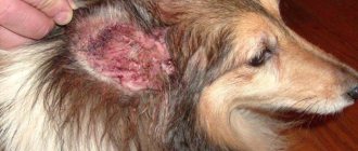
Pyotraumatic dermatitis
Pyotraumatic dermatitis, or acute wet dermatitis, or “hot spot” is mistakenly called weeping eczema by animal owners, and sometimes by doctors.
Pyotraumatic dermatitis is a self-induced traumatic skin disease that is quite common in dogs and very rare in cats. Lesions develop quickly and are soon complicated by secondary bacterial infection. The basis, as a rule, is a disease that causes itching (allergies, ectoparasites). In this case, the characteristic lesions are the result of licking, scratching or gnawing. Similar problems can also occur against the background of other skin injuries - sometimes after grooming or injury by another animal.
Clearly limited inflamed foci appear, often isolated. Severe exudation occurs, hair sticking together with sticky exudate. In most animals, the lesions can be quite painful. In many cases, a minor injury to the skin is sufficient to provoke such inflammation, which is surprising, since in many animals even more serious injuries never lead to the development of pyotraumatic dermatitis.
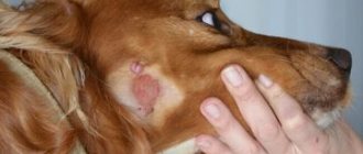
In most cases of pyotraumatic dermatitis in dogs in the trunk area, the primary cause of itching is a parasitic disease. First of all, this is allergic flea dermatitis. Sarcoptic mange or lice may also be the cause. For flea allergic dermatitis, the appearance of foci of pyotraumatic dermatitis in the croup area is more typical. In cats, lesions have been described that are clinically and histologically similar to similar skin inflammation in dogs, also associated with allergy to flea bites.
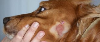
Lesions that occur in dogs in the cheek area and lateral neck are usually associated with otitis media, so ear otoscopy should be performed.
| Photo 1. Severe exudation with pyotraumatic dermatitis | Photo 2. Pyotraumatic dermatitis in a dog with itching in the muzzle area |
| Photo 3. Pyotraumatic dermatitis in a dog with otitis media | Photo 4. Pyotraumatic dermatitis at the site of injury during play with another dog |
Dogs of large breeds with dense coats, such as German shepherds, golden retrievers and Labradors, collies, Bernese Mountain dogs, etc., are more prone to developing pyotraumatic dermatitis. No age or gender predisposition was identified. There is a pronounced seasonality in many cases of such skin inflammation - more often in the warm season, with high humidity.
Pyotraumatic dermatitis clinically manifests itself as very characteristic lesions, so differential diagnoses in classic cases are not usually considered. Although it was noted that such exudation was sometimes observed in skin neoplasias accompanied by severe inflammation and ulceration (lymphoma, sweat gland adenocarcinoma). Cytological examination of exudate helps to assess the type of bacteria and assess the degree of complication of secondary infection.
The longer pyotraumatic dermatitis exists, the more likely it is that inflammation involves not only the surface of the skin, but also the follicles, epidermis, and dermis, i.e. pyoderma, superficial or deep, already appears. It is not always possible to recognize this by clinical signs, since the surface of the lesion is highly inflamed and the depth cannot be assessed unless there are clear signs of pustular dermatitis or fistulas. Most often, it becomes clear that the inflammation is not as superficial as it seemed at first glance when assessing the response to treatment. Inflammation of the surface of the skin, as a rule, can be brought under control fairly quickly, but deeper pyoderma requires long-term treatment with systemic antibiotics. One study (Holm, 2004) found folliculitis on histological examination in 45% of lesions clinically characterized as pyotraumatic dermatitis.
When treating pyotraumatic dermatitis, it is important to clear the lesion of hair and exudate to make it easier to treat and dry the inflamed area. In some cases, the pain is so severe that the treatment is performed under sedation, or general anesthesia, or at least local anesthesia. A course of nonsteroidal anti-inflammatory drugs is often prescribed at the beginning of treatment. Cleaning the surface with antiseptic agents is one of the first stages of treatment. Chlorhexidine 1–2% or povidone-iodine solution is most often used as an antiseptic. During initial treatment, a solution of hydrogen peroxide can be used to eliminate the main exudate, but then the above-mentioned antiseptics are used. If possible, the affected area is cut off, including the surrounding unaffected area. Difficulties arise with inflammation in show dogs, which need to preserve their coat as much as possible. In such a situation, shampoos based on benzoyl peroxide or chlorhexidine 4% can be used to wash the lesion.
Glucocorticoids are often used instead of non-steroidal anti-inflammatory drugs, locally or systemically. If there are no contraindications, then you can apply locally an ointment or spray with glucocorticoids or a course of prednisolone 0.5–0.25 mg/kg for 7–10 days.
This helps well in situations where a secondary bacterial infection has not yet significantly complicated the inflammation, or it is truly pyotraumatic dermatitis, and not folliculitis or furunculosis. However, as mentioned earlier, external signs can rarely distinguish inflammation of the surface of the skin from deeper types of pyoderma that may have developed at the site of the lesion. If there are signs of papules or pustules around the lesion, it is recommended to regard the inflammation as pyotraumatic folliculitis or even furunculosis. In such cases, the use of glucocorticoids may, on the contrary, favor bacterial infection rather than help eliminate inflammation. Also, if inflammation exists for a long time, it should also be treated as folliculitis or furunculosis.
In addition to eliminating the skin inflammation itself, it is necessary to pay attention to identifying and eliminating the root cause of pyotraumatic dermatitis, otherwise relapses are very likely.
| Photo 5. Pyotraumatic dermatitis after active grooming | Photo 6. Pyotraumatic dermatitis after active grooming, enlarged image |
| Photo 7. Pyotraumatic dermatitis during treatment |
Intertrigo (inflammation of skin folds)
Intertrigo develops in areas of skin folds due to friction of skin surfaces against each other, heating, maceration, and poor air circulation. Inflammation of the folds is classified according to anatomical location (Table 2).
Table 2. Types of intertrigo
| Intertrigo |
| Lip folds |
| Facial fold (nasal or infraorbital) |
| Tail fold |
| Vulvar fold |
| Obesity folds |
| Neck fold (ventral part in breeds such as Shar Pei, Basset Hound, Mastino Napolitano, etc.) |
Dogs of certain breeds, for which folds are a characteristic feature, are more often susceptible to inflammation of the folds (Shar Pei, Pekingese, English and French bulldogs, etc.). But even among cats there are cases of inflammation of the facial folds (Persian breed). If there are various types of discharge in the fold (tears, saliva, urine), the chances of developing inflammation increase significantly. In such favorable conditions, not only bacteria, but also yeast fungi multiply. With intertrigo, such infections extremely rarely go deeper into the skin, so the use of the previous term “pyoderma of the folds” is not entirely correct.
In many cases of intertrigo in animals there may be no other skin symptoms: the inflammation is limited only to the fold area. In cases where the animal has other diseases, for example, allergies or endocrinopathies, which also contribute to the proliferation of secondary microflora, inflammation in the fold may occur more often or be more difficult to control until the underlying disease is brought under control.
| Photo 8. Inflammation of the nasal fold | Photo 9. Inflammation of the folds on the lips |
| Photo 10. Inflammation of the folds under the eyes | Photo 11. Inflammation of the folds on the face of a cat with atopic dermatitis |
Diagnosing intertrigo is not difficult. Obvious inflammation or exudation limited to the area of the folds is unlikely to be observed in other pathologies. Cytological examination of the exudate collected in the fold reveals bacteria and/or fungi, allowing you to select the necessary treatment. Often, not only the usual cocci appear in smears, but also rods. However, bacteriological culture is usually not necessary because systemic antibiotic therapy is not usually used. There is a greater chance of observing rod flora in places where saliva or discharge from the eyes can get in, since they are frequent inhabitants of the mucous membranes.
Treatment requires regular drying and cleansing of exudate from the inflamed folds. For these purposes, various antiseptic solutions can be used, selected on the basis of cytology: chlorhexidine 1–4% in gel or solution or octenisept for bacterial microflora, 2.5–3% vinegar solution for yeast fungi and bacteria. Creams and ointments are used less frequently, since they can create a sticky layer in the folds, sometimes helping to maintain moisture and maceration. But for some animals such remedies are suitable. Often, for coccal and rod microflora, a cream based on silver sulfadiazine 1% is used, for fungal flora - clotrimazole or miconazole. After eliminating the excess amount of microbes, they remain on antiseptics in the form of solutions for preventive treatments. Also, in some cases, alcohol-based gel hand sanitizers are successfully used. Chronic use of antibiotics is not recommended to prevent the development of resistance in bacteria.
In the neck area or body folds, shampoos based on antibacterial (benzoyl peroxide or chlorhexidine) or antifungal (ketoconazole, miconazole) components can be used 1-2 times a week.
In some cases, inflammation does not resolve or often returns when using only antimicrobial agents. In such situations, topical glucocorticoids (hydrocortisone aceponate, for example) or tacrolimus creams for continuous use may be required. When using glucocorticoids, it is necessary to monitor the development of skin atrophy, which can occur with long-term continuous use.
However, there are animals in which the folds cannot be controlled with medication due to too great a depth, for example. In these cases, surgical treatment is performed to remove the fold.
Conclusion
Inflammation of the surface of the skin is quite common. Intertrigo, complicated by bacteria or yeast, is usually seen in certain breeds of dogs, but sometimes the folds form in other dogs, for example, those who are overweight. Pyotraumatic dermatitis occurs as an acute inflammation at sites of injury and requires additional diagnostics to identify the cause of the primary itching.
Literature
1. WHMiller, CEGriffin, KLCampbell. Small Animal Dermatology, 7th edition, WB Saunders Company, 2012.
2. TLGross, PJIhrke, EJWalder, VKAffolter. Ulcerative and hyperplastic diseases of the epidermis. Skin diseases of the dog and cat: clinical and histopathological diagnosis, 2nd edition. Oxford, UK, Blackwell Science, 2005.
3. BRHolm. A prospective study of the clinical findings, treatment and histopathology of 44 cases of pyotraumatic dermatitis. Veterinary dermatology, 2004, 15, 369–376.
4. P. Ihrke. Bacterial skin disease in the dog: a guide to canine pyoderma. Bayer, 1996.
5. APFoster, CSFoil. BSAVA Manual of small animal dermatology, 2nd edition. BSAVA, 2007.
SVM No. 3/2015
Rate material
Like Like Congratulations Sympathy Outrageous Funny Thoughtful No words
Diagnosis and treatment of dermatitis
Treatment for dermatitis depends on the causes of its occurrence. The greatest difficulty is in diagnosing atopic (allergic) dermatitis, since it requires identifying the specific allergen.
At the appointment, the veterinarian examines the dog’s skin, determines the timing of the appearance of rashes, and conducts research.
The medical history is compiled based on information:
- First appearance of the rash. The time of appearance of skin irritations is an important diagnostic factor, since atopic dermatitis manifests itself in the spring and autumn, while food allergies occur year-round.
- Heredity.
- Which areas of the body are most irritated?
- Color and nature of the rash.
- Under what conditions did dermatitis develop?
- Laboratory tests are used:
- Culture the smear for the fungus.
- General and biochemical blood test.
- Blood serum analysis for hormones.
In the treatment of dermatitis, local remedies, oral medications and physiotherapeutic methods are used. To relieve irritation, bandages with antiseptic drugs are used, which regenerate tissue, relieve inflammation, and destroy fungal infections. For these purposes, applications of ozokerite and paraffin are used. Painful sensations are relieved by novocaine blockade.
If the dog experiences severe pain, it is given novocaine blockades.
The course of treatment for dermatitis includes the following:
- Antibiotics – for the development of a secondary infection or infectious dermatitis. Drugs from the group of penicillins, cephalosporins, and carbapenems are prescribed. The dosage and duration are prescribed by the doctor. To relieve inflammation, ointments containing antibiotics are simultaneously prescribed.
- Fungal dermatitis requires the use of drugs with fungicides: Fungin, Zoomikol. Additionally, shampoos Clotrimazole, Ketonazole, Nizoral are used.
- Itching is relieved with the help of drugs Allervet, Diphenhydramine, Tavegil, Fenistil.
- In case of intoxication, Furasemide is prescribed.
- Together with medications, drugs are prescribed to simulate immunity: Hemobalance, Gamavit, Gamapren, Glycopin, ImmunolVet. To quickly restore the animal’s body and increase resistance, injections of vitamins A, group B, E, PP are prescribed.
- For parasitic dermatitis, it is necessary to treat the dog's skin with insecticidal and acaricidal preparations: Praktik, Sanofly, Certificate, Spot-on, Scalibor.
- The wound surface is irradiated with ultraviolet or infrared lamps. The procedure accelerates skin healing, relieves inflammation, and has an antibacterial effect.
- For atopic dermatitis, an anti-allergenic diet is prescribed.
Folk remedies for dermatitis
A compress of raw potatoes will help relieve inflammation.
Drug treatment of the disease can be combined with folk recipes used to treat wounds and relieve redness:
- Potato compress. Grated raw potatoes are placed on gauze and applied to the affected area.
- Herbal ointment. Chamomile, fireweed tea, a tablespoon, two glasses of hay dust are poured with water and boiled for 5 minutes in a water bath. Strain the broth, add 1 tablespoon of butter and cook until a homogeneous mass is formed. The mixture is mixed with glycerin in equal proportions. The ointment is applied to the affected areas 4 times a day for 30 days.
- Pear lotions. A glass of crushed pear leaves is boiled in half a liter of water for 5 minutes, infused for 12 hours. Used in the form of lotions.
Treatment
You cannot self-medicate, since the choice of treatment method depends on the cause of the disease. The veterinarian will prescribe the appropriate medication.
Main methods of treatment:
- Trim the affected area, which is then treated with an antiseptic.
- Ozocerite and paraffin dressings can be applied to the affected areas.
- Treatment with novocaine blockade will help relieve pain. To do this, a 0.25% or 0.5% solution of novocaine is usually administered.
- If a secondary infection occurs, the veterinarian may prescribe antibiotic therapy.
- Hormone therapy will help relieve itching and improve the general condition of your pet. Hormonal drugs are administered intramuscularly or intravenously.
- Treatment with antihistamines will relieve itching and other allergic symptoms.
- Immunotherapy treatment is effective in 60% of cases. However, it has one drawback - at the beginning of taking immunostimulants, the itching intensifies, which needs to be relieved with antipruritic drugs. The effect of immunotherapy will become noticeable only after 5-6 months.
- Vitamin therapy helps strengthen the dog’s body. Typically, intramuscular administration of vitamins B, A, PP, and E is prescribed.
- Treatment with Furosemide will help remove toxins from the body.
- Crusts can be removed using hydrogen peroxide. If peroxide cannot cope with this task, then you can try applying 10% salicylic ointment.
Symptoms of dermatitis
Common signs of the disease that appear in most cases include:
- hypermia (redness) of the affected area;
- intense itching;
- impaired hair growth, local or complete baldness;
- insomnia;
- severe swelling of the skin;
- loss of appetite or its complete absence;
- bleeding from small vessels, resulting in the formation of dried crusts;
- the formation of erosions on the surface of the epidermis and secondary wounds due to scratching the itchy area.
The disease cannot be left to chance; there is a possibility of infection, which will complicate the overall picture and may worsen the prognosis. When the first individual symptoms appear, you should contact your veterinarian.
Fighting the disease at home
Traditional medicine is used as alternative treatment methods. However, this method does not exclude the professional prescription of a specialist. Rather, this is regarded only as additional measures to alleviate the condition of a mustachioed patient, but certainly not as a panacea for the disease.
- Soap solution. One of the most popular techniques to relieve itching and remove skin rashes. Essential oils of cedar, tea tree and lavender are mixed in equal proportions with soap foam. The resulting composition is used to treat the animal’s fur before and after bathing. The owner will have to be patient, because the results of such treatment will not appear soon.
- Lotions. Infusions of chamomile, fireweed, celandine, burdock, and plantain will help reduce unpleasant symptoms. Usually a compress is applied to the affected area or the surface of the skin is wiped with a cotton swab soaked in the broth.
- Medicinal ointments. You can also make your own special paste. Take one tablespoon of all the above plants and mix with 400 ml of hay dust brew. The resulting mixture is poured with boiling water and steamed in a “bath” for about 5-7 minutes. Then filter the cake and add about fifteen grams of butter to the liquid and keep it on the fire until the mixture thickens. Then add another 15 grams of glycerin and apply to the damaged areas for a month four times a day.
- Chlorhexidine. Based on this antiseptic, various sprays and shampoos are produced that relieve inflammation, prevent infection and have an antimicrobial effect. It is allowed to use a regular 1-2% pharmaceutical solution. It is enough to gently wipe the inflamed area with a moistened cotton pad.
- Hydrogen peroxide. Popular antimicrobial agent. It is better to buy a 3% solution.
- Nutrition. During the acute stage and until stable remission occurs, the animal can be given hypoallergenic food. But it is better to exclude beef, wheat and fermented milk, which provoke allergies. Suitable natural foods would be: pumpkin, turkey, potatoes.
- Vitamins. Be sure to support the dog’s immunity, which is weakened by the fight against the unfortunate allergen. Here you will still have to consult a doctor, since prescribing supplements to your pet at your own discretion is highly not recommended.
In conclusion, I would like to remind you: if you develop hypersensitivity to flea saliva, it will be impossible to completely get rid of this type of dermatitis. The problem will be relevant until the onset of the first cold weather. Then the temperature outside will become unsuitable for fleas and the insects will go into hibernation.
During the warm season, it is recommended to constantly treat dogs prone to this infection. Try to avoid areas with tall, dry grass, where these obnoxious insects like to settle.
And finally, I would like to note that properly functioning anal glands in a pet directly affect the barrier function of the skin
It is important to regularly maintain and keep the area clean. If it is difficult to carry out this procedure on your own, seek the help of specialists.
If these glands work well, it will be easier for your pet to tolerate flea dermatitis.
Be attentive to your four-legged friends. Do not neglect taking care of their health and bring your animal to the veterinary clinic for examination on time.
Diagnostics
The primary diagnosis of interdigital dermatitis in a dog is made by a veterinarian after examining the animal. But to choose a treatment method, it is necessary to determine the cause of its development. Therefore, the doctor carefully examines the medical history (the animal’s diet, the presence of concomitant diseases, heredity, etc.). Then the dog is prescribed a series of laboratory tests: a test of blood, feces, urine, smears, scrapings or punctures taken from the affected area. This makes it possible to identify possible pathogens of the disease (infectious agents, fungi or subcutaneous parasites).
To clarify the diagnosis, it may also be necessary to conduct hardware studies (tomography, radiography) or consult specialists in related professions - an orthopedist, oncologist, allergist.
It is important! To ensure high-quality diagnostics, the skin on the dog’s paws should not be treated with anything for 2-3 days before the veterinarian’s appointment. Otherwise, the conclusions drawn during the examination of the animal may turn out to be unreliable.
Prevention
Preventive measures include:
- Timely treatment of irritations on the animal’s skin.
- Haircut in summer.
- Washing with special shampoos.
- Regular treatment against parasites.
- Regular wet cleaning of the apartment.
- Balanced diet.
- Regularly inspect the animal for irritation.
Skin diseases in dogs are common. This is mainly a reaction to parasites or an allergy. Young animals are more susceptible to dermatitis. To make their life easier, you need to conduct regular examinations and follow all the veterinarian’s recommendations.
Symptoms
First of all, you need to suspect this disease in your pet if it itches all the time.
Flea dermatitis has the following characteristic symptoms:
- Itching. The main sign that worries the dog. In mild cases, the dog itches all the time. The symptoms are much worse and more complex if the disease is severe. In this case, the animal can tear the skin until it bleeds, bite its paws - it does everything to get rid of the annoying itching.
- Redness and swelling of the skin. The skin at the site of the lesion will be hyperemic and swollen. These are signs of an inflammatory process.
- Hair loss. In areas where fleas live, hair may fall out. In some areas it is completely absent.
- Secondary symptoms that are associated with long-term indirect effects on the nervous system. The dog becomes irritable, aggressive, restless, and sleeps poorly.
Local decrease in immunity (on the skin) and constant damage to the skin (scratching, scrapes, open wounds) create the preconditions for the attachment of bacterial flora. Bacterial skin lesions are most often characterized by purulent inflammation. In this case, symptoms of general intoxication (fever, lethargy) may appear.
Other Treatment Approaches
Despite the ambiguity of clinical trials, drugs containing omega-3 fatty acids, in some cases, can relieve itching , which is very characteristic of chronic dermatoses. Be that as it may, one important circumstance has been repeatedly proven - these medications really improve the condition of the skin of sick pets, which in the case of the disease we are describing is far from superfluous. Cyclosporine also significantly alleviates the animal’s condition, and its effect is quite comparable to glucocorticoids. In addition, in practice it has been proven that this drug gives much fewer side effects, and they are not so severe. In mild cases, it is possible to maintain the pet’s health at an acceptable level using exclusively drugs based on polyunsaturated fatty acids and Cyclosporine.
Tacrolimus ointment has worked well in treating dogs whose skin lesions are more localized and do not have severe itching. The only problem, perhaps, is the cost of this drug. Oclacitinib may be a good alternative: the drug is much cheaper, and the effectiveness of its use is no worse than that of more “renowned” veterinary drugs.
Differential diagnosis
Thus, the specialist will always focus on those symptoms that can indicate the exact cause of what is happening to the animal. Must be taken into account: age, breed, gender
The owner is required to have as complete a medical history as possible: when, how and after what alarming signs began to appear in his pet. Various allergies (atopic dermatitis) are a more likely underlying cause if the pet is between one and five years of age, while neoplasia (especially cutaneous lymphoma) is more likely if seborrhea begins in older animals, or even middle-aged pets.
The intensity of itching plays an important diagnostic role. If it is minimal, then the animal may have some problems with the endocrine glands. Demodicosis or sebaceous adenitis is also possible. If the itching is significant, it is necessary to first consider the possibility of allergies and ectoparasites. It should be taken into account that many infections, malassezia or pyoderma also cause severe “pruritus”. Thus, the absence of itching is the first “bell” that may indicate seborrhea.
It is necessary to identify the presence/absence of polyuria, polydipsia or polyphagia (increased urination, thirst and appetite). These signs do not indicate seborrhea, but serious kidney pathologies. It is also useful to take into account the presence/absence of a dog’s pregnancy or sexual heat, that is, seasonality is important. The veterinarian should know what food and in what quantities your pet consumes, whether you are giving him corticosteroids, antibiotics, antifungals, antihistamines, etc.
In addition, it is important to take into account environmental factors (is there a possibility of the dog coming into contact with household chemicals, for example). Such a scrupulous approach to diagnosis is due, as we have repeatedly repeated, to the presence of many diseases, the clinical manifestations of which are very similar to seborrhea
Genital dermatoses and their types in dogs
Depending on which sex hormones are missing, there are:
- Dermatosis of bitches occurs against the background of secondary (consequences of the disease) ovarian dysfunction and with a lack of estradiol (false pregnancy, pathologies of the adrenal glands, malfunction of skin androgen receptors). Dermatosis is also possible after removal of the ovaries and in case of metabolic disorders;
- hyperestrogenism in bitches is considered as primary ovarian dysfunction in the presence of tumors and cysts. Often – as a consequence of an overdose of estrogen-containing drugs in older and older dogs;
- hyperestrogenism in males is diagnosed in the presence of neoplasms of the testes and malignancy of Sertoli cells. Sometimes - with interstitial tumors, seminoma. Hyperprogesteronymism is often diagnosed. Cryptorchidism (unilateral or bilateral) contributes to the degeneration of the testes (tumors), which is why cryptorchid dogs must be castrated and the testicles that have not descended into the scrotum must be removed.
Males of the following breeds are predisposed to hyperestrogenism: Sheltie, Boxer, German Shepherd, Collie, Pekingese. Dogs from 7 years old are affected. In mini-schnauzers, the disease is combined with pseudohermaphroditism.
Idiopathic feminization syndrome in males can also cause dermatosis. The cause has not been established. As a result, androgen receptors in the skin are blocked, which leads to the binding of testosterone. At the same time, the level of sex hormones in the blood serum is normal.
According to the observations of veterinarians at the RosVet VC, dermatosis in dogs can be caused by hyperandrogenism, testicular tumors, interstitial tumors that produce androgens.
The cause of testosterone-relieved dermatosis in males is hypoandrogenism, otherwise a defect develops in the system of cutaneous sex hormone receptors in dogs. It is diagnosed more often in old, non-castrated animals, with cryptorchidism, testicular tumors, and often in male dogs castrated at a young age (up to 9 months).
Dermatosis, the spread of which can be stopped by castration, is observed in males from 1 to 3 years of age with no abnormal development of the testes. But there is a problem with the content of estradiol, progesterone and testosterone.
Dermatosis in dogs with an imbalance of sex hormones and enzymes produced by the adrenal glands cannot be ruled out. This is due to a disruption in the production of progesterone, male sex hormones (androgens), which is more common in Spitz dogs.