Definition of pathology
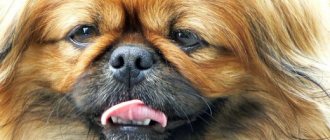
Entropion in dogs is also called entropion. This is a transformational change in the position of the eyelid, as a result of which the eyeball comes into contact with the eyelid and eyelashes.
Entropion of the eyelid in a dog (photo in the text) is a very dangerous disease. As soon as the owner notices symptoms, it is necessary to immediately take the animal to a veterinary clinic. If the pathology is not treated, the dog may lose an eye and even vision.
Therefore, you should always carefully monitor the health of your beloved pet, and if suddenly he notices clouding and redness of the eyes, lacrimation or purulent discharge, then the dog may need surgical intervention to correct the eyelid, which cannot be postponed.
Treatment
To choose the right treatment, it is very important to correctly determine the area of the disease. Lesions in Horner's syndrome are conventionally divided into:
- Postganglionic , which has a more favorable prognosis in treatment.
- Preganglionic , which requires more thorough treatment.
Unfortunately, there are no specific drugs for the treatment of this disease, so treatment is aimed at eliminating the causes of the disease . To reduce the clinical signs of idiopathic Horner's syndrome, the dog is treated with finilephrine (the animal is instilled in the eyes 1-2 times a day), as a rule, after 5-7 weeks a significant improvement in the animal's condition is observed.
If the cause of the syndrome is a tumor, then surgery , possibly even chemotherapy and radiation, sometimes for malignant tumors of the eye, the organ is completely removed.
There are cases when, despite all efforts, Horner's syndrome cannot be cured, but this does not affect the dog's life; in this case, it is very important not to forget to moisturize the dog's eye in order to avoid drying out of the eyeball.
Causes
The occurrence of entropion in dogs has a number of reasons:
- Genetic predisposition. Sometimes this reason is called by specialists, but this is only an assumption, since identifying the cause can be quite difficult. The gene that would be responsible for the occurrence of this pathology has not been identified. According to the observations of experts, this disease mainly affects purebred animals bred by crossing genetic relatives.
- Features of the structure of the skull.
- Location of the eyeballs.
- Elasticity and length of the eyelid.
- Trauma to the eyelid or eye.
- Squinting. The habit of squinting is very rare in animals, but sometimes it still happens, which can lead to an inversion of the eyelid in dogs.
Entropion can also develop after severe conjunctivitis or scarring of the eyelid.
Signs and symptoms
Entropion in dogs is accompanied by the following symptoms:
- Feeling of sand in the eyes, pain. The dog often rubs its eyes with its paws.
- Squinting.
- Frequent blinking.
- Anxiety in behavior.
- Increased tear production.
- Purulent discharge.
- Dark tracks under the eyes.
Examining the cornea with such symptoms is very problematic. The dog often looks askance, the animal’s eyes are either tightly closed or completely closed. The dog cannot look at the light.
Entropion of the third eyelid in dogs
This form of the disease is typical for shepherd dogs, pinschers, and mastiffs. Other breeds very rarely suffer from entropion of the third eyelid. It develops either as a consequence of degeneration of the cartilaginous part of the eyelid, or as a complication of the follicular form of conjunctivitis.
This type of disease causes deformation of the third eyelid and red eye syndrome. When lacrimation occurs, serous-mucous discharge from the eyes appears. Some affected individuals experience a characteristic eyelid spasm or tic, which always goes away when treated.
Entropion of the lower eyelid in dogs most often occurs in Shar-Peis and Chow Chows, which have excessively pronounced skin folds on the face. Excess skin hangs over the eyes, causing drooping eyelids.
Dog Breeds That Lose Eyes
Animals with weak fixation of the eyeballs, held solely by the eyelids, and a short, slightly upturned muzzle, which increases the risk of mechanical damage, are susceptible to proptosis.
Owners of the following breeds will have to demonstrate high vigilance:
Unlike the listed breeds, most dogs do not suffer from this problem due to:
- correct structure of the skull;
- deep eye sockets;
- brow ridges.
Pekingese's eye fell out
Various degrees
With entropion of the eyelid in dogs, there are several degrees of the disease:
- very tight fit of the eyelid;
- inversion of the eyelid, accompanied by contact with the cornea at an angle of 90°;
- touching the cornea with the hair of the eyelid and its skin at an angle of 180°.
At each degree of the disease, the animal experiences discomfort, rubs its eyes, and behaves restlessly.
Entropion in dogs can be central or lateral. In the first case, the central part of the eyelid is inverted and droops, and in the second, the skin sags from the middle to the corner of the outer eye.
It may happen that the problem goes away on its own. What happens when the disease is found in a puppy, and as the puppy grows, the problem with the eyelid disappears. Or if the bones of the skull do not grow at the same time as the skin. In these cases, the puppy is generally not harmed by the entropion of the eyelid. However, consultation with a specialist is still required.
Diagnosis
Diagnosis is carried out by examining the patient. In order to reduce the unpleasant pain from the examination, drops with an anesthetic are instilled into the dog's eyes.
To identify ulcers or erosions on the cornea that have arisen during the course of the disease, veterinarians can use fluorescent solutions, after treatment, with which the damaged areas of the cornea begin to glow under ultraviolet light.
Non-surgical therapy
Drug treatment for entropion in dogs is also possible. This occurs in mild cases of the disease. The veterinarian prescribes antiseptic drops that relieve inflammation and kill pathogenic microflora.
Treatment of the skin around the eyes with various antiseptic gels and ointments can also be prescribed. In addition, the doctor prescribes anti-inflammatory drugs internally.
There is also a temporary non-surgical procedure performed in a clinic using autohemotherapy. Here the animal's blood is injected into the thickness of the eyelid using a medical syringe. This procedure can be carried out by a strictly qualified specialist and with the use of additional medications.
The effect is possible within 10-14 days. Then, if necessary, the procedure is repeated. When using this method, the deformed eyelid unfolds, moving to its normal position. Before starting the procedure, the patient needs a 12-hour diet.
Is it possible to straighten the eye yourself?
If a dog's eye has fallen out, it is contraindicated to return it to its natural position on your own; this should only be done by a veterinarian. Before taking your pet to the clinic, it is recommended to take a number of actions:
- Rinse the eye with saline and artificial tears or plain water, brought to a boil and cooled. This can be done by douching or using a soft cloth or gauze. Cotton wool products should not be used as the lint will cause further irritation.
- Cold must be applied to the swollen area around the affected organ, without touching the cornea. To make a compress, pieces of ice are covered with plastic film, wrapped in cloth and applied for 10 minutes to eliminate swelling and bleeding.
- Then oletethrin, hydrocortisone, tetracycline ointment or a special moisturizing gel is applied to the affected area.
- To prevent the dog from injuring the eye, a protective collar is put on it.
Surgery
But still, in the vast majority of cases, surgery for entropion of the eyelid in a dog plays a leading role in the treatment process. Even if the animal has serious complications from the disease in the form of keratitis and conjunctivitis, surgical intervention will allow for the highest quality sanitization of foci of infection and will alleviate the dog’s general condition.
The technique of eyelid entropion surgery in dogs involves removing the folded part of the eyelid by straightening and cutting it. After this, supporting sutures are applied to fix the ligaments in the required position. Absorbable sutures are used that do not need to be removed. In adult individuals, additional operations are often required to adequately strengthen the ligamentous apparatus.
In puppies that have reached the age of 6 months, such operations are easiest. Their ligamentous apparatus has not yet hardened, so surgical intervention will be minimal. Here only supporting sutures are applied to fix the eyelid in the required position.
If wounds, ulcers, conjunctivitis and keratitis appear during the course of the disease, they are treated in the general manner. Dogs with hereditary inversion of the eyelids are not allowed for breeding.
The postoperative prognosis is very positive. If, of course, the treatment was carried out in a timely manner, before the appearance of irreversible processes in the cornea of the eye. In cases where it is not possible to avoid severe injuries, the outcome will depend only on the severity of the injuries received by the animal during the course of the disease. In particularly severe cases, the dog may be advised to remove the eye.
Eyeball luxation
Prolapse of the eyeball, or dislocation (Proptosis of eyebal) is a pathology associated with the protrusion of the eyeball beyond the topographic boundaries of the bony orbit.
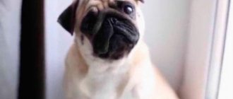
The disease can occur in any animal. Most often recorded in dogs and cats. Among canines, loss most often occurs in pugs, Pekingese, Chihuahuas, Shih Tzus, and Yorkshire terriers. Other breeds are also susceptible to pathology, but greater physical effort must be applied.
The main cause of the pathology is trauma in the temporal fossa. The bony orbit in some dog breeds is not closed. The ocular process of the temporal bone is poorly developed and the orbit is limited by the fibrous ligament. At the time of injury, the ligament is stretched and the eyeball leaves its anatomical location. The eyeball is also held in place by the rectus muscle. At the moment of dislocation, rupture of muscle tissue of varying intensity occurs, and hematomas, injuries to the optic nerve, blood vessels, and vitreous are also possible.
The extent of the pathology is influenced by the force of mechanical action during injury, breed characteristics, the reactivity of the body, and the period of time from the moment of injury to the moment of assistance.
Immediately after an injury that leads to dislocation of the eyeball, it is pinched by the eyelids and rapid swelling of the conjunctiva occurs.
The eyeball has left its anatomical boundaries completely or partially, and is restrained by the eyelids. The conjunctiva is swollen and covered with hemorrhages.
The cornea quickly loses its shine and becomes dry. If the process is left for more than a day, degenerative changes in tissues begin.
The prognosis of the disease depends on the speed of assistance.
An examination is performed for diagnosis. To predict the further functioning of the eye, pupillary-motor reactions are assessed and ophthalmoscopy is performed.
If there is a reaction to light, there is no blood in the anterior chamber of the eye and the vitreous body, and there are no more than two ruptures of the extraocular muscles, then the prognosis is favorable. If retinal detachment occurs, a lack of pupillary-motor reactions, scleral rupture, and vitreous hemorrhage occurs, then the prognosis is unfavorable.
Surgery begins immediately. Delay contributes to necrotic lesions of the cornea and conjunctiva, and timely treatment in 80% of cases allows preserving the organ of vision.
Surgery is performed under general anesthesia. To irrigate the eyeball and eyelids, use a 0.25% solution of novocaine or a solution of diocide. Washing is carried out until the tissues are completely cleansed.
The area around the eyeball is injected with a solution of novocaine 1% concentration with the presence of hydrocortisone.
Next, a centotomy is performed (dissection of the external commissure of the eyelids) by an amount of 0.5 to 1 cm to the bony edge of the eyelids. This allows the eyelid tension to be released and repositioning to be carried out quickly. The eyeball is covered with a napkin soaked in an antibiotic-based emulsion and, with light pressing movements, is pressed into the eye orbit. A knotted suture is placed on the incision line. The eyelids are closed with a purse string suture. The entire surgical field is generously lubricated with tetracycline ointment and a bandage is applied over it.
Postoperative period
Proper and high-quality care for the animal after surgery is very, very important for positive dynamics and a speedy full recovery. This cannot be taken lightly. Otherwise, the operation will have to be repeated, which means extra stress for the pet and extra wasted money for the animal owner.
Basically, for such operations, veterinary surgeons use thin suture material. Its use will not leave traces of intervention on the dog’s eyelids. However, it is very easy for an animal to tear off such light stitches before the healing process is complete, so you will have to use a special collar that will not allow the dog to waste the treatment.
After surgery, the veterinarian prescribes special eye drops and antiseptic ointments, the use of which is highly discouraged.
Prevention
Preventative measures should include the following:
- preventing injury to your pet's eyes;
- careful attention to the hygiene of the animal’s visual organs and face;
- Regular visits to the veterinary clinic for periodic examinations;
- promptly consult a doctor if symptoms and signs of entropion occur in a dog.
The health of a dog is the sole responsibility of its owner. And how healthy and well-groomed a pet is is an indicative factor for assessing the personal qualities of its owner.
Urgent medical care and treatment
If you do not visit a doctor in a timely manner, secondary phenomena develop in the injured organ, such as severe inflammation, suppuration, and drying out of the cornea. This is fraught with fatal consequences. The protruding eye is supported only by the rectus externus muscle, and is pinched by the eyelids. If the optic nerve ruptures, then, most likely, the function of the damaged eye will not be restored - the prognosis is disappointing, including removal.
After an initial examination of the injured pet, the veterinarian, under anesthesia, sets the missing eye, sutures the muscles and partially (or temporarily completely) the eyelids, and applies a bandage. To prevent the dog from rubbing the muzzle, wear a high, hard collar. Throughout the postoperative period, until the sutures are removed, sanitary and hygienic treatment and other procedures are carried out:
- washing with saline solution;
- applying special eye ointments;
- drug therapy (injections, tablets).
The sutures are removed after about a week (the attending physician will tell you more precisely). Continued medical care may be required for some time after the sutures are removed.
Forecasts
After the operation, you should protect your pet from physical exertion, from falls, from conflict situations with other animals, and from active games. Please remember that the injury may recur.
We can talk about the best prognosis only if there is a minimum period of time from the injury to the visit to the clinic. It is impossible to guarantee one hundred percent return of vision after a lesion, but the risk of complications up to the complete loss of the ability to see is real.
Complications after surgery are:
- strabismus (corrected by filing the eyelids);
- corneal sensitivity disorder and keratoconjunctivitis (insufficient hydration);
- atrophy of the eyeball (the organ must be removed);
- non-closure of eyelids.
If your dog's eyes are falling out or there is a risk of developing proptosis, cosmetic surgery to close the eyelid incision can minimize the threat. Knowing about this peculiarity of your pet’s breed, you should protect it as much as possible from possible traumatic situations.