The eyes of the affected animal should be immediately washed with holy water or, if available, with a 2% solution of silver nitrate, a 10% solution of sodium albucid and consult a doctor.
Quite rare, but chemical burns do occur in cats and dogs . They are caused by acids, alkalis, salts of heavy metals and other substances that have a cauterizing effect.
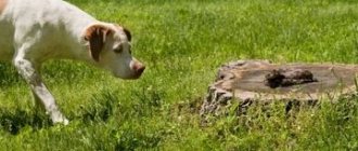
The circumstances under which chemical burns occur vary. In the presence of animals, their owners in their dachas or apartments carry out work using quicklime, acids, and alkalis. The mucous membranes and skin of animals can also be affected by the erroneous use of cauterizing substances.
The degree of damage depends on the concentration of the chemical and the duration of its contact with tissue . Low concentration solutions and strong solutions usually cause superficial skin damage when used for short periods of time. Under the influence of undiluted substances that remain on the body for a long time, tissues become dead.
Acids and salts of heavy metals form insoluble compounds with proteins and clot the blood. The tissues are quickly dehydrated, resulting in the formation of a scab: black - with a burn with sulfuric acid, yellow - with a burn with nitric acid, brown - under the influence of silver nitrate. Changes develop more often within the skin. The scab dries out and turns into a dense crust.
The destructive effect of alkalis is longer. They do not form insoluble compounds with proteins, do not clot blood in the vessels of the damaged area, and enter into compounds with fats and destroy nitrogenous compounds. As a result, a loose whitish scab appears. Its value increases as the tissue disintegrates until the entire amount of the substance that has penetrated the tissue is consumed. Rejection of the scab is often accompanied by bleeding.
Emergency care for chemical burns should be aimed at removing or reducing the concentration of the chemical substance as quickly as possible, or at neutralizing it. Therefore, the damaged area is continuously doused with a stream of water, which washes away the chemical, dilutes its residue and thereby reduces its harmful effect on the tissue. Then neutralization is carried out.
A 2% aqueous solution of acetic acid (vinegar - 3-15% solution of this acid) is used as neutralizers if the tissues are affected by caustic alkalis or quicklime; 2% aqueous solution of bicarbonate, baking soda for acid burns.
Oily caustic substances are removed from the surface of the skin by dissolving them with gasoline.
When a cat or dog is exposed to electric current, so-called electric marks appear in their mouth. Usually these are third-degree burns; they are small, but deep and involve the tongue, the mucous membrane of the palate, and often the maxillary bone. Such burns are treated in the same way as thermal burns.
And the last type of burns is eye damage from ultraviolet rays . It occurs when erythema or quartz lamps are turned on for a long time, which animal owners use to treat and prevent their own diseases or to treat their pets.
A few hours after exposure to ultraviolet rays, cats and dogs try to go to a dark place, rubbing their eyes on various soft objects. Tears flow from the eyes, the connective membranes of the eyes turn red and swell. It is difficult to open the eyelids. A cat or dog or other mammals with conjunctivitis should be placed in a dark place where there is no natural light or artificial lighting. Napkins moistened with holy water are placed on the burns. When the swelling subsides a little, the same water is instilled into the eyes, 1 - 2 drops 4-5 times a day. You can also instill a 10% solution of sodium albucid.
Source: L. Stishkovskaya. “1000 tips on how to treat pets”
What eye diseases occur in dogs?
Eye diseases in dogs can be of 3 types:
- Infectious – occurs when infected with viruses or bacteria as a primary disease or complication of infection of another organ.
- Non-infectious - mechanical damage, neoplasms, inflammation due to improper growth of eyelashes, eversion of the eyelids.
- Congenital - characteristic of some breeds, for example, Shar Peis. Includes eversion, entropion of the eyelids, deformations of the eyes and lenses. They are usually corrected surgically.
Each disease has a characteristic description, diagnostic methods and treatment.
Blepharospasm
Blepharospasm is a voluntary rapid contraction of the muscle tissue of the eyelids. It is quite simple to recognize this disease in a dog by rapid blinking, almost without stopping. In addition, the area around the eyes and the eyelids themselves swell noticeably. Touching them causes pain in your pet.
Exudate—liquid—accumulates in the corners of the eyes. Photophobia is observed - the animal begins to hide from light sources, go into the shadows on the street, constantly lowers its head and squints.
The pathology is not dangerous in itself, but indicates the presence of infection or inflammation in the body. Often the cause is spasm of the trigeminal nerve. Treatment is selected based on the original source. You cannot choose therapy for blepharospasm on your own.
Keratitis
Keratitis is inflammation of the eye cornea. The disease causes significant discomfort to the dog and threatens serious complications on the visual system. Keratitis occurs due to mechanical injuries, burns, acute vitamin deficiencies, infectious diseases (hepatitis, enteritis, plague), and allergic reactions. They can also affect dogs with diabetes mellitus and weakened immune and endocrine systems. In rare cases, it develops at the genetic level.
Keratitis can be identified by swelling of the eye and conjunctiva, cloudy cornea and lens, purulent discharge of white, yellow and gray shades. There is profuse lacrimation and photophobia. The white becomes red, the eye membrane becomes rough. There may be frequent blinking in an attempt to clear vision. Symptoms appear within 2-4 hours of infection or injury.
Keratitis threatens the development of glaucoma, cataracts, and loss of vision. Treatment depends on the cause of the appearance. General procedures for all forms of pathology are washing with a solution of furatsilin or another antiseptic, drops of levomecithin, application of erythromycin ointment, a course of oral antibiotic. Pain relief injections and a hypoallergenic diet may be prescribed. On average, treatment gives effect in 1-2 months.
Check out the full article on the eye disease in dogs - keratitis.
Third eyelid prolapse
Third eyelid prolapse is often called “cherry eye.” With this disease, the eyeball turns red and swells, becoming like a ripe cherry. At this time, the third eyelid loses its tone and gathers into a “rag” towards the corner of the eye. Bilateral prolapse is extremely rare; the disease usually affects one eye. The causes may lie in infections, but more often they are hereditary. Spaniels, bulldogs and hounds are especially susceptible to the pathology.
Prolapse is dangerous due to inflammation of the conjunctiva and corneas, as it interferes with the normal hydration of the eye. Prolapse of the third eyelid can only be corrected surgically. Moisturizing drops are prescribed as maintenance therapy.
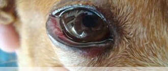
Conjunctivitis
Conjunctivitis is the most common eye infection in dogs. It is expressed in dysfunction of the conjunctival mucosa and is often infectious in nature. In addition, the causes may be allergies, viruses, clogged tear ducts, foreign body trauma, irritation due to drooping of the eyelids or ingrown eyelashes.
The symptoms are pronounced: the conjunctiva turns red, swells, discharge of tears and pus appears, the third eyelid may swell, mild squinting and frequent blinking may occur. The dog begins to rub his eye with his paw, becomes restless and whines.
Treatment includes antibacterial drugs, application of tetracycline ointment and instillation of special drops.
We recommend reading the full article on conjunctivitis in dogs.
Cataract
Cataract is expressed in severe clouding of the lens of the eye, its swelling and increased intraocular pressure. The disease may appear due to exposure to toxic substances, tissue wear and tear with age, or be congenital. Cataracts are dangerous due to complete or partial loss of vision as a result of rupture of the tissues of the eyeball.
Drug treatment is practiced (drops of viceine, catachorm, vitamin compounds), but is considered ineffective. The result is achieved only by surgical intervention followed by maintenance therapy.
Learn more about the symptoms and treatment of cataracts in dogs.
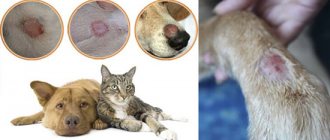
Blepharitis
Blepharitis is an inflammation of the eyelids that affects both the skin and subcutaneous layers. In this case, the eyelids noticeably swell, become covered with pus and scabs, turn red, and glands protrude from the corners of the eyes. The disease occurs due to infectious infections, prolonged contact with allergens, mechanical injuries, and hereditary predisposition. Also, the reason may lie in the bite of a parasitic tick.
Blepharitis is often combined with other eye pathologies and reflects serious infections. It must be treated urgently in accordance with the cause. The veterinarian may prescribe antibiotics, antimicrobial or antiallergic medications, compresses and saline washes. Therapy is individual in each case.
Read full information about blepharitis in dogs.
Ectropion and entropion
Ectropion and entropion are the medical names for eversion and entropion of the eyelids. Both diseases usually develop in parallel, so they are practically not separated. They are expressed in a change in the shape of the eyelids (turning outwards or inwards), loss of their natural functions. When eversion occurs, the eyeball is left without moisture and protection from germs. When curled, the eyelashes irritate the pupil and can grow into the protein tissue. In both cases, the dog begins to blink constantly, profuse lacrimation occurs, and the animal experiences pain.
Treatment is surgical when the dog is an adult, when its growth has stopped. In the early stages and during recovery after surgery, medical support is provided with hormones, antiseptics, and moisturizing drops.
Shar-Peis, Staffordshire Terriers, Dachshunds, Great Danes, Newfoundlands, St. Bernards, Basset Hounds, Great Danes, Spaniels and Ridgebacks are particularly susceptible to the disease.
Dermatitis of the century
Dermatitis is not considered an independent eye disease, but often develops into more serious pathologies. At its core, dermatitis is an inflammation of the skin on the outside of the eyelids. They turn red, become covered with moist areas (sometimes with pus with an unpleasant odor), and peel off.
The eyes gradually turn sour, swell, and the conjunctiva becomes infected with pathogenic organisms. It often appears in dogs with long hair, overhanging folds of forehead skin and floppy ears.
Treatment with antibiotics in the form of tablets or injections and ointments, antimicrobial drops is necessary. Rinsing with sterile solutions is also carried out. The fur from the ears, forehead and eyelids is neatly trimmed; some pets require a collar to prevent eye scratching.
Corneal ulcer
One of the most common eye diseases in dogs. Also called ulcerative keratitis. It is expressed in inflammation of the upper layer of the epithelium of the eyes. The tissue becomes thinner, wears out, becomes covered with ulcers, gradually growing into one large wound. All layers of the epithelium are affected.
Watery eyes appear, pus is released, the outline of the pupil blurs, and the white of the pupil turns red. The color of the cornea may change. The dog begins to squint, close its paws and lower its eyes. It may develop into a chronic form.
Occurs due to physical damage to the skin, infection, burns with chemicals, as a complication of acute conjunctivitis, volvulus and neoplasms. It is rarely hereditary.
Treatment ranges from surgical excision of the affected tissue to conservative treatment. Medication methods take up to six months, drugs are selected strictly individually.
Lens luxation
Lens luxation (or luxation) involves displacement of the corresponding part of the eye from the hyaloid fossa. Dislocation can be partial or complete. It occurs for genetic reasons, due to glaucoma, cataracts or as a complication of severe injuries and infections. It can lead to loss of visual function and is therefore considered a serious disease. Different types of terriers are most susceptible.
Dislocation occurs after the ligaments of the lens and ciliary line are torn. The pupil becomes deformed, moves away from the center and swells, and the shape of the apple may change. The movement of fluid in the ocular body is disrupted.
Treatment is carried out by surgical correction. The earlier the stage of the disease, the more positive the prognosis for saving the eye. After removal of the lens, an intraocular lens implant is placed. In rare cases, implantation of the entire eyeball is indicated.
Eyeball luxation
It consists in the exit of the eyeball from the orbit behind the eyelid completely or partially. Caused by mechanical damage to the bones of the head and temples, large muscle tension as a result of the shallow depth of the bone orbit.
The apple extends far beyond its natural boundaries, the conjunctiva swells, dries out, and takes the form of a hanging cushion. Such a dislocation threatens with blindness, ulcers, and necrosis of eye tissue. Often found in Pekingese, Japanese Chins and similar breeds.
As first aid for loss, it is suggested to carefully irrigate the apple with a solution of furatsilin or novocaine to reduce pain and prevent drying out. It is corrected surgically under anesthesia after excision of the eyelid adhesion. A fixing suture is applied, which is removed after a few days. Then corticosteroid therapy is used to relieve inflammation.
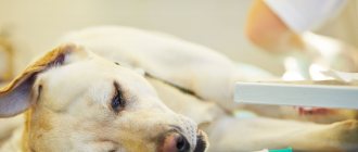
Treatment
Each disease has its own characteristics, so a veterinarian must prescribe treatment. Most often, to accurately determine the cause and choose a treatment method, it is necessary to conduct a thorough diagnosis of the pet. However, it is possible to identify general principles in the approach to treating eye problems in dogs.
What medications should I give?
When a pet is prescribed antibacterial therapy, injections must be administered intramuscularly.
Attention! Before using this or that antibacterial drug, make sure that sensitivity tests to the drugs of the selected group have been carried out.
If a pet has an acute form of inflammation, then in addition to antibacterial therapy, anti-inflammatory drugs are also prescribed.
Before instilling special preparations into your eyes, you need to rinse them and remove excess discharge and pus. It is best to use saline solution or tea leaves.
Antiviral and astringent eye ointments must be applied using a special bandage.
Make sure your dog has a special collar for his neck and socks for his paws, especially if he is actively trying to scratch his eyes. Do not allow him to do this, this will protect your eyes from additional germs.
For rinsing, you need to use gauze (if you use, for example, cotton pads, the lint from them can get into the dog’s eyes).
Vision is of great importance for a dog, and many eye diseases can lead to partial or even complete blindness, so it is necessary to seek qualified advice from a specialist as soon as possible and provide immediate assistance to our little brothers.
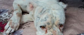
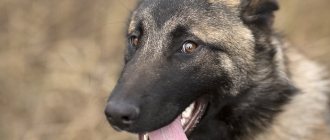
Symptoms of eye burns in cats and dogs
Symptoms of eye burns in dogs and cats are difficult to miss:
- This is severe lacrimation, redness in the conjunctiva, the white is filled with blood.
- The pet is worried, tries to scratch its eye on soft objects or rubs its muzzle with its paw.
- The eyelids do not open, there may be frequent blinking. If you look at the cornea, damage will be noticeable (you will not be able to determine its integrity at home).
If you don’t see that your pet has been in contact with chemicals, you may not even immediately understand that these are all the symptoms of a chemical burn to the eye of a cat or dog. They can be mistaken for signs of conjunctivitis, corneal ulcers or other eye diseases.
Keratitis or diseases of the cornea of the eye
One of the reasons why a dog's eyes are watery may be an inflammatory process in the cornea. Destructive changes in this area of the eyeball are called keratitis. They can be:
- catarrhal;
- purulent;
- serous;
- pigmented;
- deep and superficial;
- one- and two-sided.
When the cornea becomes inflamed, the animal's eyes begin to water and acquire a cloudy, ashy tint. In addition, mucopurulent discharge, redness and swelling are characteristic. A sick pet feels discomfort and tries to eliminate it by friction, avoids sunlight, becomes nervous or shows apathy.
The disease can be triggered by a wide range of factors, ranging from genetic predisposition to a weakened immune system. Advanced cases of the disease are often accompanied by a corneal ulcer and loss of vision in the dog.
Symptoms of burns in dogs
Despite the numerous types of burns, the symptoms of injury have similar signs - swelling and redness, pain, and subsequently the development of vesicles, their opening and the formation of scars (or granulation of damaged tissue).
If the chemical eye
One of the dangerous types of injury is a chemical burn to a dog’s eyes. Contact with alkali or acid on the mucous membrane is accompanied by the development of severe pain. The owner observes severe photophobia and anxiety.
The pet tries to hide in a dark place, since daylight has an irritating effect on the eye environment damaged by aggressive chemicals. The dog rubs its face with its paws and whines. With a chemical burn, swelling and redness of the eyelids and conjunctiva are observed.
In case of severe trauma, when there is damage to the structure of the deep layers of the retina, the dog may experience clouding of the lens and ulceration of the cornea. In advanced cases, when the wound becomes infected with pathogenic microorganisms, panophthalmitis may develop, which results not only in vision impairment, but also in the animal’s blindness.
If boiling water
A common thermal injury in dogs is scalding from boiling liquids. The disease is characterized by redness of the skin and pain due to deep heating of the tissues. The first aid in such a situation is to cool the damaged area.
If a burn of the esophagus
When an aggressive chemical (acid, alkali, alcohol solution of iodine) enters the pet’s digestive tract, the esophagus is the first to be affected. The animal exhibits anxiety and refuses to feed. If a dog has a burn to its esophagus, it may whine and stand with its mouth open. In some cases, drooling is observed.
If it's sunny
Pets with short hair, such as bulldogs, Chinese crested dogs, and American Staffordshire terriers, are usually susceptible to sunburn. A characteristic feature of thermal injury is redness of the skin.
The dog is worried, itches, and licks its body. Pets with fair skin are most often affected. The lesion is typical for areas around the nose and groin area.
Adenoma of the third century
Adenoma of the third eyelid , or correctly said – prolapse (prolapse) of the lacrimal gland of the third eyelid.
Usually observed in young dogs aged from 6 months to 2 years, most often in brachiocephalic breeds (Shih Tzu, cocker spaniels, pugs, bulldogs, etc.), beagles, etc. This occurs due to genetically determined weakness of the ligaments that fix the gland to the edge of the eye socket. Symptoms:
— An oval red formation appears in the inner corner of the eye, protruding from under the edge of the third eyelid. It is most often observed in one eye, but can also be bilateral.
- There may be moderate lacrimation and squinting of the eyes (blepharospasm).
Treatment:
1. The gland must be returned to its place surgically.
============================================================================================================================================================================================
Cataract
This eye disease is associated with aging of the lens, a violation of its transparency - subnuclear (nuclear) sclerosis.
The development of cataracts is most likely at the age of 8 years, when the elasticity of the lens decreases, it hardens and ceases to adjust vision to different distances. It is more common in Golden Retrievers, Poodles, Yorkshire Terriers and Boston Terriers. The disease progresses over the years.
Cataract photo:
Causes
Less commonly, the causes of cataracts are heredity. Acquired cataracts occur after trauma, after inflammatory diseases, diabetes, etc.
Symptoms, treatment and diagnosis
An external sign of cataract is clouding of the lens. The cloudiness of cataracts is uneven. It is possible to recognize cataracts on your own. When a beam of light hits the affected eye, the clouding will shrink in size. In low light, on the contrary, it increases.
In some cases, this dangerous disease can develop for a long time without visible symptoms. But a dog owner may notice decreased vision by analyzing the dog's behavior.
The most reliable way to diagnose an eye disease called cataracts remains to be examined by a specialist.
Treatment without surgery
How to treat cataracts without surgical intervention at home? But no way. The use of medicinal drugs does not allow us to hope for the recovery of our four-legged pet. At best, the progression of the disease will slow down. It is possible to use drugs only at the very beginning of the development of cataracts, when surgery is not yet required. Vitamins and enzymes are usually prescribed.
Cataracts cannot currently be prevented. Do not fall for the tricks of deceivers who recommend Smirnova eye drops, vitaiodurol, vicein, catachrome, vitafacol for the treatment of cataracts . These drugs cannot prevent, slow down, or even cure clouding due to the fact that the nature of the disease is not clear.
When is cataract surgery necessary?
The only effective way to treat cataracts is surgery to replace the clouded lens with an artificial one. From a technical point of view, this operation is very complex. Its result largely depends on the stage at which the application was made to an ophthalmologist. Obviously, earlier treatment guarantees a better result.
The modern technology of lens removal using ultrasound - phacoemulsification - is finding more and more fans. But it is also effective in the early stages.
Prevention
The need for surgery is determined by the animal’s ability to navigate in space using vision. If decreased vision does not allow the dog to move without the threat of harm to health, we can talk about the need for urgent surgical treatment.
- When purchasing a puppy, you should ask whether any of the parents have been diagnosed with cataracts.
- After 6 years, it is necessary to regularly visit a veterinarian.
- Maintain eye hygiene for your pet: wipe and apply eye drops if indicated.
- A nutritious diet and adherence to the vaccination schedule are also important.
Diseases and problems of the choroid and cornea
The choroid and cornea of the eye may suffer mainly from the progression of inflammatory processes. Failure to promptly contact a veterinary ophthalmologist can lead to complete blindness of the dog. Moreover, this can happen in a short period of time, since such pathologies have intensive development dynamics.
Ulcerative keratitis
In the eyes of an animal, ulcerative keratitis develops as a result of sun or thermal burns, when exposed to mechanical forces during impacts, or when foreign objects enter the eye. In addition, ulcerative keratitis is a secondary disease against the background of allergic abnormalities, vitamin deficiency, bacterial and viral infections. Another reason for this pathology is endocrine diseases (for example, diabetes mellitus).
With such a lesion, tearing develops. In this case, the dog rubs its eyes with its paws, which indicates itching, discomfort and the presence of foreign bodies on the cornea. The eye may be very sore. Blue eye syndrome also occurs when the pigmentation of the pupil changes under the influence of pathological factors.
In these circumstances, veterinarians prescribe drug therapy with antimicrobial, antihistamine, painkillers, as well as external agents to localize the inflammatory process.
Uveitis
Uveitis is an inflammatory ophthalmological disease. It is accompanied by damage to the choroid of the eye and disruption of the blood supply to its tissues.
Signs of intense inflammation of the irises are changes in their color, fear of bright light, half-closed red eyelids, and decreased visual acuity. Uveitis occurs due to injuries to the head and eye area, viral and bacterial infections.
If a dog's eye is inflamed in the area of the iris, anti-inflammatory drugs are mainly used to treat uveitis, as well as drugs to reduce pain.
Fundus disease
Retinal atrophy
The disease is inherited. Signs are: deterioration of vision, especially at night, deterioration of vision during the day and slow blindness, pallor of the pupil.
Retinal atrophy
Retinal detachment
Occurs due to injury, high eye pressure, retinal atrophy, or a neoplasm in the eye. Manifested by hemorrhage, rapid loss of vision, lack of pupillary response.
Glaucoma
A number of diseases with increased intraocular pressure. Characterized by redness of the apple, dilated pupil, high intraocular pressure. The dog is afraid of light, loses appetite, and becomes lethargic. An ophthalmologist treats glaucoma.
Keratitis
Keratitis is an inflammation of the cornea (the most superficial, transparent layer of the eye in contact with the external environment), manifested by the appearance of individual areas of cloudiness or clouding of the entire cornea, which can lead to decreased vision and, in advanced cases, to blindness.
There are:
- Shepherd dog keratitis (the disease is typical for shepherd dogs, but it is also often found in Staffordshire terriers, Labradors and mixed breeds, somewhat less often in other breeds: collies, St. Bernards, Siberian huskies, huskies, Airedale terriers, Russian greyhounds, greyhounds).
- Boxer keratitis (boxers, Boston terriers, French bulldogs, Pekingese, pugs).
- Dachshund keratitis.
Shepherd keratitis, or pannus, is a superficial, usually without ulceration, chronic inflammation of the cornea.
Symptoms:
- On the conjunctiva (normally pink) - extensive brown pigmentation appears.
— Gray-white clouding of the cornea and the formation of a red shaft, due to the appearance of blood vessels, which spreads over the surface of the cornea over time and is prone to pigmentation.
— Often combined with changes in the third eyelid: plasmoma of the third eyelid (depigmentation of the edge of the eyelid is observed, it thickens).
— Erosion of the cornea from the nasal part of the eye is observed.
Treatment:
1. Lifelong. Local preparations with antibiotics and anti-inflammatory components.
2. Many dogs have a good effect when using ointment with cyclosporine (Optimmune) or tacrolimus (Protopic).
3. In case of loss of vision due to damage to the cornea and severe cases that cannot be treated, surgical intervention (superficial keratotomy) is required.
Boxer's keratitis, or chronic non-healing corneal erosion, Boxer's ulcer is a chronic superficial ulcerative keratitis.
Symptoms:
— At the beginning, a minor defect of the cornea appears, invisible to the naked eye, later it becomes whitish, and erosion appears.
— The dog squints its eye (blepharospasm), it becomes watery (less often, purulent discharge).
— Vessels appear, due to which in some cases the surface of the eye appears red.
Treatment:
1. Long lasting. Local preparations with broad-spectrum antibiotics at least 6 times a day.
2. Surgical removal of lagging epithelium of the cornea.
Dachshund keratitis.
Symptoms:
— At an early stage, local irritation and pinpoint opacities are observed, which are colored green with a special diagnostic dye—fluorescein. Sometimes transparent pits are visible on the surface of the cornea (these are ulcers covered with epithelium).
— As the disease progresses further, blood vessels appear on the cornea (vascularization) and the cornea becomes red in color.
- In the later stages, there is a gray-pink cloudiness on the surface of the cornea, the cornea loses transparency, pigmentation of the cornea appears (it becomes brown), and dry eye syndrome often develops.
Treatment:
1. Long lasting. Local preparations with broad-spectrum antibiotics at least 6 times a day;
2. Preparations for healing and moisturizing the cornea at least 6 times a day.
============================================================================================================================================================================================
Ocular fundus
Retinal atrophy
It is considered a hereditary disease. Main symptoms:
- initially, an increasing decrease in visual acuity at dusk and night blindness,
- later deterioration of daytime vision,
- gradually - blindness,
- paleness of the pupil.
Retinal detachment
The causes of retinal detachment can be trauma, high blood pressure, collie eye abnormalities, progressive retinal atrophy, and neoplasms. Symptoms:
- rapid or sudden blindness,
- impaired pupillary reflex,
- hemorrhages.
Ectropion and entropion
This is what scientific terminology calls inversion and entropion of the eyelids. Both diseases are considered “canine” because they are much less common in other pets. Due to the structure of the eyelids, Great Danes, Basset Hounds, St. Bernards, Dachshunds, Newfoundlands and Spaniels are particularly susceptible to ectropion and entropion.
Both pathologies often accompany each other and develop in parallel, but differ in their consequences: eversion of the eyelids less often leads to serious problems than entropion. With ectropion, drying and inflammation of the conjunctiva, lacrimation, suppuration, and pain when touching the eyes are observed. With entropion, the symptoms are the same, but they develop faster. If the entropion is not treated in a timely manner, it can be complicated by the ingrowth of eyelashes into the eyeball.
Sources:
- https://zooclub.ru/dogs/vet/19-2.shtml
- https://sobaki-pesiki.ru/bolezni-glaz-u-sobak-siptomy-lechenie-foto.html
- https://usatiki.ru/ozhog-glaz-u-koshek-i-sobak/
- https://zootvet.ru/ozhog-u-sobaki/
- https://sobakus.com/zdorove/glaza/mutnye.html
- https://pets-expert.ru/bolezni-glaz-u-sobak-klassifikacija-i-lechenie/
- https://.ru/zabolevaniya-glaz-u-sobak-shhenkov-simptomy-lechenie-foto/
- https://doggiedog.ru/bolezni-sobak/bolezni-glaz-u-sobak.html
Blepharitis
Blepharitis is inflammation of the eyelids. In dogs, the disease is often chronic and bilateral, less often - unilateral and acute.
Symptoms:
- Itching, redness and thickening of the edges of the eyelids (pronounced to varying degrees).
- Blepharospasm (squinting).
- Discharge, the nature of which varies from tears to mucous and purulent.
Blepharitis can be focal or diffuse.
- Focal blepharitis (hordeolum).
There are:
- External stye is an abscess of the sebaceous gland of the edge of the eyelid on the outside. Occurs predominantly in young dogs.
- internal stye, or meibomitis, is a purulent inflammation of the glands on the inner surface of the eyelids (meibomian glands), most often in middle-aged dogs.
- Chalazion (hailstone) is a rough formation on the inner side of the eyelid, formed as a result of meibomitis due to blockage of the outlet with masses prone to calcification.
- Diffuse blepharitis . Often this form is a concomitant disease with allergies, as well as with chronic eye diseases (conjunctivitis, keratitis, etc.), with parasites (demodex), fungi (microsporia, trichophytosis), and sometimes with general diseases.
Treatment:
1. Put the dog on a protective collar to prevent self-injury and aggravation of inflammation.
2. Carry out hygienic treatment of the eyelids (chlorhexidine solution (removal of scales, etc.)).
3. For blepharitis caused by bacteria, apply ointments with antibiotics and corticosteroids.
4. In the case of fungal (microsporia, trichophytosis) and parasitic (demodex) blepharitis - systemic treatment with antifungal and antiparasitic drugs.
============================================================================================================================================================================================