What are eosinophils
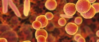
Eosinophils are a subtype of leukocytes, white blood cells. These cells are responsible for phagocytosis of antibodies, that is, the fight against foreign pathogenic proteins. Eosinophils are most active during the period of animal illness. But there is a norm for their quantity, when owners do not need to worry about the physical condition of their animal.
Veterinarians consider the norm for cats and dogs to be from 2 to 6% of eosinophils included in the total number of leukocytes in the blood. But the upper indicator already serves as a reason for an additional check by a specialist. It is noteworthy that the absence of these cells is not completely normal. An animal with ideal health and strong immunity can have such a result in a blood test, but a minimal amount ensures a quick response of the antibody mechanism.
After maturation, eosinophil cells remain exclusively in the blood for approximately 4 hours. After which they enter the body tissues, where they remain at the appropriate level for about 2 weeks. In this case, fluctuations occur constantly. So at night the level of these cells increases and decreases during the day.
What are they researching for?
In almost any pathological process, a clinical blood test is performed first, which includes:
- study of the morphological features of formed elements (red and white blood cells), their qualitative and quantitative composition;
- determination of physical and chemical properties: density, color, viscosity, osmosis, alkaline reserve, etc.;
- analysis of biochemical composition: glucose, protein, albumin, urea, creatinine, etc. (we will consider this item in a separate article).
In the first case, uncoagulated (whole) blood is delivered to the laboratory; in the second case, serum is used (in other words, the upper layer formed when biological fluid settles).
According to indications, other types of blood or serum tests are also carried out, the purpose of which is:
- detect a specific pathogen, for example, with hemobartonellosis or;
- determine the presence of antibodies to microorganisms or toxins (ELISA, PCR, serology);
- isolate the pathogen by bacteriological culture;
- study hormonal levels, etc.
Causes and symptoms
Diseases that cause an increase in eosinophils in the blood of cats belong to different groups, but each of them can be dangerous for a cat.
- allergic dermatitis;
- rhinitis;
- asthma;
- parasitic infection;
- tumor development;
- inflammation in the respiratory tract, gastrointestinal tract or uterus;
- eosinophilic granulomas;
- hypereosinophilia;
- clinical leukemia;
- meyloleukemia.
But in any case, if ALT and eosinophils are elevated, the cat has a clear suspicion of an internal irritant, an allergy.
Cat owners should closely monitor the condition of their pets and know the reason for the increase in eosinophils. Some symptoms may not be pronounced, but if there are persistent problems or a relapse, you need to show the cat to a doctor. Eosinophilia occurs with improper feeding, colds, and chronic diseases.
Approximate forecasts depending on the blood picture
Scientists, and then practicing veterinarians, have learned to predict the outcome of the disease using the leukoformula. We will try to convey this information, maybe it will be useful to someone.
- A moderate increase in neutrophils (NE) with a slight shift in the presence of eosinophils (EOS) in smears indicates a simple infection. A gradual improvement in the picture indicates a speedy recovery.
- An increase in the total white blood cell (WBC) count with a mean shift with a decrease in EOS and lymphocytes (LYM) with further progression indicates infection.
- A significant increase in WBC with a strong shift to the left against the background of a decrease in LYM and EOS (up to their disappearance) makes it possible to judge a very serious condition, but there are still chances to get out. But if too many young cells appear (there are much more of them than rod cells), then the picture is disappointing.
- A constant decrease in WBC with a shift to the left, absence of EOS and a significant decrease in the number of LYM - death is guaranteed. At the same time, a progressive decrease in EOS against the background of an increasing WBC indicates an increase in infection, and the same decrease against the background of a fall in WBC indicates that the microbes have overcome the body’s resistance.
- The appearance of EOS and a decrease in NE in situations where the former were absent and the latter were too much – recovery is guaranteed.
- A sharp drop in LYM with existing clinical signs of infection is an unfavorable sign.
- A sharp decrease in LYM with increased NE indicates the spread of inflammation. The prognosis is poor when the WBC falls amid a strong shift to the left.
- An increase in LYM, which is followed by an increase in NE and an increase in EOS against the background of a gradual restoration of the amount of NE, indicates both an improvement in the general condition and a rapid recovery.
KotoDigest
Thank you for subscribing, check your inbox: you should receive an email asking you to confirm your subscription
Allergic dermatitis
In addition to the painful general condition and refusal to eat, cat owners will notice:
- Constant scratching of the animal, attempts to bite the tip of the tail are signs of itching;
- the fur loses its shine and begins to fall out a lot;
- a dream where the cat was itching, swelling, turning red;
- the cat begins to show aggression, is reluctant to approach, and is noticeably nervous;
- temperature increase.
Treatment of the infection takes place after a full examination by a doctor. The itching may be caused by the appearance of fleas; you can wash the cat yourself with a special shampoo. Even after recovery, it is necessary to ensure that the animal does not continue to actively scratch itself due to inertia. He needs to be kept in isolation from other animals.
The cat is having a seizure. + EOSINOPHILES 8!
Standard biochemistry/Blood test Date of issue of results 09/08/2019 Standard biochemical profile ALT (U/l) 55.2 total 10-55; cat 19-79; horse 4-30 AST (U/l) 23.8 total .10-55; cat.9-59; horse.100-290 Albumin (g/l) 37.4 collection 25-37; cat.25-37; horse.27-42 Amylase (U/l) 1000, 0 total 280-942; total 280-942; total 9-34 total bilirubin (μmol/l) 3.4 total 1.0-20.5; total 1.0-18.0; total 10-40 direct bilirubin (μmol/l) l) 2.2 eq.0.01-5.5; cat.0.01-5.5; l.4-15 Gamma-GT (U/l) 8.8 eq.1-9; cat.1-8; l.9-25 Glucose (mmol/l) 5.0 event 2.5-6.0; cat.2.5-6.3; horse.4.1-6.9 Creatinine (µmol/l) 67.8 event. 68-124; cat 68-160; horse 40-168 Creatine kinase (CPK) (U/l) 73.0 event.0-458;cat.0-458;horse.113-333 LDH (U/l) 408.0 event.50-1000;cat. 55-1000; hp.45-1000 Uric acid (µmol/l) 27.0 e.g.0-160; e.g.0-150; hp.4-150 Urea (mmol/l) 6.1 e.g.2.1-8.3 ;cat.2.0-10.0;hp.3.3-9.2 Total protein (g/l) 76.6 total 58-78;cat.54-77;hp.55-73 Triglycerides (mmol/l) 0.8 total. 0.1-2.0; cat.0.4-1.1; horse.0.04-0.5 Alkaline phosphatase (U/l) 53.0 event.5-150; cat.15-130; horse.102-257 Globulins (g/l) 39, 2 26-46 Albumin-Globulin coefficient. 1.0 0.7-2.0 De Ritis coefficient 0.4 0.5-1.5 Indirect bilirubin (µmol/l) 1.20 1.5-17.0
Standard biochemistry/Blood test Date of issue of results 09/08/2019 General blood test Leukocytes (WBC) (number x 109/l) 4.2 total. 6-17; cat 5.5-19.5 Lymphocytes#(number x 109/l) 2.7 count. 1.2-4.0; cat 1.2-4.2 Monocytes#(number x 109/l) 0.2 inc. 0.1-0.8; cat 0.2-0.9 Granulocytes#(number x 109/l) 1.3 ecc. 2.5-15; cat 2.1-15.0 Lymphocytes -% 64.5 inc. 12-30 ; cat 12-45 Monocytes -% 4.7 ecc. 2-9; cat 2-9 Granulocytes -% 30.8 events. 60-83; cat 35-85 Hemoglobin (HGB)(g/l) 70 sob. 115-190; cat 115-180 Red blood cells (RBC) (number x 1012/l) 7.5 inc. 5.5-8.5; cat 4.6-10.0 Hematocrit (HCT) 31.6 event. 39-65; cat 35-59 Average erythrocyte volume (MCV)(fL) 42.4 events. 42-75; cat 39-72 Average hemoglobin content in erythr.(MCH)(pg) 9.3 event. 20-25; cat 13-21 Average concentration of hemoglobin in erythr.(MCHC)(g/l) 22.1 reports 30-38; cat 30-38 Red blood cell distribution width (RDW) 14.6 ecc. 11-15.5; cat 14-18 Platelets (PLT) (number x 109/l) 61 counts. 110-460; cat 110-460 Platelet volume (MPV)(fL) 6.6 events. 7-12; cat 5-9 ESR 13 events. 1-11; cat 1-12 Leukocyte formula Band 1 sob. 0-4 ; cat 0-4 Segmented 21 events. 60-70; cat 60-70 Lymphocytes 65 coll. 20-70; cat 20-70 Monocytes 5 coll. 1-8; cat 1-8 Eosinophils 8 sob. 1-5; cat 1-5 Basophils 0 esp. 0-1 ; cat 0-1 Notes
Allergic rhinitis
At its core, rhinitis is a runny nose, but in cats it is more severe and can have unpleasant consequences with an increase in eosinophils. The symptoms are :
- labored breathing;
- copious nasal discharge;
- frequent sneezing;
- inflammation, redness of areas near the eyes and nose;
- temperature increase.
Treatment of allergic rhinitis primarily involves identifying the allergen itself and eliminating it. The doctor may prescribe a course of antibiotics.
Elevated eosinophils in a child
Depending on the age of the child, the following factors may be the cause of excess cell content:
- In newborns, a high rate of eosinophils can be caused by Rh conflict, staphylococcus, hemolytic disease, dermatitis and allergic reactions to medications or food.
- Between the ages of one and a half to three years, high eosinophil counts can be caused by atopic dermatitis, drug allergies, and angioedema.
- In children over three years of age, eosinophils increase in the presence of bronchial asthma or allergic rhinitis, during exacerbation of skin allergies, chickenpox, scarlet fever and helminthiasis. Malignant tumors can also cause an increase in eosinophils in a child.
We suggest you read: How to collect urine from a cat for analysis, basic principles
Elevated eosinophils in the blood are not an independent disease; all efforts should be aimed at finding the main cause of their increase and, if possible, eliminating it.
Parasites in the body (helminths)
Parasites, entering the body, quickly cause dulling of the coat, constant problems with constipation and diarrhea. Subsequent symptoms :
- eye discharge;
- severe, frequent vomiting;
- unpleasant odor from the mouth;
- bloating and tightness of the abdomen;
- loss of appetite;
- temperature increase.
In this case, the cat may either suffer from gluttony or completely refuse to eat. A change in the animal's behavior should cause concern among the owners.
General clinical blood test (CBC)
Test material: venous blood.
Blood is taken into a clean disposable tube with an anticoagulant (EDTA) (tube with a green or lilac cap). Blood is stored for no more than 6-8 hours at room temperature or 24 hours in the refrigerator.
Factors influencing results:
- a decrease in hemoglobin and red blood cells can occur due to the action of drugs that can cause the development of aplastic anemia (antitumor, anticonvulsants, heavy metals, antibiotics, analgesics);
- Biseptol. vitamin A, corticotropin, cortisol - increase ESR.
Hematocrit
Hematocrit (Ht, HCT) is the ratio of the volumes of erythrocytes and plasma (volume fraction of erythrocytes in the blood).
Tumor
The tumor does not develop instantly, so the symptoms in the first stages will not be pronounced.
- At the first stage of the appearance of swelling, the cat’s appearance and behavior will remain virtually unchanged.
- With further maturation of the tumor, everything depends on its location.
- The cat will begin to quickly lose weight and become passive and drowsy.
The tumor is almost impossible to detect with the naked eye. Therefore, it is worth periodically contacting a veterinarian if your cat begins to behave unusually, changes its rhythm of life, or refuses to eat.
Prevention of eosinophilic granuloma in cats
Is it possible to prevent the development of eosinophilic granuloma? Preventive measures consist mainly of eliminating the cause that could provoke the appearance of ulcers and plaques.
The most common allergens are feed components, detergents, household chemicals, plant pollen, and insect bites. If a kitten is prone to allergic reactions, then from a young age you should accustom it to hypoallergenic food and let it go outside as little as possible. It will not be possible to completely eliminate the risk of the disease, but it is possible to reduce the likelihood of its occurrence to zero using these methods.
After each walk, the cat should be carefully examined for ticks and fleas. If an animal constantly leaves the house, it must be vaccinated and wear an anti-flea collar. Before going outside, wool and skin are treated with anti-tick sprays.
Eosinophilic granuloma usually responds well to treatment, especially if therapy is started early. However, it is better to prevent the appearance of ulcers and plaques than to treat skin inflammation later.
Eosinophils in a general blood test. Leukocyte formula
A general blood test is one of the main diagnostic methods
state of the animal. It determines such indicators as the total number of leukocytes, erythrocytes and platelets, as well as hematocrit, hemoglobin, average volume of erythrocytes, average content and concentration of hemoglobin in an erythrocyte.
Leukocytes
- white blood cells. These are cells of the blood vascular system, diverse in morphology and functions. White blood cells are produced in the bone marrow.
Leukocytes protect the body from infections through phagocytic activity, that is, they engulf foreign cells. They are also involved in the formation of humoral immunity (the formation of antibodies by lymphocytes) and in the recovery process in case of tissue damage.
According to morphology, leukocytes are divided into 2 groups, the cells of which differ in appearance and functions:
Granulocytes
– cells whose cytoplasm contains specific granularity. These include neutrophils, eosinophils, and basophils.
Agranulocytes are cells characterized by the absence of specific granularity in the cytoplasm and non-segmented nuclei. This group includes lymphocytes and monocytes.
In a general blood test, the total concentration of blood leukocytes and the percentage of the main subpopulations of leukocytes are determined.
Leukocyte formula (leukogram) is the percentage (or absolute) ratio of different types of leukocytes. The leukocyte formula is calculated in a stained blood smear under a microscope.
In clinical practice, the leukogram is of great importance, since with changes in the body, the content of some types of leukocytes increases or decreases due to changes in the number of others.
Eosinophils
– blood cells measuring 8 – 20 microns. Their entire cytoplasm is filled with large pink granules, since when stained according to Romanovsky, eosinophils are intensely stained with the acidic dye eosin. The eosinophil nucleus consists of 2 lobes. Eosinophils are granulocytic leukocytes. The pellets vary in appearance among different animal species. For example, in dogs they are round in shape and vary in size and number in the cytoplasm. In cats, the grains are rod-shaped and fill the entire cytoplasm.
Eosinophils have some phagocytic and motor activity and are involved in allergic reactions
. They are microphages, which means they absorb small foreign particles and cells. Eosinophils are capable of active amoeboid movement. They penetrate beyond the walls of blood vessels. Also, a property of these cells is chemotaxis - movement towards the site of inflammation or damaged tissue. Most eosinophils do not remain in the blood for long. Then they migrate to tissues, where they remain for a long time.
The main function of eosinophils is to fight parasites and participate in allergic reactions. They neutralize excess histamine
, which is released in large quantities during allergies. They participate in the transfer of breakdown products of proteins that have antigenic properties and prevent the local accumulation of a large number of antigens. Consequently, during allergic reactions, eosinophils bind and transport antigens and histamine to neutralizing organs (liver). Eosinophils are also capable of releasing histamine when necessary to prevent an allergic reaction.
The blood contains a small number of eosinophils. An increase in their level is called eosinophilia, a decrease is called eosinopenia.
Parasitic diseases
(for example, ascariasis, opisthorchiasis, trichinosis).
Allergic diseases (bronchial asthma, allergic dermatitis, drug allergies, food allergies).
Malignant neoplasms ( lymphogranulomatosis
, chronic myeloid leukemia and others). This is especially true for tumors accompanied by metastases and necrosis.
Connective tissue diseases (rheumatoid arthritis).
Eosinopenia is a relative concept. May be observed in healthy animals. In some cases it occurs with sepsis
, injuries, burns. Also, a decrease in eosinophils is characteristic of the initial phase of the infectious-toxic process. A favorable symptom is the appearance of eosinophils in the blood during an acute infectious disease. This is a sign that recovery is beginning.
If you notice a change in your pet's condition, please consult your primary care physician.
Leukocytes: norm and pathology
Leukocytes – white blood cells; the main role is to protect the body from pathogenic agents by absorbing and destroying them. The following types are distinguished: neutrophils, lymphocytes, basophils, monocytes, eosinophils.
- Norm: 5.5-18.5*103/l.
- Above normal. The increase can be physiological and reactive. Physiological occurs after eating, stress, pain, during pregnancy. As a rule, the physiological increase in the number of leukocytes is short-term. A true increase occurs during infections, inflammation, and young forms of cells predominate.
- Below normal: radiation exposure, infectious process, shock, long-term use of certain medications.
Neutrophils are live creatures that strive to destroy microbes, foreign particles and destructive cells in the body. In addition, they contain antibodies that neutralize microbes and foreign proteins.
- Normal: 0-3% band-nuclear and 35-75% segmented from the total number of leukocytes.
- Above normal: sepsis, any infection, oncology, leukemia, poisoning, long-term administration of corticosteroids and antihistamines.
- Below normal: impaired immune response, bone marrow tumors, long-term use of certain antimicrobial and other medications.
Eosinophils are another destroyer and neutralizer of foreign protein and toxins.
Basophils - synthesize heparin and histamine, both of these substances accelerate the process of resorption and healing of the inflammation.
- Normal: not detected.
- Above normal: allergies, inflammation in the intestines, administration of hormones, leukemia.
Lymphocytes produce antibodies, taking a direct part in the formation of immunity against infections; they also reject foreign protein after transplantation.
- Normal: 20-25% of the total number of leukocytes.
- Above normal: viruses, toxoplasmosis, lymphocytic leukemia.
- Below normal: immunodeficiency, long-term use of corticosteroids, liver and kidney diseases.
Platelets are blood platelets that vary in shape and size depending on their location: in the bloodstream they are round, in capillaries they are stellate. The main role is blood clotting. They are sticky and, in contact with a foreign object, the cells stick together and immediately disintegrate into fragments, releasing lamellar substances, which are involved in coagulation.
- Norm: 300-600 million/l.
- Above normal: physical activity, food intake, pregnancy, bleeding, surgery, long-term administration of corticosteroids.
- Below normal: anaphylactic shock, some acute infections, bone marrow diseases.
We invite you to read: Predatory dinosaurs - theropods: description, lifestyle
What are eosinophils?
It should be noted that eosinophils are cells that engage in phagocytosis of the antigen-antibody complex.
Eosinophils are cells.
Upon completion of maturation, they circulate in the blood for some time—about four hours—and then enter the body’s tissues, where they remain at the same level for about twelve days.
What else does a high eosinophil count mean?
During pregnancy, a cat needs more of its own, internal defenses of the body. The level of eosinophils during pregnancy and breastfeeding may be elevated. Also, young, healthy, but overly active cats have such indicators after a blood test.
Cats during heat or when under stress are susceptible to changes in the chemical composition of their blood. Taking medications may increase the number of eosinophil cells. Repeated analysis is often required.
Eosinophils are elevated in a cat: what does this mean?
Reasons why the level may increase:
- allergic dermatitis;
- allergic rhinitis;
- asthma;
- parasitic infestations: nematodes, trematodes;
- tumors;
- inflammatory processes in the lungs, intestines, uterus;
- eosinophilic granulomas;
- hypereosinophilic syndrome;
- leukemia;
- myeloid leukemia.
Eosinophils may be elevated in a cat due to asthma.
Regardless of the disease, an increased number of these cells is always associated with the presence of an allergen in the animal’s body.
Symptoms of causes
- With allergic dermatitis, the animal constantly itches; they often try to bite the tip of the tail, which indicates incessant itching.
- The fur may become dull and fall out.
- The scratched areas are red and swollen.
- Inflammatory foci are visible.
- The cat gets nervous, becomes aggressive, and is reluctant to make contact.
- In some cases, the temperature may rise.
With allergic dermatitis, the cat is constantly itching.
Allergic rhinitis
- The presence of allergic rhinitis is manifested by the pet’s difficulty breathing and copious nasal discharge.
- The cat is constantly sneezing.
- There is redness and inflamed areas around the eyes and near the nose.
- Often the inflammatory process provokes an increase in body temperature.
Allergic rhinitis is accompanied by constant sneezing.
Asthma
Signs of asthma in cats are similar to heart disease, so comprehensive diagnosis in the early stages of manifestation is very important.
- The pet breathes frequently and difficultly with an open mouth, wheezing, coughing, and sniffling can be heard.
- In advanced cases, shortness of breath and attacks of suffocation begin.
With asthma, a cat breathes often and with difficulty with its mouth open.
Helminths (parasites)
The presence of helminths is accompanied by dull fur, eye discharge, weakness, alternating constipation and diarrhea.
- Severe vomiting often begins.
- The abdomen is hard, swollen and painful, and there is an unpleasant odor from the mouth.
- Loss of appetite - gluttony or complete refusal.
Eye discharge often occurs when helminths are present.
Tumor development
The development of a tumor occurs in stages and the appearance of symptoms depends on this.
- During the early period of maturation, the neoplasm, as a rule, occurs without symptoms, and the animal’s behavior practically does not change.
- Symptoms in later stages depend on the location of the tumor.
- A common symptom is fatigue, chronic drowsiness, and weight loss.
Fatigue in a cat may be a sign of tumor development.
White blood cells.
Hematocrit or the volume of red cells in a certain volume of blood.
- Norm: 25-50%.
- Below normal: anemia, kidney failure, chronic inflammation, malnutrition, oncology.
- Above normal: indicates an increase in the number of red blood cells in the blood due to their increased formation, which happens with oxygen starvation, problems with the kidneys and liver, and can also increase with dehydration.
Red blood cells - consist of hemoglobin and protein, covered with a thick membrane. They participate in the processes of gas exchange, transport of nutrients, removal of toxins from the body, and affect blood clotting.
- Norm: 5-10x106/l.
- Below normal: anemia, severe blood loss, last days of pregnancy, chronic inflammation, severe edema.
- Above normal: hemolytic anemia.
Hemoglobin - the main function is the transfer of oxygen and carbon dioxide, so it is directly involved in the gas exchange process.
- Norm: 8-15 gd/l.
- Below normal: anemia, large blood loss, internal bleeding, tumor, bone marrow disease, administration of large amounts of fluid through IVs.
- Above normal: hypochromic anemia.
Color indicator - shows how much hemoglobin is contained in one red blood cell. Its main role in clinical diagnosis is to determine the type of anemia. Norm: 0.6-0.9.
The indicator of erythrocyte anisocytosis is a determination of the size of erythrocytes. Normal cells, large and small, usually circulate in the blood. So, the norm for the last two should not exceed 14-18%. Deviation mainly indicates some type of anemia or oncology.
ESR – erythrocyte sedimentation rate. Usually, this indicator is used to judge the severity of the disease process.
- Norm: 0-12 mm/h.
- Below normal: .
- Above normal: pregnancy, chronic inflammation, infection, oncology. In principle, almost any pathology in a cat’s body leads to an increase in this indicator.
To conduct a general blood test, it is best to take blood from an animal on an empty stomach or no earlier than 2-3 hours after the last meal. Feeding may cause a temporary (physiological) change in the blood picture, which will lead to false conclusions about the pet’s condition.
The main function of red blood cells (erythrocytes) is to deliver oxygen to the tissues of the cat's body. When the number of red cells decreases too much, the cat becomes anemic because the blood cannot carry enough oxygen for normal functioning.
Red blood cells (or cells) are formed by the bone marrow. In the bone marrow, all blood cells begin to form from a single type of cell - so-called stem cells. Stem cells divide to produce immature forms of cells that produce red blood cells, white blood cells, or platelets.
These immature cells continue to divide, mature, grow, and eventually become mature red blood cells, white blood cells, or platelets. The total number of red blood cells in the blood of a healthy cat always remains approximately constant. Mature red cells have a limited lifespan - their production and destruction must be carefully balanced, otherwise the cat will begin to develop various diseases.
A decrease in the number of red cells in a cat's blood (anemia) can be caused by blood loss, destruction of red blood cells (hemolysis), or decreased production of red blood cells. With a large loss of blood, the death of a cat, however, usually does not occur due to anemia, but from a decrease in the total volume of blood in the body. Hemolysis can be caused by toxins, infections, respiratory problems, or antibodies that attack red blood cells.
Some medications, such as acetaminophen, can also cause hemolytic anemia in cats. A decrease in the production of red blood cells by the bone marrow can be a consequence not only of bone marrow diseases, but also other reasons, for example, infection with the feline leukemia virus, kidney failure, the use of drugs, poisoning, etc.
The main function of white blood cells ( leukocytes) is to protect the cat's body from infections. There are two main types of white blood cells: phagocytes and lymphocytes.
Phagocytes.
Phagocytes are blood cells that surround and destroy foreign particles that enter the body - particles and bacteria. Their main task is protection against invading microorganisms.
Phagocytes are also divided into two types - granulocytes and monocytes. Granulocytes, primarily neutrophils, protect the body from bacteria and fungi. Others, known as eosinophils and basophils, are involved in allergic reactions. Monocytes become macrophages and destroy large foreign particles and cellular debris in the tissues of the cat's body.
Unlike red blood cells, which constantly circulate in the blood, phagocytes use blood vessels as a route to body tissues. Therefore, the number of phagocytes in the blood can serve to assess the condition of the body. For example, the number of neurophils increases in the presence of inflammation. In cats, neutrophils are typically the most abundant type of white blood cell.
Lymphocytes.
Lymphocytes are a type of white blood cell that produces antibodies against infectious microorganisms. In addition, they destroy foreign particles and cancer cells. There are two types of lymphocytes: T cells and B cells. T cells are responsible for eliminating foreign particles and cancer cells.
B cells produce antibodies that help destroy harmful microorganisms and formations, such as viruses or cells infected by them. Antibodies can also attach to bacteria, making them more vulnerable to phagocytes. If there are fewer lymphocytes than normal (see Lymphopenia), the cat's immunity decreases, and the risk of contracting various infections increases.
Antibody molecules are called immunoglobulins. They include several classes, each of which performs different functions. For example, some of the classes are commonly found in the cat's lungs and intestines; others are located mainly in blood vessels; still others are the first to produce antibodies for new foreign microorganisms; the fourth are involved in allergic reactions.
We invite you to read: Why do cats lie on the owner’s body, why does a cat sit or lie down between people: 5 reasons why cats love to lie and sit on people
As a rule, lymphocytes react to foreign agents entering the cat's body that can cause disease. A false reaction also occurs, in which antibodies are produced against the cells of one’s own body. This may be the result of autoimmune diseases (literally, immune diseases directed against oneself), such as immune-mediated hemolytic anemia.
Lymphocytosis, an increase in the number of lymphocytes in a cat's blood, can develop in response to the release of epinephrine (a hormone also known as adrenaline). A decrease in the number of lymphocytes in the blood can be caused by the use of corticosteroid drugs.
Features of the study of eosinophils in the blood of a cat
It is worth noting that the percentage of eosinophils can vary significantly in females during pregnancy and lactation, in young and overly active cats.
During pregnancy, the percentage of eosinophils can vary significantly.
But the indicators will also fluctuate in animals during sexual heat or stress, while taking medications. In order to determine the exact percentage, a re-analysis should be done after these circumstances.
Physiological reasons
The content of eosinophils varies depending on the action of various factors:
- The highest levels of these cells can be observed exclusively at night, when a person is sleeping, and during the day, accordingly, the lowest.
- The analysis reveals variations in the number of cells in women throughout the menstrual cycle: in the initial stages their number increases, after ovulation it gradually decreases;
- Treatment with certain medications may affect the indicator: medications for tuberculosis, penicillins, aspirin, diphenhydramine, sulfonamide and gold preparations, vitamin B complexes, chymotrypsin, imipramine, miscleron, papaverine, aminophylline, beta blockers, chlorpropamide, hormonal medications, etc. d;
- Diet: sweets or alcohol increase the likelihood that the analysis will be incorrect.
Elevated eosinophils detected in a blood test for the first time require a repeat study and study of changes in their number over time (several sequential tests).