Ear diseases in cats
Ear diseases in cats can be non-contagious or contagious in origin.
The most common non-contagious ear diseases in cats include:
- Inflammation of the middle and inner ear - otitis media.
- Inflammation of the external ear is an inflammation of the skin of the auricle and external auditory canal.
- Hematoma is an accumulation of blood under the skin of the ear.
- Lymphoextravasate is an accumulation of lymph under the skin of the auricle.
- Auricular necrosis is the death of the ear cartilage.
- Foreign bodies in the ear canal.
- Neoplasms.
Ear hematoma in cats
Hematoma in cats occurs as a result of mechanical damage to the auricle - blows, bites from other cats, insects, scratching.
With a hematoma, blood flows from the blood vessels of the auricle into the tissue under significant pressure, pushing these tissues apart and forming a cavity. The size of the hematoma depends on the strength of blood pressure in the damaged vessel, as well as on the degree of compliance of the tissues located near it.
A hematoma occurs quickly and its volume increases until the pressure from the stretched tissue becomes equal to the pressure in the damaged blood vessel. After this, the spilled blood clots, and a blood clot forms in the blood vessel.
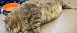
Most often, hematomas in cats occur on the inner surface of the ear and much less often on the outside. The damaged ear increases in size, hangs down, and the swelling is painful and hot to the touch. If the hematoma is not treated, the pain only increases, and the hematoma itself can become infected with secondary microflora, which can ultimately lead to necrosis of the ear cartilage.
During a clinical examination of such a cat, a veterinarian notes the following symptoms:
- We observe anxiety and nervousness in the cat.
- The cat almost constantly shakes its head from side to side.
- He constantly scratches his damaged ear with his paws.
- When trying to stroke the cat's head, it becomes aggressive.
Treatment. Treatment of auricular hematoma is not very difficult. If no more than 48 hours have passed since the formation of the ear hematoma, then the cat owner fixes the ears with a bandage on the back of the head and applies cold. In the future, in order to resolve the hematoma, it is necessary to use heat and apply locally irritating ointments.
If the hematoma cannot be cured at home, the owner needs to contact the nearest veterinary clinic. In a veterinary clinic, the veterinarian will open the resulting hematoma, remove blood clots from it, wash the resulting cavity with a solution of novocaine with antibiotics and give recommendations to the owner so that the hematoma resolves safely.
Possible causes of ear swelling
Tumors in a cat's ear can lead to complete deafness and sometimes death. Reasons that can cause them.
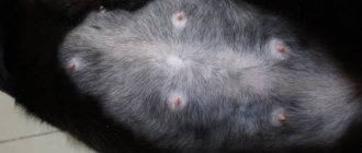
Ticks
They dig into the thin skin on the ear, causing inflammation;
Otitis
This is an inflammatory process that can lead to a tumor in the cat's ear;
© shutterstock
Hematomas
This is a tumor that may look like a swelling filled with blood. The cat's ears will appear droopy and the area of the hematoma will appear hot. The cat becomes nervous and aggressive. He constantly scratches his swollen ears and shakes his head. Hematomas can lead to serious complications, and in some cases to a disease such as cartilage necrosis;
Abscess
Any injury received may be accompanied by the penetration of a pathogenic infection into it. Those. if a cat’s ear swells due to injury, then most likely pus has accumulated inside, which can result in an abscess;
Lymphextravasate
In cases of injury, lymphatic extravasation may also develop. However, in this case, a bubble containing blood and lymph forms on the swollen ear.
© shutterstock
Neoplasm
These include papillomas, sarcomas and fibromas. They are quite dangerous, as they grow in the cat’s ear and block the entrance to the auricle, which can lead to deafness.
If any tumors appear in your cat, you should immediately consult a doctor. The clinic will conduct an examination, take the necessary tests, based on the results of which they will offer treatment options.
If the situation is not critical, then it will be suggested to be treated with antibiotics, as well as rinsing the swollen ear with disinfectants, etc. Don't be afraid that your cat's ears are swollen. Perhaps it’s just otitis media, but you should always play it safe and give your cat all the help you can.
If the tumor threatens the health and life of the pet, the doctor will suggest surgical intervention. If all goes well, the cat will recover within a year with proper treatment.
Otohematoma in a cat
Lymphatic extravasation of the auricle in cats
Lymphatic extravasation is an accumulation of lymph in a cavity formed as a result of tissue dissection and rupture of lymphatic vessels.
Lymphatic extravasation of the auricle in a cat occurs for the same reasons as a hematoma.
It develops slowly in a cat and is characterized by the development of a contouring swelling in the ear area; there is no local increase in temperature.
The diagnosis is made by clinical signs. To clarify the diagnosis, a puncture of the resulting swelling is performed.
Treatment. With this disease, unlike a hematoma, it is strictly forbidden to use cold or heat. In case of this disease, the owner must contact a veterinary clinic. Where the drained lymph will be aspirated using a syringe. If this procedure does not lead to the desired results, then the veterinarian resorts to surgery, which boils down to making a skin incision and more thoroughly removing the contents of the cavity and applying small sutures.
Symptoms of hematoma
The ear hangs, swelling is visible, the animal does not touch it, and reacts aggressively to touch. When the ear is examined, a dark red or black spot is visible. The hematoma is soft to the touch, fluctuation is audible, and skin tension is felt. Later, the pain disappears or decreases, but the swelling remains and the blood clots.
First aid should be provided at home. At the first sign of a hematoma, apply a pressure bandage to stop the bleeding. The skin is first treated with antiseptics; it is rational to apply a cold compress (ice in bags) to the bandaged ear.
If it is possible to stop the spread of the hematoma, then the operation is not performed, since the clotted blood will resolve itself. Otherwise, it is necessary to remove blood clots, for which surgical intervention is resorted to.
[custom_ads_shortcode2]
Necrosis of the auricle
Necrosis of the auricle in a cat can occur as a result of:
- Transfer of purulent processes from surrounding tissues to the auricle.
- With prolonged squeezing of the auricle.
- Infection of hematomas, lymphatic extravasates with pathogenic microflora and with an abscess in the ear area.
When a purulent process develops in the area of the auricle and in the absence of proper and necessary treatment, the resulting abscess opens, forming areas of skin necrosis (necrosis), resulting in ulcers appearing on the auricle.
During a clinical examination, the ear cartilage begins to be visible through the areas of damage, and its blood circulation is disrupted. The cartilage itself becomes black in color and emits an unpleasant putrid odor. With necrosis, the cartilage tissue rots, and the ear becomes deformed.
Treatment. Treatment of auricular necrosis should be carried out in a veterinary clinic. A veterinary specialist performs either a complete amputation of the auricle or a necrotic part of it, followed by a course of treatment with antibiotics.
Etiology
A hematoma is a consequence of severe trauma and often appears in cats as a result of a fight with other animals. Another cause is scratching in the presence of ear parasites: mites, fleas, scabies.
In this case, treatment for an ear hematoma in a cat should include therapy against skin parasites. Scratching may be a consequence of the presence of debris in the ear (grains, twigs).
The formation of a hematoma begins with damage to blood vessels without compromising the integrity of the skin. As a result, the flowing blood begins to stretch the surrounding tissues due to pressure. This process occurs until the pressure inside the vessel is balanced by the resistance of the stretched tissues.
Small hematomas resolve on their own, but large ones without treatment form a subcutaneous thrombus , which is later replaced by connective tissue. Further, calcium salts may be deposited in this area, causing a cyst to form. When infection penetrates, an abscess or phlegmon forms at the site of the hematoma.
[custom_ads_shortcode1]
Foreign body in the external auditory canal
Foreign bodies can get parts of plants, insect larvae, sand, lice into the ear canal, wax plugs and other objects can form.
Sometimes the presence of a foreign body in a cat’s ear does not cause any concern and may go unnoticed by the animal’s owners. Most often, a foreign body causes irritation and inflammation in the external auditory canal.
Treatment. Treatment should be aimed at removing the foreign body from the ear. After removing it, the ear canal is washed with a solution of soda or a 3% solution of hydrogen peroxide. In order to reduce the cat's pain reaction, it is necessary to drop a few drops of camphor oil into the ear canal.
Neoplasms in the external auditory canal
In cats, the most common neoplasms in the external auditory canal are sarcoma, fibroma and papilloma. As they grow, they cause the cat to become deaf.
If a cat has neoplasms, the main signs are:
- The cat's head is lowered towards the affected ear.
- We note uncoordinated and manege movements in the cat.
Treatment. Treatment of tumors in the external auditory canal is only surgical, which must be carried out in a veterinary clinic.
Dermatitis and eczema of the ears
With dermatitis, the cat's ear turns red and a rash appears on the skin. A sick cat begins to scratch its ears due to severe itching, increasing the symptoms of dermatitis. With dermatitis, hair begins to fall out from the damaged area of the skin. Food allergies cause dermatitis in cats. Streptococcosis (Streptococcosis of dogs and cats) can lead to dermatitis.
Parasitic otitis.
Parasitic otitis in cats occurs with otodectosis and notoedrosis (Otodectosis in cats, notoedrosis (pruritic scabies).
Symptoms and disease identification
Animals with an ear hematoma have fluid-filled swelling in the entire ear or just part of the pinna. On palpation, the tumor may appear hard, sometimes soft and fluctuating. This can lead to blockage of the ear canal or death (necrosis) of the tip of the ear. A veterinarian can diagnose this condition during a physical examination. To make a diagnosis, it is important to tell your veterinarian about symptoms of infection (your pet scratching its ears or shaking its head excessively). The veterinarian will likely examine the ear canal with an otoscope and take a swab from the ear to examine under a microscope to determine if parasites or infection are present. Allergic skin diseases (including reactions to food) are the most common conditions under which this condition occurs in dogs. No dog or cat is immune from ear hematoma. Allergic skin diseases, provided the animal is prone to allergies, are extremely common circumstances for the development of hematomas. Dogs with heavy floppy ears are most susceptible to the disease because such ears hit the skull quite hard when shaking their heads.
[custom_ads_shortcode2]
Otitis in cats
Otitis in cats can be due to inflammation of the outer, middle and inner ear.
Causes of otitis. The cause of otitis in cats can be food allergies, the presence of parasites (ticks, fleas), ear injuries, foreign bodies, etc. Otitis of the middle and inner ear can be the result of complications of inflammation of the outer ear.
Signs of otitis media . During a clinical examination of a sick cat, the skin of the sore ear is reddened, the cat rubs the sore ear with its paw, and tries to keep the sore ear folded and pressed to its head. If the pain in the ear is acute, “shooting,” the cat suddenly jumps up, looks around in fear, and screams. With constant pain, the cat does not allow the sore ear to be touched, avoids stroking the head, and presses the sore ear to its bedding.
With purulent otitis media, inflammatory exudate is released from the cat's ear; when touched, the ear gurgles and squelches. An unpleasant odor emanates from the diseased ear.
In advanced cases, in the absence of proper treatment, the cat’s body temperature rises, the cat becomes depressed, and there is no appetite. If timely treatment measures are not taken, the eardrum may be perforated, and the inflammatory process may spread to the brain.
Treatment. Treatment of otitis media depends on the severity and severity of the inflammatory process in the ear. In the initial stage, the cat is prescribed special drops (Bars), Stop-Itching spray, antibacterial drugs (tylosin, etc.)
Prevention of otitis. Prevention of otitis in cats should be based on compliance with the rules of care and maintenance of cats. To help keep your pet healthy:
- Timely cleansing of the ear canal from accumulated wax. To do this, the pharmacy chain has recently recommended using a special lotion - “Dewdrop for the ears”, which is used to remove wax and inflammatory products from the auricle and external auditory canal.
- When bathing your cat, be careful not to let water get into its ears.
- Avoid keeping your cat in damp and cool areas to prevent hypothermia.
- In order to prevent infection with ear mites, try to avoid contact with stray cats.
Periodically disinfect and decontaminate the cat's place of residence and cat care items.
Surgical treatment of auricular hematoma
An animal ear hematoma is an accumulation of a mixture of blood and lymph in the area of the auricle between the skin and cartilage tissue. This is a fairly common occurrence in cats and dogs. The Best veterinary clinic in Novosibirsk provides services for surgical treatment of auricular hematoma on the day of treatment.
The formation of otohematoma is associated with damage to blood vessels in the ear area. This may happen as a result of:
- bruises;
- insect or other animal bites;
- scratching (including those caused by chronic otitis, allergies or otodecosis);
- chronic dermatitis.
Otohematoma looks like a hemispherical tumor on the surface of the auricle. The formation is soft to the touch, painful, hyperemic. Possibly mild fever and anxiety. The animal constantly tilts its head towards the damaged ear, obsessively tries to scratch it, or simply shakes its head.
Treatment of auricular hematoma
Uncomplicated cases are treated by evacuating accumulated blood and lymph by puncture with a needle, followed by aspiration of the fluid and injection of anti-inflammatory drugs into the resulting cavity. Typically, a solution of novocaine (0.5%), antibiotic and hydrocortisone is used for these purposes.
Surgery to remove blood
Suction does not always give results, so if after 5-7 suctions no effect is observed, then an operation is prescribed to open the hematoma and suture the vessel. Surgery is also recommended to remove a blood clot when the blood in the hematoma has already clotted. Surgery is performed under general anesthesia.
Anesthesia Before anesthesia, a fasting diet is required for 8-12 hours; 2-3 hours before surgery, the cat should not be allowed to drink. There are two common anesthesia mixtures:
- Premedication . A 1% solution of atropine sulfate is injected subcutaneously in an amount of 0.5-1 ml/kg of animal weight. After 15-20 minutes, cats are injected intramuscularly with a 2% solution of Rometar in an amount of 0.1 ml/kg body weight. In the same dosage, the ketamine-containing drug ursotomin can be used intramuscularly. When using Rometar, it is recommended to administer a 1% ketamine solution intramuscularly at a dose of 0.1 ml/kg.
- Thiopental sodium anesthesia . Fast-acting type of anesthesia. A thiopental sodium solution is used in an amount of 0.025-0.03 g/kg of cat weight, the drug is administered intravenously at a concentration of 5-10%. It is especially important to administer the drug slowly to preserve respiratory and heart function. The action occurs instantly - after the introduction of the first drops. The anesthesia itself lasts 20-40 minutes, and post-anesthesia sleep lasts 1-4 hours.
Operation technique First, treat the skin with any available antiseptic. Then an incision is made along the ear in the center of the hematoma. If there is heavy bleeding from a damaged vessel, then it is blocked by pinching it with your fingers. Liquid blood is removed using a syringe, and blood clots are removed using tweezers; however, blood clots should not be torn off by force; you must act carefully so as not to create a new wound.
If necessary, the vessel is sutured with a ligature. After all manipulations, the hematoma cavity is washed with an antiseptic solution (0.02% furatsilin). Usually the doctor stitches the ear through, this leads to complete healing of the ear, but it may lose its natural shape and become thicker than normal. Therefore, it is recommended to use a rolled suture, especially for competition cats. The stitches are removed after 10 days.
Rolled suture technology Used on tissues with high tension, such as stretched skin due to a hematoma. For stitching, short gauze rolls, one centimeter long and half a centimeter thick, should be prepared in advance. There should be at least twice as many of them as the required number of stitches.
The skin is pierced with a needle at a distance of 0.5-1.5 cm from the edge of the incision; with strong tissue tension, the distance can be doubled. In this case, the needle is not passed simultaneously through two edges of the wound, but first the first edge is pierced, the needle is taken out, and only then the other edge is stitched. It is especially important that when puncturing the wound surface, the thread passes over the very bottom of the wound.
To create a beaded seam, use a thick silk thread, threaded so that the two ends of the threads are of equal length. When the seam is created, the needle is removed, resulting in a loop at the first edge, and hanging threads at the opposite edge. At both edges, rollers are inserted between the threads, and then tied tightly.
Symptoms and Disease Identification An otohematoma, or pinna hematoma, is a blood “pouch” that forms on the outside of the pinna, between the skin and cartilage in your pet's pinna. As a rule, otohematoma is caused by damage to the ear due to scratching or occurs as a result of too intense and frequent shaking of the head, which leads to injury. Both dogs and cats suffer from ear hematomas, although dogs (especially those breeds that are prone to allergies and ear infections) are more susceptible to the disease. Otohematomas are usually caused by some kind of self-inflicted trauma - for example, when the pet aggressively scratches and scratches the ears, or shakes its head, causing the ears to be injured when they hit the head. This injury can lead to bleeding, vascular damage, and blood pooling in the space between the skin and cartilage. In general, there are underlying causes of ear scratching and excessive head shaking:
- ear mites
- bacterial and/or fungal infections in the external auditory canal (otitis media)
- allergic skin diseases are considered one of the main causes of bruises - dogs that suffer from allergies are much more susceptible to ear infections
[custom_ads_shortcode1]
Causes and treatment methods for ear hematoma in cats
Due to their curiosity and mobility, furry pets are often subject to injuries to various organs, including the ears, which result in hematomas. This pathological formation is a hemorrhage of varying degrees into the space between the cartilage and the skin of the ear.
The danger of a hematoma lies in the risk of developing unpleasant complications in the form of inflammatory processes, abscesses and even blood poisoning. After a hematoma, deformation of the auricle often develops.
Read in this article
Causes of auricular hematoma
The development of the disease occurs due to mechanical effects on blood vessels. The cat's ear has thin and delicate skin, is equipped with a numerous capillary network, and therefore often suffers from hemorrhages. Damage to a blood vessel leads to the fact that blood, having no outlet, fills the interstitial space.
In the case of an ear hematoma, blood accumulates in the space between the cartilage tissue of the organ and the skin. The result is swelling, compression of nerve endings, and pain.
The structure of the ears of cats and kittens
The main cause of pathology is mechanical damage. Free-ranging cats have an increased risk of ear hematoma. On the street, such an animal faces many dangers. A cat can injure its ear as a result of a showdown with its relatives, or in fights with its eternal enemies – dogs.
Sometimes the auricle is injured when an animal falls from trees, roofs of houses, or balconies. Often the cause of mechanical damage and subsequent development of hematoma is a collision with a vehicle.
A cat can get a hemorrhage in the ear area not only on the street due to injury. A common cause of the development of the disease are all kinds of non-contagious, infectious and parasitic diseases.
Common otitis media, fungal infections of the auricle, the presence of diseases such as otodectosis and other diseases accompanied by itching in this area force the animal to itch and damage the blood vessels. Self-injury occurs when excessive head shaking occurs due to ear pathologies.
The presence of fleas in a cat, an allergic reaction, or bites of blood-sucking insects is also the cause of the development of hemorrhage due to the mechanical impact on the ear when scratching with its paws.
The cause of a hematoma in a domestic cat can also be surgery in the area of the auricle. In this case, the damage may be completely normal as a consequence of the intervention. A serious postoperative complication develops, as a rule, when the rules of asepsis and antisepsis are not followed, the cauterization of the wound is poor, as well as when the sutures are damaged as a result of violation of the rules of animal care.
Why does pathology occur?
In a cat, the outer part of the ear consists of 2 layers of dermis, supplied with blood vessels and a cartilage pad. Under the influence of unfavorable factors, the ear is injured or compressed, causing blood vessels to rupture and blood to accumulate under the skin. Most often, a cat injures itself with its claws if it is infected with ear mites (otodectosis disease). Hematomas also appear on the ear, paw or back due to bites of subcutaneous parasites. In addition, the etiology of otohematoma is as follows:
- hitting your ear on a heavy object while playing;
- decorative items falling on the pet's head;
- allergic reaction;
- autoimmune failure;
- chronic ear inflammation.
Stages of development
Veterinary specialists note the following stages of the pathological process in the development of hemorrhages in the area of the outer ear in animals.
This leads to a change in the color of the blood - it turns blue. The animal has a red or purplish-bluish swelling and mild pain when touched. The degree of pain reaction largely depends on the size of the damage.
As a rule, the first stage of hematoma develops within 24 hours after mechanical damage to the soft tissues of the organ.
Such stages are typical for hemorrhages uncomplicated by infection. In case of extensive release of blood from the vessels, failure to provide assistance to the animal in a timely manner at any stage of the disease, an inflammatory process, infection and suppuration of the injury may develop.
Treatment of the auricle
The choice of treatment tactics for the disease largely depends on the size of the hemorrhage and the stage of the pathological process. For small hematomas, cold is indicated in the first hours after injury. Frozen foods from the freezer wrapped in cloth are suitable for this purpose. You can cool the damaged ear for 10 - 15 minutes.
If there is an open wound, it is treated with disinfectants. To reduce blood leakage, a pressure bandage is applied to the ear.
If the hematoma occurs as a result of an allergy, an infectious or parasitic disease, or otitis media, then priority attention is paid to the underlying disease.
For extensive hematomas, due to the fact that there is a high risk of infection, the most effective method of treatment is surgery. In addition, when the pathology progresses spontaneously in the final stage, fibrinous tissue grows at the site of damage. This leads to deformation of the ear cartilage, which is undesirable for show animals.
During the operation, the veterinarian opens the hematoma using an incision at the base of the ear. Blood clots and fibrin are removed. At the end of the manipulation, a fixing suture and bandage are applied. Surgery is usually performed under local anesthesia. After the operation, the animal must be in a special collar that prevents the paw from scratching the ear.
Pumping out blood with a syringe
The positive aspect of the surgical treatment method is the minimal risk of relapse, as well as the preservation of the natural shape of the ear cartilage. The disadvantages of surgery include a small stitch in the cat's ear.
In addition to surgical treatment, a veterinarian can use special dressings for show animals. A special feature of this method of treating hematoma is the absence of postoperative scars. The disadvantage is that the owner cannot apply the bandage himself; constant visits to the veterinary clinic are required.
A common way to treat small, fresh hematomas is to drain the blood with a syringe. The method is often used when the blood that has escaped from the blood vessels has not yet clotted.
Anti-inflammatory and antibacterial ointments are widely used in the treatment of the disease: levomycin, syntomycin, levomycin. For suppuration, ichthyol and Vishnevsky ointment are used. Additionally, if the hematoma becomes infected, the animal is prescribed a course of antibacterial therapy. For this purpose, penicillin antibiotics, tetracyclines, and cephalosporins are used.
Blood suction
The operation should be performed immediately after detection of a large hematoma, while the blood has not yet clotted. The blood is sucked out using a syringe. Due to the low invasiveness, the cat is not subjected to anesthesia, it is not even necessary to perform anesthesia, but it is important to firmly fix the animal so that the needle does not cause additional damage.
The needle is inserted into the upper part of the ear, and the blood is sucked out carefully so as not to create excessively low pressure in the hematoma cavity. After suctioning the blood, the needle is not removed, but the syringe is disconnected from it and a solution of antibiotics and novocaine is injected through the same needle. After the operation, a pressure bandage and collar are applied to prevent the animal from trying to remove it.
[custom_ads_shortcode3]
Prevention of hemorrhages in cats
In order to prevent such an unpleasant illness as hemorrhages on the outer ear of animals, veterinary experts recommend that owners adhere to the following rules:
- Prevention of injuries and mechanical damage to the ear. Sterilization and castration of pets and restricting the cat’s free access to the street help reduce their risk.
- Timely treatment of ear diseases, including tick-borne, fungal and infectious etiologies.
- Identification of allergen and treatment of allergic manifestations in cats.
- Prevention of infection of an animal with parasites (use of special collars, long-term drops, etc.).
Hematoma of the ear in domestic cats is not such a rare phenomenon. Thin skin and many blood vessels contribute to the development of hemorrhage even with minor organ trauma. The characteristic clinical signs of the disease, however, require confirmation from professionals.
One effective treatment is surgical removal of blood clots. For show animals with minor injuries, special bandages are applied. The owner should be aware that hematomas can become infected and lead to deformation of the ear cartilage. In this regard, self-medication is unacceptable.