Symptoms Prognosis Treatment
Thromboembolism is a pathology characterized by acute circulatory disorders (ischemia) of tissues and organs, and resulting from blockage (embolization, thrombosis) of a section of an artery.
Thromboembolism is classified according to the site of blockage: thromboembolism of the abdominal aorta, pulmonary artery, and cerebral vessels.
Most often in cats, thromboembolism of the abdominal aorta occurs at the site of its branching into the femoral arteries (aortic trifurcation).
Thromboembolism is most likely as a complication against the background of heart pathologies, sepsis, infectious pathologies, or trauma (mechanical compression) of one or another part of the vessel.
This pathology is most common in cats with asymptomatic cardiomyopathy. Thromboembolism develops against the background of enlarged left chambers of the heart and pronounced congestion in the vessels of the lungs, impaired blood flow, decreased blood flow speed and high platelet aggregation in cats.
In addition, this pathology is a complication due to prolonged compression of the abdominal wall, for example, if the cat is stuck in a narrow passage (window for “winter ventilation”).
Symptoms of thromboembolism in cats
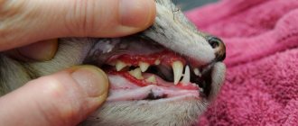
Symptoms of thromboembolism in cats depend on the location of the process. With aortic thromboembolism, the following symptoms are observed: lameness, altered gait of the animal. When examining and palpating the hind limbs, paresis or paralysis of both hind limbs is revealed. The muscles on them become very hard and painful. Externally, you can notice the pallor of the paw pads and claws.
Lower back pain, vomiting and a rapid increase in the amount of nitrogenous metabolic products in the blood, due to impaired renal function, all this may indicate thromboembolism of the renal arteries.
Sharp pain in the abdominal area without a specific localization, vomiting and diarrhea, often mixed with blood, these symptoms are characteristic of thromboembolism of the mesenteric arteries.
Thromboembolism of the blood vessels of the brain can be determined due to clear signs: seizures, coma, lesions of the vestibular apparatus.
Thromboembolism of the pulmonary artery; at the first glance at the animal, breathing problems, coughing, weakness and an anxious state of the animal are noted. A detailed examination of the pet can reveal rapid shallow breathing, weak pulse, swelling of the jugular veins, and pale mucous membranes.
The danger of thromboembolism lies in the high degree of mortality in cats, primarily due to blockage of blood vessels, and ischemic toxins cause significant damage to the cat’s body when they enter the bloodstream, they have a pathogenic effect on vital organs and systems.
For a positive outcome of thromboembolism, the most important thing is to contact highly qualified veterinarians in a timely manner. As soon as possible, you need to make a correct diagnosis and prescribe the necessary treatment measures. All this will help save lives and minimize damage to your pet’s health.
What it is?
Typically, thromboembolism refers to aortic thromboembolism, which means that a blood clot (usually originating in the heart) is lodged in the aorta. Of course, this pathology can also be venous, but still it is the aortic variety that leads to the most severe consequences.
It usually begins with a blood clot forming in the left atrium. Of course, in the case of a normal and healthy heart, this situation is unlikely, but with cardiomyopathy this often happens. Soon it begins to move down and reaches a place called bifurcation. There the aorta divides into two branches. If the clot is small, then it passes in one of the directions, after which (as a rule) it tightly clogs one of the vessels of the extremities .
It’s even worse when the blood clot remains at the bifurcation site and completely blocks the blood flow to both paws. This is generally fraught with the sudden death of the pet.
The mechanism of formation of a venous thrombus is the same, only it all happens much faster. The fact is that the blood flow in the arteries is very strong, their intima (inner lining) is very smooth. All this leads to the fact that under normal conditions the formation of a blood clot there is very, very rare. But Vienna is a completely different case. Blood moves under relatively low pressure, slowly. If there are at least some predisposing factors (vascular disease, poisoning, parasitic infestations), then a blood clot will almost certainly appear. Another thing is that such pronounced consequences (sudden death), which occur with aortic thromboembolism, still occur infrequently.
Diagnosis of thromboembolism
With pronounced clinical signs, diagnosing thromboembolism is not difficult. If the symptoms of this disease are unclear, a series of diagnostic measures should be urgently carried out.
Blood is taken from the cat for a biochemical blood test, and a study is also carried out to determine the blood clotting time. In addition to blood diagnostics, it is necessary to perform an ultrasound of the heart (echocardiography), this procedure allows you to visualize the heart, determine anatomical and topographical features, reduction or enlargement of the atria and ventricles, and assess myocardial contractility.
To get a complete picture of the animal’s condition, a veterinarian performs electrocardiography.
When diagnosing thromboembolism, radiography (angiography) is mandatory.
Angiography is a contrast X-ray examination of blood vessels, which allows one to study the functional state of the vessels and the extent of the pathological process.
Clinical signs and initial diagnosis
Diagnostic Notes
Additional diagnostic methods
In addition to the location of the thrombus itself on CT, it is necessary to examine other tissues and organs for the presence of contrast defects. In our practice, in animals with TKA, we found small infarcts of the renal cortex, which previously could not be detected on ultrasound (Fig. 8), and a segmental defect in the distribution of contrast in the spleen parenchyma.
Visual diagnosis of animals with thrombosis gives us not only a diagnosis and topographic orientation of the pathology, but also an algorithm for further treatment of such patients and life prognosis.
⦁ Laboratory diagnostics (general clinical, biochemical blood tests, electrolyte studies) can reveal a variety of biochemical disorders. Most cats exhibit stress hyperglycemia, prerenal azotemia (which may also be associated with renal artery thromboembolism), hyperphosphatemia, and a sharp increase in serum creatine kinase. There are reports of hypocalcemia and hyponatremia. A potentially dangerous complication of thromboembolism is an increase in potassium concentration, often occurring suddenly as a consequence of restoration of tissue perfusion, although potassium levels may be low at the time of the initial study. Additionally, coagulation tests may be performed, although they are often normal.
Treatment of arterial thromboembolism
Any treatment that results in sudden reperfusion of ischemic tissue carries the risk of life-threatening complications of reperfusion injury, so the prognosis is usually guarded to poor. Surgical treatment (embolectomy using a balloon catheter or surgery) is rarely used because cats are at increased risk and often die during surgery or subsequently develop another blood clot. One of the publications of foreign colleagues mentions the successful removal of a blood clot from the arteries in five out of six cats using rheolytic thrombectomy. Therapeutic treatment. Currently, most veterinarians prefer medications to treat arterial thromboembolism.
Treatment of thromboembolism in cats
Treatment of thromboembolism in cats directly depends on how much time has passed since the occurrence of this complication and the visit to the veterinary clinic.
Taking measures to restore blood flow through a blocked blood vessel. This problem is solved by surgical intervention. The aorta is opened to remove the blood clot, thereby freeing blood flow and preventing further continuation of coronary artery disease.
Infusion therapy is carried out to preserve the liquid part of the blood in the vascular bed.
Thrombolytic drugs are prescribed to prevent the formation of new blood clots. The use of these drugs should be carefully monitored by a veterinarian to avoid bleeding and hemorrhagic complications.
Causes
Blood clots can form in blood vessels due to the following factors:
- infection and sepsis;
- poisoning of an animal with toxic substances;
- pathologies of the cardiovascular system;
- oncological diseases;
- the presence of enzymes in the blood;
- mechanical damage to blood vessels;
- previous operations.
It is important for cat owners to know that, according to statistics, these animals more often than others suffer from diseases of the cardiovascular system. Therefore, the formation of clots in arteries and veins is not uncommon for them.
Treatment
Hospital treatment is recommended for cats with aortic thromboembolism, as most require treatment for concomitant heart failure.
Physical activity should be limited and rest and protection from stress should be ensured. Most cats become anorexic. It is necessary to monitor nutrition to avoid the development of liver lipoidosis. You should limit your salt intake for a long time.
Thrombolytic therapy with streptokinase and tissue plasminogen activator is widely used in humans and less commonly in cats.
The drugs are dangerous due to the possibility of bleeding.
It is preferable to use heparin, preferably low molecular weight. It does not affect already formed blood clots, but prevents further thrombus formation. The first injection is intravenous at a rate of 200–300 IU/kg, subsequent injections are administered subcutaneously at a rate of 100–300 IU/kg every 8 hours. The dose is determined by the activation time of thromboplastin.
Aspirin is useful during and after a thromboembolic episode as an antiplatelet agent. 81 mg tablets are given every 2nd or 3rd day.
Clopidogrel has fewer side effects on the gastrointestinal tract; it is used in a dosage of 18.75 mg orally every 24 hours.
Butorphanol is used as an analgesic at a dose of 0.5–1.0 mg every 6–8 hours, acepromazine at a dose of 0.1–0.5 mg as a sedative and vasodilator.
Warfarin (prescribed with caution), a vitamin K antagonist, long-acting anticoagulant, is prescribed to prevent recurrent episodes of thromboembolism. The initial dose is 0.25–0.5 mg orally once a day. Provide an overlapping course of heparin administration for 3 days. The subsequent dose of warfarin is determined by an increase in prothrombin time by an average of 2 times from the initial level or, according to the international system, by 2–4 times.
In case of heart diseases, they are simultaneously treated.
It should be borne in mind that the use of anticoagulants or thrombolytic drugs can lead to hemorrhagic complications. Non-selective beta-blockers (eg, propranolol) should not be used due to the possibility of peripheral vasoconstriction.
Many cats experience recurrent embolism, but most animals survive. They need to carry out the same anticoagulant therapy, which often requires examination and home lifestyle. In this case, the function of the limbs is practically restored, but neurological disorders may remain.
Treatment and prognosis
Prevention and treatment of animals with suspected thromboembolism involves the use of anticoagulant drugs (heparin, dalteparin, warfarin, aspirin, etc.) and the use of painkillers to reduce pain that occurs due to neuromyopathy. Surgical treatment is usually ineffective.
The prognosis for cats with thrombosis of the vessels of the extremities is cautious, often unfavorable. Most cats die early after thrombosis, or owners are forced to use euthanasia in order to relieve the animal from excruciating pain.
It is worth remembering that early diagnosis of hypertrophic cardiomyopathy (HCM), renal failure or thyroid pathologies allows for timely prevention of further thrombus formation.
Diagnostics
Pain and acute paralysis are the most common symptoms. Lameness, changes in gait, tachypnea or respiratory distress, vocalization, and anxiety are also observed.
Upon examination, paraparesis or paralysis of the hind limbs is revealed, and less commonly monoparesis of the forelimbs. Palpation of the limbs is painful. The calf muscle often remains hard several hours after the embolism. The femoral artery pulse is absent or significantly reduced. Nails and paw pads are cyanotic or pale. Abnormal galloping heart rhythm, arrhythmias, tachypnea, or dyspnea are observed.
In the differential diagnosis of paresis of the hind limbs, one should first assume a spinal cord neoplasm, trauma, myelitis, ischemic lesion, or intervertebral disc herniation.
Laboratory and other research methods
In response to muscle damage, creatine phosphokinase activity increases. When the liver and muscles are damaged, the activity of alanine aminotransferase and aspartate aminotransferase increases.
As a reflection of the stress response, glucose concentration increases. With embolism of the renal vessels, the content of blood urea nitrogen and creatinine increases slightly.
Blood clotting tests usually do not reveal abnormalities, since hypercoagulation is a consequence of platelet aggregation. Blood clotting indices may be useful in determining heparin and warfarin doses.
X-ray reveals cardiomegaly in 85–90% of patients, pulmonary edema or pleural effusion in 66% of cases.
Echocardiography. In most cats, pathological hypertrophy is often found of the left ventricle without expansion of its cavity, as well as expansion of the left atrium, and increased contractility of the heart. Restrictive cardiomyopathy should then be ruled out.
Dilatation of the cavities of the heart and thrombosis of the left atrium may be detected. Among heart diseases, aortic thromboembolism often reveals left atrial hypertrophy by echocardiography (>50%).
The atrium/aorta ratio is 1:2 or more.
Abdominal ultrasonography and/or angiography can be used to determine
thrombus in the caudal part of the aorta.
Electrocardiography. The most commonly recorded is sinus tachycardia, much less frequently - atrial fibrillation, ventricular and supraventricular arrhythmias, sinus bradycardia, left ventricular hypertrophy or conduction disturbance of the anterior branch of the left bundle branch.
