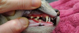
Often, dog owners turn to veterinarians with complaints that their pets have health problems - the dog’s eyes are red, purulent crusts form, tears flow heavily. One of the most common causes of this pathological condition is canine dry eye syndrome (DES).
Symptoms of the disease
The symptoms of the pathology at the initial stage are in many ways similar to conjunctivitis, so breeders are in no hurry to seek help from a veterinary clinic, considering the condition to be harmless.
Among the first manifestations are:
- redness, swelling of the conjunctiva;
- occasionally appearing purulent discharge;
- intense lacrimation.
Determining the development of a dangerous disease based on these signs can be problematic. Symptoms are in many ways similar to other ophthalmological pathologies .
After some time, the animal's condition worsens.
The following changes are noted:
- copious discharge of pus, with the eyelids becoming glued together;
- dryness around the eyes;
- the cornea becomes cloudy;
- blepharospasm;
- ulcerations in the cornea;
- the third eyelid falls out;
- keratitis.
Symptoms of the disease
The clinical picture of dry eye syndrome varies depending on the form and severity of its manifestation.
Mild severity
Paradoxically, at the first stage of development of dry eye syndrome, increased tear production is observed. In this way, up to a certain point, the animal’s body tries to compensate for the lost ability of the eye to maintain the protective tear membrane. But gradually, discharge from the corners of the eyes takes the form of translucent mucous threads, the pet begins to blink frequently, hits itself in the eyes, and the conjunctiva becomes hyperemic and swollen.
Moderate severity
Characteristic signs of the second phase of keratoconjunctivitis sicca are:
- dullness and lack of specularity of the cornea;
- catarrhal-purulent discharge from the eyes in the form of strands;
- adhesion of the conjunctiva to the cornea;
- constantly slightly open palpebral fissures;
- photophobia;
- erosions on the cornea.
Severe form
In advanced cases, the following phenomena are observed:
- the cornea becomes lumpy due to the proliferation of blood vessels in it;
- erosions develop into ulcers;
- Dried crusts of pus constantly accumulate in the corners of the eyes;
- blepharospasm is observed.
Next, cloudy areas form on the cornea, which then merge, and the dog completely loses vision.
Reasons for development
For normal functioning of the eye, tear fluid must be produced, which moisturizes the cornea.
A tear not only wets the surface of the eyeball, but also washes away microorganisms and foreign bodies. When this process is disrupted, dry eye syndrome develops.
The surface of the eye is moistened by the work of a number of structures. The tear film is produced by the lacrimal glands, when their activity is disrupted, the mucous membranes become dry.
Causes of pathological changes:
- congenital anomalies of the lacrimal gland (underdevelopment, complete absence);
- chemical, thermal burns of the conjunctiva;
- mechanical damage;
- age-related features affecting tear synthesis;
- chronic inflammatory process of the ciliary margin;
- removal of the third eyelid;
- use of certain groups of medications;
- improper nutrition (lack of vitamins, starvation);
- herpesvirus;
- neurological diseases;
- neoplasms;
- the presence of systemic pathologies (diseases of the skin, kidneys, thyroid gland, diabetes).
Some dog breeds are more predisposed to developing the disease, including:
- Yorkshire Terrier;
- Cocker Spaniel;
- poodle;
- English bulldog;
- pug;
- Shih Tzu.
Treatment of the disease
Treatment of dry keratoconjunctivitis in dogs is based on the use of tear replacement agents that provide the missing moisture to the cornea and the use of drugs that dissolve mucus. But such treatment is now considered outdated, especially when the cause of the pathology is autoimmune destruction of the lacrimal gland.
According to the study, a real breakthrough in the treatment of the disease was the use of cyclosporine locally. The drug acts by stimulating the functioning of the lacrimal gland, in addition to having an immunosuppressive effect (suppressing the body's immune response). The product is used in the form of ointments and drops; it can suppress the process of destruction of the lacrimal gland and partially restore normal tear production.
Cyclosporine preparations are used 1-2 times a day for 3 weeks. After this, the Schirmer test is repeated. If there is no improvement, then increase the frequency of cyclosporine use to 3 times a day. If the dynamics are positive, reduce gradually to 1 time per week.
According to the observation of specialists, the effectiveness of drug treatment is effective even in animals with low Schirmer test scores. At 2 mm, 8 out of 10 dogs are cured (80%).
The use of antibiotics locally (ointments, solutions) is necessary to prevent bacterial complications. Glucocorticoids are used to reduce inflammation, they somewhat inhibit the growth of blood vessels into the cornea, and reduce abnormal pigmentation. But with prolonged use they can provoke ulcerative lesions, and are also not used for ulcerative keratitis. To liquefy dense and viscous deposits, mucolytics (acetylcysteine) are used.
If your dog has problems with vision, there are signs of inflammation or discharge from the eyes - do not hesitate! Call the RosVet VC by phone, 24 hours a day. Make an appointment with a veterinary ophthalmologist and bring your pet for an examination. The sooner treatment is started, the higher the chance of preserving the dog’s vision.
Diagnostic methods
Diagnosis begins with collecting anamnesis, a comprehensive examination, and identifying the causes of the development of the pathological process.
The veterinarian conducts a visual examination, assesses the condition, position of the eyelids, frequency of blinking, and the nature of the discharge.
The following diagnostic measures are carried out:
- staining the cornea with a one percent fluorescein solution;
- Schirmer test;
- Norn test;
- bacteriological culture;
- biochemical, general blood tests;
- determination of hormone levels.
How is the diagnosis carried out?
Diagnosis begins with fairly obvious external signs:
- discharge from the eyes;
- red eyes;
- darkened cornea.
The doctor must conduct an ophthalmological examination.
And in order to confirm the diagnosis he does:
- Schirmer test;
- Norna sample.
Nornu sample
The tear film breakup time test (Nornu test) evaluates the time it takes for a drop of fluorescein placed in the eye to begin to break. The norm is 10 seconds. If this indicator is lower, then this confirms the presence of keratoconjunctivitis in the dog.
Important! Do not use over-the-counter drops intended for humans, such as Visine, to treat eye problems in your dog. They are not suitable for animals.
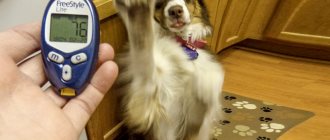
Schirmer test
A test to evaluate lacrimation was first described by German ophthalmologist Otto Schirmer in 1903. The modern test is represented by a strip of special paper measuring 5 × 35 mm. It is impregnated with a special dye.
Once removed from the packaging, the rounded tip of the curved strip is placed in the lower part of the conjunctival fornix near the junction of the middle and temporal third of the eyelid. He remains in this position for 1 minute. The amount of tear produced is visualized by the movement of the dye down the strip. The normal tear rate in dogs ranges from 18.64 ± 4.47 mm/min to 23.90 ± 5.73 mm/min. A deviation from the norm indicates confirmation of the diagnosis.
Methods of therapy
Treatment is selected taking into account the cause that led to the pathological changes. Most often, medications are used, but it is not always possible to fight the disease with this method.
In severe cases, surgery is performed. Sometimes traditional medicine recipes are used during therapy, but before using them you need to consult a doctor.
Drug treatment
Pathology should be treated comprehensively. The therapy is aimed at replacing tear deficiency in a puppy with the help of special solutions, eliminating autoimmune and inflammatory processes.
Treatment is carried out in the following areas:
- Stimulation of tear synthesis . For this, ointments, eye drops Tacrolimus, Cyclosporine are used. Medicines have a stimulating, anti-inflammatory effect. When they are used, the cells are partially restored and begin to produce fluid. The effect is not immediately noticeable. Positive changes are observed after a few days.
- Elimination of the inflammatory process . For this purpose, topical drugs are prescribed, corticosteroids and ophthalmic antibiotics are prescribed.
- Use of tear substitutes . Replenishing fluid deficiency is the main direction of therapy. Artificial substitutes are used, produced in the form of gels and drops.
- Antibacterial treatment . Produced to control secondary microflora. Antibiotics are prescribed in the form of drops, which have a wide spectrum of action.
- Corneal protectors . The drugs help to activate metabolic processes in affected tissues, improve trophism, and stimulate tissue regeneration.
- Antihistamines . They help prevent and relieve allergies characteristic of certain forms of pathology.
Surgery
Among the surgical techniques, the following stand out:
- occlusion of lacrimal openings;
- transfer of ducts of the parotid salivary gland;
- partial tarsorrhaphy.
Surgery is resorted to when drug therapy is ineffective.
When the duct of the parotid gland is transferred to the eye, saliva begins to flow into the visual organ. It replaces tears, but leaves a small amount of mineral deposits, so your pet will need to periodically instill eye drops.
The disadvantage of the technique is that while eating, the animal intensively produces saliva. It begins to flow down the face.
With partial tarsorrhaphy, the palpebral fissure decreases. The technique is effective in the development of the syndrome against the background of the optic fissure that does not completely close when blinking.
Removal of the superficial corneal layer is carried out when the pigment formed on it causes vision problems. The operation is permissible only if the synthesis of tear fluid is restored.
Treatment with folk remedies
You can use folk remedies at home only after consulting a doctor.
It is worth understanding that self-medication can worsen the situation, and the chances of a favorable outcome will decrease.
If the disease is bacterial in nature, it is permissible to wash the visual organs with chamomile decoction.
To prepare it, 1 tbsp. raw materials are poured with a glass of boiling water, left for about half an hour, and filtered.
A cotton pad is moistened in the product, one eye of the animal is washed, and a new pad is taken for the other.
The procedure is carried out twice a day. It will not be possible to cure the dog in this way, but the development of the pathology will stop and the pain syndrome will be relieved.
It is necessary to properly prepare your dog's diet. The menu should be complete, balanced, rich in nutrients. Malnutrition and starvation during this period are unacceptable.
Keratoconjunctivitis sicca in dogs and cats
Keratoconjunctivitis sicca in a dog
Keratoconjunctivitis sicca (dry eye syndrome) is a pathology in which the function of the lacrimal glands is disrupted, which leads to disruption of the production of the aqueous part of tears. In turn, this leads to dryness and inflammation of the conjunctiva and cornea of the eye.
Normally, tears in animals provide nutrition to all the surface membranes of the eye and perform a protective function. Tears contain many ocular immune factors and antibacterial enzymes. They protect the eyes of animals from the penetration of foreign pathogenic microorganisms.
When the amount of tears decreases, the eye becomes very susceptible to various infections and small irritating particles from the external environment. Against the background of impaired immunity of the eye, purulent conjunctivitis first develops, then inflammation affects the cornea - keratitis occurs with many newly formed vessels.
In the later stages of dry eye syndrome, due to hypoxia, deterioration of tissue trophism, autoimmune damage to the cornea and conjunctiva, the animal becomes completely blind due to total pigmentary keratitis.
This disease can occur with varying degrees of severity of clinical signs and lead to complete loss of vision. Its diagnosis in the early stages is difficult due to the lack of characteristic symptoms. The development of the syndrome is caused not only by pathology of the organ of vision, but also by a number of other factors: general health, genetic predisposition, unfavorable environmental conditions.
Factors contributing to disruption of tear fluid production:
- Injuries to the glands, muscles and nerves of the eye responsible for the function of the lacrimal glands.
- Congenital underdevelopment of the lacrimal glands.
- Atrophy of the lacrimal glands.
- Autoimmune processes in the body.
- Anesthesia (causes temporary dryness of the eye, reducing tear secretion).
- Congenital absence of lacrimal glands (very rare, but sometimes occurs in some toy breeds).
- Removal of the third eyelid or the lacrimal gland that is attached to it.
- Damage to the facial nerve innervating the lacrimal gland.
- Use of sulfonamide drugs.
- Infectious diseases (canine distemper, leishmaniasis, herpes virus).
Regardless of the cause of the disease, it remains for life and requires lifelong care for the eyes of the sick animal. With proper care, the animal is provided with normal, full vision, while in the absence of treatment and proper care, the result can be complete blindness and chronic inflammatory reactions in both eyes, which cause suffering and discomfort to both the animal and its owner.
Among the dog breeds that have a congenital tendency to dry keratoconjunctivitis are the American cocker spaniel, miniature schnauzer, and West Highland white terrier. Purebred and brachycephalic cat breeds are also predisposed to the appearance of such inflammatory processes.
Symptoms
Keratoconjunctivitis sicca can develop due to many reasons, therefore the clinical manifestations will depend on the nature of the underlying process. Distinguish between acute and chronic; one- or two-sided; temporary or permanent dry keratoconjunctivitis.
Characteristic signs of keratoconjunctivitis sicca are:
- Blepharospasm.
- Corneal ulcers (possibly with perforation).
- Pigmentation and dullness of the cornea.
- Mucous or mucopurulent discharge
- Hyperemia (redness) of the conjunctiva
- The cornea exhibits dryness, loss of shine, clouding and swelling.
- Pigmentous keratitis (the cornea is replaced by an opaque black opacification) - begins from the periphery and spreads to the center of the cornea, closing the pupillary zone.
- Keratitis (inflammation of the cornea with vascularization, pigmentation).
- Deterioration of visual function with severe keratitis.
Bilateral keratoconjunctivitis sicca in a cat
The most characteristic sign of keratoconjunctivitis sicca is a sticky, thick discharge that adheres to the inner corner of the eye. The purulent component of the discharge can be sterile and develop due to infiltration of the cornea and conjunctiva with inflammatory cells, and it can also be septic when a secondary bacterial infection is attached.
Also, dry keratoconjunctivitis of dogs and cats is characterized by blepharospasm and protrusion of the third eyelid, the severity varies significantly depending on the remaining sensitivity of the ocular surface. In severe or acute cases, there is loss of corneal epithelium with the formation of ulcers (especially in the center); in rare cases, corneal perforation and anterior uveitis develop.
In chronic cases of keratoconjunctivitis sicca, superficial as well as deep vascularization and pigmentation of the cornea are noted. These changes are the main cause of visual impairment in this disease.
Dullness of the cornea in dogs is observed only in 25% of cases. Dry nasal planum is more often observed in the neurogenic form of keratoconjunctivitis sicca.
Diagnostics
The diagnosis of keratoconjunctivitis sicca is established based on the medical history, characteristic clinical signs and special tests.
The stability of the tear film can be determined using the Norn test: 1 drop of 0.2% fluorescein sodium is instilled into the lower conjunctival sac. The time from the last blink to the appearance of a break in the tinted tear film in the form of a black spot or gap on the surface of the cornea is determined. The time it takes for the tear film to break down is an important indicator of its stability.
Another important method for determining the function of the lacrimal glands is the Schirmer test, which establishes the total tear production. To perform this test, some pharmaceutical companies produce special strips of filter paper. The strip is bent at the marked end at an angle of 45° and placed in the lower conjunctival fornix in the outer third of the palpebral fissure: the bend should lie on the edge of the eyelid, and the bent part of the strip should not touch the conjunctiva. The animal's eye is closed, after 1 minute the strip is taken out and the result is taken into account by measuring the length of the moistened area from the fold line.
Also, corneal damage is identified by staining with Rosebengal and fluorescein, and systemic diseases of animals (diabetes mellitus, hypothyroidism, polyarthritis and polymyositis, immune-mediated skin diseases) are also excluded.
Treatment
Complex treatment of dry keratoconjunctivitis comes down to replacing the lack of tears with artificial solutions and relieving inflammation and autoimmune processes in the eye and lacrimal glands.
In general, treatment includes several main areas:
- Stimulation of tear production. It is achieved by prescribing cyclosporine and tacrolimus in the form of eye drops and ointments. Cyclosporine and tacrolimus have anti-inflammatory and stimulating effects on the lacrimal gland. Thanks to this, the epithelial cells of the lacrimal gland begin to partially recover and produce fluid. It is important to remember that these drugs may not begin to work immediately, but may take several days, and are not effective in all animals.
- Anti-inflammatory therapy. To treat the infectious and inflammatory components of dry eye syndrome, ophthalmic antibiotics and corticosteroid drugs are used topically.
- Use of artificial tear substitutes. Replacing tear deficiency is one of the main areas of treatment for keratoconjunctivitis sicca. In practice, it involves the use of artificial tear substitutes in the form of drops and gels.
- Antibacterial therapy - to control secondary microflora (eye drops with broad-spectrum antibiotics).
- Purpose of corneal protectors. They activate metabolism in the tissues of the cornea and conjunctiva, improving trophism and stimulating regeneration processes.
- Antiallergic drugs - to prevent or relieve allergic reactions characteristic of some forms of dry eye syndrome.
Surgical methods include occlusion of the lacrimal openings, transposition of the parotid duct into the inferior conjunctival sac, and partial tarsorrhaphy.
When treating dry eye syndrome, it is important to correct the general condition of the animal based on the examination results obtained. For example, with hypothyroidism, the symptoms of keratoconjunctivitis sicca can significantly decrease, and in mild cases, disappear during replacement therapy with thyroid hormones.
The main general signs of conjunctivitis in dogs
- discharge from the eyes (watery, mucous, purulent),
- narrow palpebral fissure,
- blinking,
- closing an eye,
- redness,
- swelling of the conjunctiva,
- formation of bubbles (chronic course).
Treatment of conjunctivitis is initially aimed at hygienic treatment of the eye and the use of various eye medications (depending on the nature of the inflammation). These may be drops or ointments containing glucocorticoids or antibiotics. The active ingredients in them are aimed at reducing swelling, itching and excessive lacrimation; have anti-inflammatory and antibacterial effects.
Diagnostics
To accurately diagnose this disease, a special test is performed to determine tear production - the Schirmer test. The test kit consists of a strip of filter paper of a certain type, which is usually impregnated with dye to make the result easy to read.
To perform this, a strip of filter paper is folded at the marking end and placed in the lower conjunctival sac beyond the edge of the eyelid. Measure the length of the moistened section of the strip in 1 minute. Before the test, the contents of the conjunctival sac should be removed with a dry cotton swab. In this case, preliminary anesthesia is not performed.
The norm is to wet a paint-impregnated filter strip to a length of 12-15 mm in 1 minute for dogs and 10 mm in 1 minute for cats. There may be discrepancies between the results of testing both eyes, but a difference greater than 27% is clinically significant.
The Schirmer I test is performed using the method described above, the results of which describe the total secretion of tears. In order to distinguish between the portion of the “reflex” (on contact of the eye with the test strip) secretion and the portion of the “basic” tear released during the same time, L. Jones made changes to the test in 1966, calling it the Schirmer II test.
To perform it, drops for local anesthesia are first instilled into the conjunctival sac and residual moisture is removed from the fornix with a cotton swab; the test is also carried out for 1 minute, after which the results are evaluated.
Purulent conjunctivitis in puppies
Occurs under closed eyelids (after birth), the pathology is also called “physiological ankyloblepharon.” The cause of inflammation can be intrauterine infections.
Symptoms:
- swelling of the closed eye,
- Characteristic discharge accumulates in the inner corner of the eye.
Treatment:
- a closed eye is opened manually or surgically,
- the sore eye is cleaned of secretions with a cotton swab dipped in chamomile decoction,
- antibacterial eye ointments are prescribed.
Symptoms of conjunctivitis in dogs
Canine conjunctivitis, regardless of the type and form of the disease, manifests itself with the following characteristic symptoms:
- Baldness of the eyelids.
- Swelling of the eyelid and mucous membranes of the eyes.
- The appearance of specific discharge, either purulent-mucous in nature.
You can also suspect the disease by paying attention to the behavior of your pet. Since conjunctivitis is accompanied by a feeling of discomfort and severe pain localized in the visual area, the dog becomes nervous, irritable, passive, often closes its eyes and rubs its eyes with its paws. Often with conjunctivitis, the animal experiences sleep disturbances and permanent pain.
Main causes
You need to prepare for the fact that sometimes you won’t be able to detect them. Then you will have to monitor the animal’s condition and constantly alleviate it with drops and eye ointments. The problem is complex, but the following reasons can be identified:
| Immunity | Immunity problems. The lacrimal gland becomes inflamed, after which its dysfunction is observed. This organ is very small, so inflammation is not visible from the outside. The problem can also be autoimmune in nature, when the body attacks itself for a number of reasons, causing a disruption in basic functionality. Usually these are the consequences of long-term drug therapy or the consequences of anesthesia. |
| Eye loss | In small breeds like pugs and French bulldogs, the eye may fall out of its socket due to injury or excessive play. This does not have to be very visible; there may simply be a slight displacement, but the lacrimal gland will be compressed. In this case, swelling is possible due to the accumulation of fluid under the eyelids. |
| Medicines | Medicines can also greatly affect tear production. This is especially true for those varieties that retain fluid in the body. Typically these are medications containing various alkaloids, as well as strong painkillers. |
| Operation | Tears disappear after complex surgical intervention in a dog’s body. There are cases when this happened after stitching a wound on the face. This is due to a disturbance in nerve stimulation; a necessary channel has been interrupted somewhere. This is a common reaction after eye surgery. |
| Systemic diseases | Lead to an atrophied lacrimal gland. Long-term conjunctivitis can lead to the complete destruction of this organ. It is simply eliminated by bacteria, turning into pus. Eye functions are not restored in dogs with advanced forms of inflammation. |
| Pathology | In the smallest breeds, this disease can be congenital as a result of pathology. Experts also often associate this with inbreeding. |