Pyometra
– accumulation of purulent exudate in the uterine cavity.
It is the most serious problem of the reproductive tract in females.
The prerequisites for the development of CGE in bitches are the regularly repeated effects of progesterone on the endometrium (a series of estrous cycles usually not accompanied by pregnancy). As a rule, this pathology occurs in middle-aged and older females. It can also occur in young animals, but less frequently. The use of contraceptives increases the risk of pyometra by up to 12%
Pyometra is less common in cats. This is due to the fact that ovulation does not occur if there has been no mating, therefore, the uterus is not exposed to progesterone and cystic changes in the endometrium do not occur. Both cats that have given birth and those that have not given birth are equally susceptible to this pathology. Older animals get sick more often than young ones.
Basic information
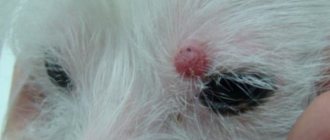
This is the name of the pathological process in which tissue cells begin to grow rapidly.
Moreover, it is to grow, and not to divide randomly, as is the case with cancerous tumors. In particular, with prostatic hyperplasia, the size of the latter can reach an orange! However, this brings little joy to a sick animal: such a rapid increase in cell volume provokes severe pain in the affected organs. In addition, at least 40% of cases of hyperplasia ultimately end in cancer. But it's not all that scary. For example, gum hyperplasia in dogs during puppyhood is normal, as teeth grow at this time. But the same process, if it begins against the background of stomatitis, can lead to oral cancer.
What tissues are most often affected by this disease? In most cases, only glandular tissue and epithelial cells undergo changes. Simply put, the same hyperplasia in dogs during estrus is a disease of the mammary glands. Sometimes there is damage to the vagina and/or uterus. Other organs suffer from this pathology much less frequently.
Adrenal glands
The congenital anomaly is accompanied by the following symptoms:
- underdevelopment of the mammary glands;
- abnormal arrangement of the fundus vessels;
- convulsions in the dark.
The disease is recognized by clinical signs, laboratory tests, radiography, and ultrasound. Treatment is lifelong hormone therapy.
Occurs when the structure of consumed feed is disrupted. It is characterized by an increase in the volume of the gum adjacent to the tooth. Pockets form and fill with food debris, and gingivitis or periodontitis develops. The dog finds it difficult to eat, darkening of the tooth enamel is observed, and there is a stench from the mouth. Gums are bleeding.
To determine whether the tumor is benign, a biopsy sample is taken. Treatment begins with ultrasonic teeth cleaning. If suppuration develops, they resort to dental surgery. To prevent exacerbation, use toothpaste, brush, and diet food.
Therapy and prognosis
What is the treatment? If your pet belongs to that same 30%, then your intervention or the help of a veterinarian will not be required: when the estrus ends, the hormonal levels will return to normal, and the swollen tissue of the vulva will return to its normal parameters. As for cases where the tissue does fall out of the vagina, it is necessary to use moisturizing and healing ointments, as well as bandages used to protect such a delicate organ from possible damage.
It is important to prevent licking, for which surgical collars are used. If it comes to inflammation (dirt gets in, or as a result of injury), the surface of the vulvar mucosa is inflamed and purulent, surgical intervention and a long course of antibiotic therapy may be required. But it’s better not to let things get to this point, and immediately take the injured animal to the veterinarians.
In most cases, the prognosis for the disease is favorable, despite the fact that hyperplasia may well return in subsequent heat cycles. Alas, if a dog is predisposed to this pathology, the only 100% effective solution to the problem is to remove the uterus and ovaries.
Purulent vulvitis and vaginitis in dogs
Inflammation of the vulva (vulvitis) and vaginal mucosa (endovaginitis) is observed in puppies, young immature females, adults and castrated females.
Etiology. It occurs as a result of the introduction of pathogenic bacteria and fungi into the tissues of the vulva and vagina. Predisposing factors in adult dogs may be injuries, and in castrated dogs - a decrease in estrogen titer.
Symptoms and course. In bitches with an undisturbed general condition, swelling and pain of the vulva, redness of the mucous membrane of the vestibule and vagina, purulent or mucopurulent discharge from the genitals are recorded. Purulent vaginitis must be differentiated from endometritis and pyometra.
Treatment of dogs consists of washing the vagina with weakly disinfectant or astringent solutions, followed by intravaginal administration of iodoglycol, sulfonamide emulsions or antibiotics, taking into account the sensitivity of the microorganisms isolated from them, as well as ointments containing corticosteroid drugs. In some cases, antibiotic therapy in the form of a course of treatment is also indicated. In immature bitches, local treatment is often ineffective and is abandoned. With the onset of the first emptying, purulent vaginal discharge usually stops. Neutered female dogs are prescribed estrogen drugs (sinestrol) in small doses.
Breast hyperplasia
This type is much more common, and its consequences are much worse than in the previous case. How does it all begin? As in the case we discussed above, the main “trigger” is a hormonal imbalance in the body, and therefore the disease develops against the background of estrus, pregnancy, imminent childbirth, or treatment during which progesterone is used.
The clinical picture is very similar to mastitis: the area adjacent to the nipples greatly increases in volume. In special cases, the nipples hang down almost to the knee joint. On palpation, soft, dough-like tissue is felt. Unlike mastitis, the animal does not experience pain during this operation. In addition, exudate does not ooze from the mammary glands, and the local temperature is not elevated.
Unlike vaginal hyperplasia, diagnosis of breast lesions is significantly complicated. While almost any experienced breeder can distinguish this pathology from mastitis, it will not be so easy to differentiate this process from the initial stages of oncology. This will require a biopsy. A piece of tissue is taken, which is then carefully examined by a veterinary cytologist.
What is the therapy? Firstly, all hormonal medications, if any, are canceled. Secondly, animals are prescribed progesterone and prolactin inhibitors. All this time, the dog should be under the close supervision of a veterinarian: if a worsening of the process is noticed, then the dog may have to be sterilized, simultaneously removing all the swollen mammary glands. The reason is simple - the possibility of developing breast cancer.
dog 10 years old Vaginal prolapse (June 2020).
Some breeds are more susceptible to the effects of vaginal hyperplasia than others, with Boerboels being higher on the list than those that do. The condition occurs when a woman is in season and it is where the soft tissue around her vagina becomes very swollen. The swelling is due to a reaction to the estrogen she produces at a certain stage during her cycle, and the problem begins when the swelling is so excessive that it causes tissue to fall out of her vulva.
Prevention
If your dog is capable of reproducing, but you do not allow him to mate, this may increase the likelihood of developing the diseases described in this article. Therefore, sterilization (or spaying) is the best form of prevention.
07.10.2016
Cystic (glandular) hyperplasia (change) of the endometrium is a hormone-dependent change in the endometrium, characterized by the formation of glandular structures.
The endometrium is the inner mucous membrane of the uterine body (mucosal layer), lining the uterine cavity and abundantly supplied with blood vessels.
Pyometra is an accumulation of pus in the uterine cavity due to the introduction of bacteria into the altered endometrium. Repeated exposure of the endometrium to high concentrations of hormones (estrogens and progesterone) in the absence of pregnancy leads to cystic endometrial hyperplasia. Bacteria enter the altered endometrium ascendingly from the vagina through the cervix, which partially opens before and during ovulation. As a rule, pathogens belong to the normal vaginal flora: Escherichia coli is the most common. Old (after 6 years) nulliparous animals get sick more often. Taking estrogen or progesterone also provokes cystic endometrial hyperplasia. Pyometra can develop in the uterine stump in the absence of ovaries. Therefore, if you decide to sterilize your animal, it is best to remove both the ovaries and the uterus during this operation.
The figure shows the normal state of the uterus and the pyometra itself.
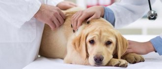
DIAGNOSIS In dogs, the disease is usually diagnosed 12 weeks after ovulation; in cats this period may vary. When the cervix is closed, general symptoms are more often observed: depression, drowsiness, anorexia (lack of appetite), polyuria (increased volume of urine) and polydipsia (extreme thirst), vomiting, bloating, fever (not always). Septicemia and shock may develop. Palpation determines the enlargement of the uterus, and sometimes its size can be determined. Rough palpation can lead to uterine rupture. If the cervix is open, the uterus may not be enlarged. In this case, bloody or mucopurulent discharge is observed. The disease is differentiated from pregnancy, other conditions characterized by polyuria and polydipsia (diabetes mellitus, hypercortisolism, kidney disease), and vaginal diseases. LABORATORY AND OTHER RESEARCH METHODS In the blood, neutrophilic leukocytosis with a shift of the formula to the left, moderate normocytic normochromic anemia, hyperglobulinemia and hyperproteinemia, azotemia, increased activity of alanine aminotransferase and aspartate aminotransferase (with septicemia or severe dehydration), and sometimes electrolyte disorders are observed. Cytological examination of vaginal discharge reveals regenerative polymorphonuclear cells and bacteria. However, with diseases of the vagina (vaginitis, tumor, foreign body, developmental anomalies), the picture may be similar. A bacteriological examination is also performed, although it rarely helps to establish a diagnosis, as it reveals normal vaginal flora, and an antibiotic sensitivity test. Serological methods can detect Brucella canis. X-rays can reveal an enlarged uterus and recognize pregnancy (45 days after ovulation or 43-54 days after mating). When pus accumulates in the uterus, an x-ray shows an oblong formation in the lower abdomen.
The photo shows an X-ray of a dog. The diagnosis is pyometra.
The size of the uterus can be determined by ultrasound, which also helps to assess the degree of endometrial hyperplasia and exclude pregnancy (starting from 20-24 days after ovulation). The normal, unchanged uterine wall is not visible. With cystic endometrial hyperplasia and pyometra, thickening of the organ wall and fluid in its lumen are observed. In dogs (rarely in cats), pyometra can be combined with pregnancy.
The photo shows an ultrasound image. Diagnosis of pyometra and cystic endometrial hyperplasia. TREATMENT The development of pyometra with a closed cervix can threaten the life of the animal. Emergency hospitalization is required. With pyometra, regardless of the degree of cervical dilatation, extirpation (removal) of the uterus and appendages is indicated. The operation is carried out only in a clinic; if necessary, the animal is left in the hospital until its condition stabilizes.
The photo shows the dog's uterine horns filled with purulent exudate. Drug treatment is carried out to preserve reproductive function in purebred animals or, with the consent of the owner, to provide lifelong treatment with non-gestagenic drugs. NB! Surgery is much more effective than drug therapy. In dogs, prostaglandin F2α (PGF2α) is prescribed. Stop taking it when the uterus acquires normal size according to palpation, radiography, and ultrasound. If the animal is in the corpus luteum phase (serum progesterone >2 ng/ml), prostaglandin is administered subcutaneously every 12 hours for 4 days. A single dose of prostaglandin promotes contraction of smooth muscles and, consequently, evacuation of the contents of the uterine cavity. Double administration also causes luteolysis and a decrease in serum progesterone concentrations. The animal is re-examined 2-4 weeks after stopping the use of PGF2α. If the uterus has increased in size again or vaginal discharge persists, the course is repeated. In case of refractoriness to prostaglandin, extirpation of the uterus and appendages is performed. Cats are given PGF2α subcutaneously once daily for 2–5 days until the uterine size returns to normal. Side effects of PGF2α include hypersalivation, vomiting, defecation and (in cats) the need for intensive vulvar care. The side effect occurs a few minutes after the injection and lasts 30-60 minutes. The frequency and severity of side effects can be reduced by diluting the drug with an equal volume of isotonic sodium chloride solution, and also by forcing the animal to move for 20-30 minutes after administration. It should be taken into account that PGF2α causes strong contractions of the uterus, which, if the cervix is closed, can lead to uterine rupture or the release of pus through the fallopian tubes into the abdominal cavity with the development of secondary peritonitis. Before prescribing PGF2α, you should make sure that there is no pregnancy, especially in purebred animals, since the drug helps to terminate it. All animals with pyometra are treated with systemic antibiotics. The course of antibiotic treatment lasts depending on the condition of the animal and is prescribed by the attending physician. FOLLOW-UP Discharge from hospital occurs when the patient's clinical condition improves. Vaginal discharge may continue for up to 4 weeks. After surgery, neutrophilic leukocytosis remains in the general blood test for some time.
Endometritis in dogs is an inflammation of the uterine cavity. As a rule, this phenomenon is caused by pathogenic microorganisms. In some cases, the disease occurs in combination with other serious illnesses, then the dog’s life is at risk and urgent medical intervention is necessary. To avoid unpleasant consequences, the owner must monitor changes in the health of his pet, because this is how the first symptoms of the disease can be suspected.
Clinical picture
Note that the symptoms of endometritis in dogs are in most cases quite standard. A sick dog develops streaks of exudate in the genital area. When drying out, the latter turns into crusts. If an animal has developed necrotic endometritis, it emits an unbearable smell of rot. With purulent inflammation, the pet's genital area is always covered with a greenish-yellow mass.
Despite the abundant discharge from the external genitalia, the general condition of the animal, as a rule, remains stable. The exception is advanced cases of purulent endometritis, as well as inflammation of the necrotic type . The latter, by the way, is always accompanied by a sharp deterioration in the animal’s condition. The dog's general body temperature rises, appetite decreases or disappears altogether, and thirst may increase. All this is due to significant intoxication of the body.
If a sufficient amount of exudate accumulates in the uterine cavity, the organ can be easily felt by palpating the abdominal wall. The latter, by the way, swells greatly and sags. In advanced cases, you may think that the dog is pregnant again. With purulent and necrotic endometritis, sepsis is likely to develop. In such cases, the animal’s overall body temperature drops, it becomes lethargic and weak. If you notice these signs in your pet, call your veterinarian immediately, as further delay could cost your dog his life!
And further. Considering that endometritis is almost always accompanied by severe intoxication, an excessive burden falls on the animal’s liver and kidneys. If the dog is no longer young and already has problems with these organs (not to mention cases of kidney/liver failure), everything can end very badly.
Symptoms of pyometra
Speaking about the manifestations of endometritis, one cannot fail to mention pyometra, which is often a consequence of this disease. Interestingly, in some cases its first manifestations can be seen during estrus.
The initial symptom of the pathology is a sharp and sudden increase in general body temperature. However, the appearance of this sign is much more typical on days 20-70 from the end of estrus (Bigliardi and Pamigiani 2004). In more than 93% of cases, the clinical picture of pyometra developed at 12 weeks from the end of estrus (Borresen 1979). The pathology develops against the background of glandular hyperplasia and cystic degeneration of the endometrium of the uterus. It is believed that the mentioned proliferative and secretive changes are an effect in response to the action of estrogens and progesterone (hyperestrogenization of the body). In addition, ovarian dysfunction in bitches often contributes to degenerative changes in the uterus, which not only contributes to the development of endometritis and/or pyometra, but also significantly aggravates their course. As for the characteristics of the exudate released from the genital tract of dogs with this pathology... most often it is not there. Only occasionally can a small amount of reddish-gray or brown exudate be seen at the root of the tail of a sick animal, from which a disgusting, putrid odor emanates.
Causes of endometritis
Veterinarians believe that the most common cause of endometrial inflammation in dogs is hormonal disorders. The imbalance negatively affects the entire body as a whole, because of it the walls of the uterus thicken and an excessive amount of secretion accumulates. Often against this background, purulent endometritis is diagnosed; the secretion leaves the dog’s body along with serous discharge.
Hormonal imbalances are possible for many reasons. In dogs, quite often they are caused by a phenomenon called false pregnancy. The animal is diagnosed with all the signs of an “interesting” situation: swelling of the nipples, enlargement of the abdomen, weight gain, and the appearance of milk. False pregnancy occurs in parous and nulliparous individuals. Hormonal disruptions are possible during a dog’s puberty, as well as due to heredity.
Endometritis appears after an infection from the vagina penetrates into the uterine cavity. Various vaginitis, diseases of the genital organs, genitourinary system - all this can cause inflammation of the endometrium.
Endometritis often occurs during childbirth. A dog can get it if it is carrying very large puppies, because in this case there is a high chance of ruptures and erosions on the walls of the uterus.
If the puppy dies at the time of birth or while still in the womb, the dog, as a rule, becomes ill. Decomposition of fetal tissue occurs, as a result of which bacteria multiply, causing inflammation. In most cases, endometritis against this background is severe, with purulent discharge.
Then, when a person helps a dog give birth, you need to remember about hygiene. Sometimes there is no time to wait for the veterinarian, then the owner decides to pull the puppies out by hand. Infection will occur if such procedures are carried out without antiseptics and hygiene gloves. Bacteria can enter the body in the event of ruptures or microtraumas of the vagina, they can also occur due to the fault of the owner who does not know the basics of obstetrics.
About some features of endometritis
It should be remembered that under normal conditions the internal cavity of the uterus is completely sterile. Of course, pathogenic and conditionally pathogenic microorganisms can still get into it, but in most cases nothing bad happens. Due to the special environment and secretions continuously secreted by the uterine epithelium, the microflora usually dies. The most dangerous period is the postpartum period , since it is at this time that the maximum number of microorganisms enter the organ cavity, and the natural defense mechanisms of the animal’s body do not always cope with them. It is believed that during the normal course of the postpartum period, sterility should be completely restored within about one week. If this does not happen, endometritis of the uterus is likely to develop. But still, one should not assume that this disease develops exclusively after childbirth. It happens that inflammation of the uterus is diagnosed in a female who has not yet given birth. There is nothing “mystical” about this: such cases are associated with serious infections and parasitic diseases, when the pathogen can enter the uterine cavity.
As for the direct “culprits” of endometritis, most often they are gram-negative anaerobic organisms. Symptoms of infection range from obvious and persistent purulent uterine effusion to asymptomatic infection. In severe cases, the uterus becomes so swollen with exudate that it can be detected by simple palpation of the abdominal cavity.
The presence of purulent effusion in the vagina does not necessarily confirm endometritis: a similar clinical picture is characteristic, as we have already said, of vaginitis, as well as cervicitis (inflammation of the cervix). Diagnosis is especially difficult in small animals, since it is not possible to use a vaginal speculum. Chronic endometritis can be even more difficult to identify, since the animal does not have any clinical manifestations. In such cases, cytology of scrapings of the uterine mucosa is indicated, and ultrasonography or intrauterine biopsy is also necessary.
The danger of all types of endometritis is that they can “mutate” into pyometra. But this disease is much more dangerous and often leads to death.
Symptoms of endometritis in dogs
The first signs that endometritis is developing in the dog’s uterus do not appear immediately. Symptoms usually appear after 3-5 days, in some cases the incubation period lasts longer, can reach up to 7 days. Sometimes endometritis is characterized by mild symptoms; its manifestation may not be noticed at all for a long time.
The disease has 2 forms:
- Spicy.
- Chronic.
In most cases, the acute form of endometritis appears after childbirth, during which bacterial infection occurred. This form is characterized by pronounced symptoms that are difficult to miss:
- The general condition and mood of the animal changes, lethargy, apathy, and reluctance to move appear.
- Serous masses are released from the genital organs; in some cases, purulent impurities are possible.
- Body temperature is increased.
- The dog often (without special need) sits in a position in which urination occurs.
- In some cases, the animal suffers from bouts of vomiting.
- Due to vomiting, the dog’s body loses a lot of fluid, so the animal may be tormented by previously uncharacteristic thirst.
- Frequent urination is possible, the dog begins to urinate in the apartment.
- The owner can independently detect abnormalities in the dog’s heartbeat by placing his ear to the animal’s chest.
- Due to the strain on the heart muscle, breathing may become difficult and becomes difficult.
- If the treatment of acute endometritis is delayed, the dog begins to lose hair and hyperpigmentation occurs - all this is a consequence of hormonal disorders.
Chronic endometritis is possible when the dog is not treated for the acute form of the disease; it also occurs against the background of a hormonal imbalance. The main symptoms include:
- Violation of estrus - its duration, time intervals from one to another.
- Reducing body weight.
- Weak puppies that often die after birth.
- It is extremely difficult to get pregnant, and in the future it is impossible, and infertility develops.
These symptoms may indicate not only chronic endometritis, but also many other gynecological diseases in dogs. Therefore, it is important to contact a veterinary clinic for help, where an extensive diagnosis will be carried out and subsequent treatment will be prescribed.
Hyperplasia and vaginal prolapse in dogs
This disease is characterized by protrusion of the vaginal wall from the genital slit outward.
Etiology. In some bitches of a number of breeds (boxer, Doberman, etc.), during emptying, the vaginal mucosa, under the influence of estrogen hormones, hypertrophies and swells so much that it protrudes beyond the genital slit.
Symptoms and course. In practice, complete vaginal prolapse is sometimes observed in bitches within a few minutes as a result of previous inflammation of the digestive tract.
When the disease is mild, only a protrusion of the infiltrated vaginal mucosa in the form of a spherical fold of pink-red color is observed. In severe cases, not only the upper and lateral walls protrude, but also partially the lower wall, the part that is normally located from the vaginal vault to the urethra. It should be noted that this disease is observed at the end of whelping, but more often during estrus, and in some - during every estrus. The mucous membrane of the prolapsed part of the vagina dries out over time, becomes injured, bleeds, undergoes ulceration and necrosis.
Treatment of dogs. In females with vaginal prolapse during estrus, they limit themselves only to washing the prolapsed part with a 2-3% alum solution, and then apply antiseptic ointments. With repeated vaginal prolapse, treatment is reduced to straightening the prolapsed part and strengthening it by placing one or two loop-shaped sutures on the vulva, or to ovariohysterectomy. In case of severe thickening, ulceration and necrosis, the prolapsed part of the vagina is tightly bandaged with silk, without capturing the ends of the ureters. The bandaged part of the vagina disappears after 6-10 days. In some cases, surgery is performed. First, a perineotomy is performed under local or general anesthesia, then a catheter is inserted into the opening of the urethra and the prolapsed part of the vagina is excised. Bleeding is stopped by ligating the vessels, and the wound edges are connected with catgut.
Diagnosis and treatment of endometritis in dogs
After contacting the clinic, the veterinarian will collect anamnesis. He will be interested in the following points:
- Symptoms of the disease.
- The course of estrus and pregnancy.
- Number of births, their course and outcome.
First of all, the veterinarian will be interested in the nature of the dog’s discharge; this needs to be discussed in more detail - to clarify its frequency, color, and consistency.
After the conversation, the animal is examined. The veterinarian will record the dog’s body temperature, examine the condition of the mucous membranes, and palpate the abdominal cavity. The condition of the vaginal walls and discharge is checked.
At this stage, the veterinarian is already able to make a preliminary diagnosis, to confirm which the dog is sent for further research. First of all, they carry out an ultrasound diagnosis, use a sensor to check the condition of the uterus and its walls, and identify the presence of neoplasms - cysts and tumors.
Blood and urine sampling is required to assess the general condition; these tests will immediately identify a possible inflammatory process in the body and evaluate the functioning of the urinary system.
If the doctor detects an abnormal heart rhythm during the examination, the dog will be sent for an EGC, where a clear diagnosis will be revealed. In some cases, an ultrasound and chest x-ray are necessary.
Based on these data, the veterinarian makes a final diagnosis and prescribes a treatment regimen for the pet.
First of all, medications are prescribed aimed at restoring the water-salt balance; this is necessary if the dog has been vomiting. At elevated temperatures, antipyretic drugs are used.
Treatment of inflammation of the uterine walls involves antibiotics and lasts at least 10 days. If the selected drug does not act properly on the pet’s body, bacteriological culture is prescribed.
In advanced cases, surgical intervention may be inevitable, during which the uterus and sometimes the appendages are removed. The operation is performed when there is a high risk of rupture due to accumulated pus, because in this case the dog will face death.
After surgery, the animal spends some time in the clinic, where health is monitored and heart function is assessed. The doctor will also prescribe antibiotics; their use will reduce the risk of postoperative infection.
Physiology and pathophysiology
Modern research has proven that endometritis is a complex pathology, the occurrence and development of which often cannot be explained only by the action of pathogenic microflora that entered the uterus from the outside. This process involves many immunological changes, as well as specific molecular mechanisms responsible for inflammation of the mucous membrane and even the cervix. It is known that the same changes can stimulate the development of pyometra. In addition, a number of foreign researchers insist on the existence of certain genetic mechanisms that also contribute to inflammatory processes in the uterine cavity. But still, all experts agree that endometritis is inextricably (which is not at all surprising) associated with hormonal cycle disorders in animals.
In particular, inflammation of the uterus in more than 70% of cases develops in animals that have a persistent (delayed) corpus luteum in the ovaries. In addition, cystic formations in the ovaries have an extremely negative effect on the condition of the uterus. Why does this happen? Everything is quite simple if you know about the basic physiological characteristics of the dog’s body. The fact is that hormonal pathologies always have a bad effect on the condition of the mucous membrane of the uterus itself. It degrades, and the natural protection of the organ cavity, which prevents the growth and development of pathogenic microflora, deteriorates. Accordingly, the likelihood of endometritis increases significantly.
The very nature of the uterus plays against the animal. This organ is characterized by the fact that during pregnancy, lymphocytes, protective blood cells, cannot penetrate into its cavity. This “miscalculation” of nature can be explained simply: the dog’s own immune system cannot be allowed to destroy unborn puppies. Of course, endometritis is a disease of animals that have already given birth, but only with hormonal pathologies (especially in the presence of a persistent corpus luteum in the ovary), the body may not “know” that there is actually no pregnancy. So the path to the uterus for lymphocytes is blocked. And one more problem. Hormones secreted by the corpus luteum greatly reduce the rigidity of the uterine muscles. This leads to subinvolution of the uterus. Simply put, the organ turns into an analogue of an old, flabby sac. Since the uterus cannot contract, practically no exudate leaves its cavity, and all decay products are absorbed into the blood.
Endogenous variant of endometritis
Cystic endometrial hyperplasia-endometritis complex (CEH/P) is one of the most serious and common uterine diseases in bitches. Moreover, scientists came to this conclusion relatively recently (Kida et al. 2006). This pathology is characterized by severe hyperplasia of the walls of the uterus and the penetration of inflammatory factors into all layers of the organ. Interestingly, this phenomenon was first described back in the 80s of the last century. It is believed that this syndrome can occur in dogs in three types, and they are still mistaken for different, unrelated diseases:
- Pyometra.
- Chronic endometritis.
- Cystic glandular endometrial hyperplasia
The exact reasons for this phenomenon have not yet been explained, but it is known that the stimulus for its development is provided by hormonal pathologies and bacterial contamination of the uterus. But the most important role in the development of this type of endometritis, scientists believe, is played by increased production of estrogen. It is caused by ovarian cysts, neoplasia and other negative factors of the internal environment (Kida et al. 2006). However, the connection between increased levels of sex hormones in the blood and endometritis has been proven relatively long ago (de Bosscher and Ducatelle 2002).
Hormones and endometritis: how are they interconnected?
The problem is aggravated by the fact that estrogens themselves are often prescribed to dogs for certain diseases of the reproductive system. This is due to the fact that they promote the opening of the cervix and increase the rigidity of the muscular lining of the organ. All this contributes to the rapid removal of exudate from the organ cavity.
Other hormones are used to stimulate the reproductive cycle and increase the fertility of animals of breeding value. Such drugs are in considerable demand among professional breeders. Unfortunately, progesterones, commonly used for this purpose, can also stimulate the rapid development of endometritis (Nolte et al., 1990). Because of this, veterinary endocrinologists strongly advise against administering hormonal drugs to dogs under at least three years of age. The consequences of such rash actions can be extremely serious. The fact is that the effect of progesterone is exactly the opposite of estrogen: the cervix closes, its rigidity decreases. If there is a pathogenic or conditionally pathogenic microflora in the organ cavity, an extremely favorable environment is created for its development. Even common E. coli (simple strains), if it entered the uterus before the introduction of hormones, can cause severe necrotizing endometritis (Johnston et al. 1985). In cases where E. coli is represented by more “complex” variants, even strong antibiotics do not always guarantee a cure for the animal.
But the most unpleasant thing is that it is progesterone that enhances the adhesive properties of the uterine membrane, which is why colonies of pathogenic and conditionally pathogenic microorganisms can easily gain a foothold on it. It is assumed that at least 20% of cases of severe endometritis, difficult to treat, have a hormonal etiology. It is also known that this outcome is often caused by unsuccessful intrauterine biopsy, attempts to take scrapings, and ... there are many cases where endometritis developed in dogs after cesarean section due to rejection of suture material by the uterine tissue (Noakes et al. 2001).
Thus, today veterinarians are confident in the hormonal origin of most forms of endometritis: bacterial contamination plays only a secondary role in the development of this pathology. As for specific microorganisms, based on a study of surgically removed uteruses, a simple conclusion can be drawn: in approximately 70% of cases, inflammation of the endometrium is caused by the action of pathogenic and conditionally pathogenic forms of Escherichia coli; various forms of streptococci and Pseudomonas aeruginosa are slightly less common. Some authors believe that acute endometritis of hormonal etiology is more often provoked by the action of hemolytic strains of Escherichia coli (Arora et al. 2006).
Age predisposition
In the relatively distant past, researchers were able to prove that canine endometritis does not have any breed “markers”; all varieties of dogs are equally susceptible to this disease (Niskanen and Trusfield 1998). But the age of animals is a completely different matter. There is a clear correlation here. It is believed that at least 25% of cases of endometritis (that is, a quarter!) of all cases of inflammation of the uterus develop after childbirth in bitches of approximately ten years of age (Egenvall et al. 2001). The average age of animals in which endometritis is even more common is six to ten years (Niskanen and Trusfield 1998). Experts note that in cases where endometritis has developed in a young dog less than five years old, the disease is more than 70% likely to be of a hormonal nature (which means it needs to be treated with special methods).
But there is another breakdown by age. Blendinger and Bostedt (1991) wrote that only 2% of affected bitches were under two years of age, another 9.3% of dogs were between two and four years of age, 28.5% were animals aged five to seven years, and 42% of affected dogs developed in females at 8-10 years of age. Another 15.5% are pets who fell ill between the ages of 11 and 13 years, and another 2.3% are “veterans” whose age is “off scale” for 13 years.
Prevention of endometritis in dogs
Preventive measures can in many cases prevent the development of endometrial inflammation. Since the disease often occurs against the background of hormonal imbalance, it is important to pay attention to your pet. If you have alarming symptoms, you should contact a veterinary clinic, where medications will be prescribed to normalize the level of hormones in the body.
It is important to monitor pregnancy in a dog; it is advisable to conduct an ultrasound diagnosis of the pelvis in the early stages to eliminate the possibility of a false condition. False pregnancy must be treated, otherwise the risk of endometritis increases several times.
It is recommended to periodically examine the dog; if the condition of its genitals or secretions has changed, it is necessary to contact a veterinarian to rule out vaginitis and other infectious diseases.
The issue of dog hygiene is often not taken into account by many owners, but you should wash your pet regularly, keep an eye on it - do not allow it to walk near trash cans, limit communication with other unhealthy-looking dogs, and take care of its coat.
Particular attention should be paid to the birth process of the dog. It is best to call a veterinarian to your home to help your pet. If the owner has to provide emergency obstetric care, be sure to use hygiene gloves, as well as antiseptics, such as Miramistin or Chlorhexidine.
You should act with extreme caution to avoid rupture of the cervix or vagina. If the owner had to provide independent assistance, it is necessary to show the dog to a doctor as soon as possible in order to make sure that the birth process proceeded without consequences.
With careful attention to your pet, the chance of serious consequences is minimal, so the basis for preventing the disease is comprehensive monitoring of the dog.
Medical treatment and at home
Depending on the stage of development of the disease, treatment of endometritis in dogs is:
In the first case, endometritis did not take a severe form , and there is a chance to cope with the help of medications. The doctor prescribes the following medications to treat endometritis in a dog:
- products that normalize water-salt balance if the dog is vomiting;
- antibiotics (if necessary, bacteriological culture is carried out, because the selection of an antibiotic is required individually), the duration of treatment is from 10 to 14 days, it cannot be canceled without a doctor’s prescription;
- drugs that relieve intoxication , and those that support the kidneys, if the dog’s pus is already absorbed into the body.
Important! Even if endometritis is completely cured, it can develop again after another estrus.
Therefore, if a pet is not planning a pregnancy, but she has suffered from the disease, she must be sterilized to avoid a relapse.
Surgical intervention is done when there is a danger of uterine rupture, i.e. Pyometra has already begun. In this case, both the uterus and ovaries are removed from the dog.
After the operation, the pet needs to stay in the clinic so that doctors can closely monitor the progress of recovery. Here they inject antibiotics and anti-intoxication agents, and also monitor to ensure that there is no post-operative infection.
Treatment at home
It is impossible to cure a dog for endometritis without visiting a veterinary clinic! This must be remembered, because the risk of developing the disease is very high, and its consequences are severe.
However, you can help a sick animal, but only after consulting a doctor. What do they usually do?
- Provide balanced, high-quality nutrition and improve diet.
- An external massage of the uterus is performed, i.e. through the stomach. Do this 2 times a day for a week.
- If the veterinarian approves, treat with tissue preparations.
Attention! The best help for a dog owner is to carefully follow the doctor's instructions.
Symptoms
Endometritis has a hidden course. The owner of the animal may not even be aware of the inflammatory process in the animal’s uterus. However, repeated inseminations that do not lead to pregnancy force us to examine the animal more deeply. Sometimes endometritis is detected during artificial insemination during massage.
A symptom of endometritis is often the discharge of various types of exudate from the uterus. Serous, fibrinous, purulent, it can alternate. The consistency of the discharge can be thick or liquid, in color from milky white to brown, depending on the microflora developing in the uterus. Discharge is usually noticed while the animal is lying down.
Cats and dogs often lick the discharge, and it is difficult to determine its nature.
Rectal examination of large animals with massage can improve discharge. In this case, the unevenness of the uterine horns, fluctuation, and the descent of one horn into the abdominal cavity are determined.
Sexual cycles are disrupted, a clear arrhythmia appears, and over time they stop altogether, resulting in infertility.
The general condition of the animal is satisfactory. Milk yield may drop slightly, but appetite and chewing gum are preserved.
When examining the vagina, slight streaky hyperemia, accumulation of mucus and exudate are determined. The cervical canal may be slightly open (by 1-2 fingers).
If the contractility of the uterus is reduced, then the exudate is retained in the cavity. When it is released, a strong stench is felt.
Purulent catarrhal endometritis can be accompanied by an increase in temperature by 1-1.5 ᵒC, a decrease or absence of appetite, loss of productivity, and progressive emaciation. Sexual cycles stop, and a creamy, foul-smelling exudate is released from the vagina.
In dogs, endometritis manifests itself clinically 1.5-2 months after estrus. Discharge may not be observed, but the female’s increased “interest” in the problem area and constant licking of the external genitalia is alarming. Body temperature can rise to 41 ᵒC. During exacerbations, the animal becomes depressed and the appetite is reduced. Endometritis occurs especially often in those animals that are constantly treated with drugs that suppress estrus.
In cats, pathology most often develops after birth, after 5-6 days. Body temperature rises, milk secretion may decrease or stop, and appetite is lost. The cat often takes a posture to urinate, arches its back, worries, and moans. The abdominal area increases and a strong painful reaction occurs.
>Diagnosis
Diagnosis is based on the clinical picture, anamnestic data and the results of rectal examinations.
For small animals, ultrasound and radiology are applicable.
Cystic endometrial hyperplasia in dogs
A little theory
First you need to tell us what kind of pathology is called “endometritis”. It's simple. This is a disease accompanied by inflammation of the uterine mucosa. In severe cases of pathology, the inflammatory process can also involve tissues located below the epithelial layer.
Endometritis can be classified according to the nature of the exudate released:
- Catarrhal type. It is relatively rare in its pure form. With this disease, a thick, mucous, viscous exudate is released from the uterine cavity. As a rule, it is completely transparent, and only sometimes the discharge is somewhat cloudy.
- Purulent endometritis. The most common, common option. As is easy to understand, it is characterized by the release of large volumes of purulent exudate.
- Necrotic variety. One of the most difficult options. It is characterized by necrotic processes not only in the thickness of the mucous membrane, but also in the underlying tissues.
- Purulent hemorrhagic endometritis. Also a severe variant of the disease. As it develops, pus streaked with bloody inclusions emerges from the animal’s uterine cavity.
According to the type of course, the disease is divided into only two types: endometritis can be acute and chronic. Below we will consider all these varieties in more detail, describing, among other things, its clinical picture.