Treatment of musculoskeletal disorders in dogs is a long and complex process that does not always end successfully. Veterinarians often make an erroneous diagnosis of dysplasia, but the prescribed therapeutic measures do not lead to an improvement in the pet’s condition. In this case, it is worth remembering Perthes disease. This is one of the severe orthopedic diseases that occurs mainly in small breed dogs.
What kind of syndrome is this?
Legg-Calvé-Perthes disease. This is the name for aseptic necrosis of the femoral head. It was first described by a Swedish orthopedist in 1909 regarding hip disease in children.
The name was adopted from the names of the doctors who studied this disease in the period from 1909 to 1910, Arthur Legg, Jacques Calvet and George Perthes. In 1937-1938 veterinarians Schnelle and Moltzen-Nielsen described the same problem in dogs, with the exception that in dogs the disease could also affect the glenohumeral joint.
The essence of the disease is the disruption of the blood supply to the epiphysis of the femur, which leads to its gradual death. Deprived of nutrition, bone tissue becomes fragile and collapses, deforming the surrounding cartilage.
How to treat Perthes disease in dogs
Avascular necrosis of the femoral head can be treated conservatively or through surgery, and the optimal method is determined solely on the basis of data obtained by the doctor as a result of studying the x-ray. The dog’s well-being, its appearance, the severity of lameness, joint pain, as well as other clinical manifestations of the disease are only partially taken into account in this case.
Thus, necrotic changes in the joint are conventionally divided into 5 stages:
| Stage | Special name | Degree of development of pathology |
| 1 | Hidden | Changes in the bone tissue of the femoral head are minor, with gradual death of the bone marrow and trabecular (spongy) substance. The cartilage is not damaged. |
| 2 | Impression fracture | Due to thinning, the bone loses its ability to bear the load. As a result, an intra-articular fracture occurs, after which the affected joint thickens and is depressed. At this stage, the animal is only able to partially lean on the affected paw. |
| 3 | Resorption (resorption) | Necrotic areas gradually disintegrate into several parts due to the penetration of the surrounding fibrocartilaginous connective tissue, including blood vessels and nerves. The dog can no longer lean on the affected paw. |
| 4 | Reparation (restoration) | The areas where necrosis occurred are replaced by bone tissue, while the deformed femoral head does not return to its previous configuration, which is externally manifested by severe limitation of movements and severe pain. |
| 5 | Ultimate | In general, the joint restores its structure, returning almost to its basic state, but retains its irregular shape: a mushroom, a saddle or a ball. Bone strength also remains low. The configuration of the articulation of the bones changes, which is why the diseased paw looks shorter. The pain may disappear at this stage, but the animal cannot move normally. |
If there is a breakdown between the pelvic bone and the femur, and the space between the femoral head and acetabulum is uneven, the animal will need surgery.
Important! Conservative treatment of Legg-Calvé-Perthes disease is possible only if the articulation of the bones in the joint remains normal and the head of the femur has retained its original round shape.
With the help of drugs
It is impossible to completely cure Legg-Calvé-Perthes disease with the help of medications - drug therapy can only be prescribed as a course accompanying the recovery process, aimed at reducing pain, stopping the inflammatory process and accelerating the formation of healthy cartilage and bone tissue instead of necrotizing tissue.
For this purpose, accordingly, the following drugs are used:
- analgesics: Metacam, Lidocaine, Bonharen, etc.;
- non-steroidal anti-inflammatory drugs: Vetalgin (Diclofenac), Phenylbutazone, Ketofen (Ketoprofen), Rimodil (Carprofen), Meloxidil (Meloxicam), Trocoxil, etc.;
- chondroprotectors - Artroglycan, Kanvit Chondro, Chionat, Felvit Chondro, Activet GLGL, etc.
Operation
Treatment of Legg-Calvé-Parthes disease by surgery in dogs can be carried out in two ways.
In the first case, the head and neck of the femur are completely removed. This method is called resection hip arthroplasty and is the most common, although it assumes that the animal will, in a certain sense, remain disabled for life. However, the advantages of resection include the relatively low cost of the operation, the possibility of performing it on an animal of any age, and a fairly rapid recovery after surgery.
Did you know? In 1932, the American surgeon Smith Peterson first designed a cap prosthesis for the femoral head, and 7 years later the English doctors Wiles and Moore replaced the entire joint with a metal prosthesis. In 2010, British veterinarian Noel Fitzpatrick successfully implanted two endoprostheses into the ankles of a cat whose hind legs were run over by a harvester.
If the operation is performed at an early stage of the disease, when the dog’s joint muscles are not yet too atrophied, after subsequent physiotherapeutic procedures, within a few months the pet will be able to partially restore motor activity and, importantly, move around without experiencing pain.
The second type of surgical treatment is the replacement of the damaged joint with a full-fledged endoprosthesis. The advantage of this method is that the dog’s motor function is completely restored after the operation, but the complexity and cost of the procedure in this case is much higher than with standard resection.
How to care for your dog during recovery
A peculiarity of the treatment of Legg-Calvé-Parthes disease is that during the rehabilitation period the animal must be provided with absolute rest, which implies complete immobility of the affected joint. For this purpose, the dog must be placed in a crate, and if the disease is detected at a relatively early stage, sometimes such a measure is sufficient to stop the progressive pathology without any additional medications or manipulations, including surgery.
Important! Conservative treatment of aseptic necrosis of the femoral head in a dog using the method of complete immobilization can last from 4 months to six months. Partial restriction of mobility does not give a positive result and almost always leads to joint destruction.
You can only let your pet out of its cage so that it can relieve its natural needs. Periodically, but at least once a month, the doctor prescribes a control x-ray examination and, based on its results, determines how the joint is being restored.
If during this period the pain goes away, and x-rays show that the pet’s condition has returned to normal, the animal returns to normal life. If destructive changes in the hip joint progress even during the period of immobilization (forced immobility), the doctor decides on the advisability of surgery.
Despite the fact that such a strict method of conservative treatment with a high degree of probability leads to the complete disappearance of clinical signs of the disease, the attitude of dog breeders and veterinarians to this type of therapy remains ambiguous, since a dog that has been sitting in a cramped cage for six months subsequently acquires serious psychological problems that are much more severe comes into contact with a person, shows signs of inappropriate behavior, i.e., essentially remains disabled, only not in the physical, but in the psychological sense of the word.
Important! Also, do not forget to monitor your pet's diet. Some types of feed, enriched with many useful elements, can prevent the occurrence of a number of diseases of the joints and bones.
Causes
There is a breed predisposition among breeds. For example, Perthes disease most often occurs in Spitz dogs, Chihuahuas, Mexican hairless breeds, Jack Russell terriers, and Italian greyhounds, and is practically never found in dogs of medium and large breeds. No specific gene was identified.
Researchers are inclined towards a genetic anomaly - underdevelopment of the neurovascular system of the hip joints and the multifactorial appearance of the disease. Also, the majority of patients had a history of shock loads on the joints during the period of active growth.
Perthes disease and its causes
As we have already mentioned, this pathology is observed in small dogs, for example, toy terrier, miniature pinscher, spitz, etc. In this case, the animal experiences necrotization of the femoral head, due to which the normal structure of the joint is destroyed and the condition of the bone tissue in it changes.
The reason for this destructive process is impaired blood circulation in this area, which significantly reduces the supply of nutrients to the tissue and bone marrow of the femoral head.
The result of insufficient blood supply is necrosis. Subsequently, secondary osteoarthritis develops in the affected femoral head, and the dead tissue is replaced by new connective tissue. But the young tissue cannot withstand the mechanical loads on the hip, so there is no talk of recovery, the process only continues to worsen.
One hip joint or both can be affected, so if your pet is limping on its front leg, then the diagnosis of “Perthes disease in dogs” is immediately excluded.
The causes of the disease have not been fully elucidated by veterinarians, but based on many years of observations, doctors have come to the conclusion that there may be several of them:
- The first and main version is a genetic predisposition to this disease in dogs of dwarf breeds.
- Weakness of the musculo-ligamentous system in the hip area, leading to increased load on the hip joint during physical activity in dogs.
- Hormonal disorders occurring in the animal's body.
In dogs, Perthes disease is common to both sexes and first appears in puppyhood, from 5-6 months.
If this disease is diagnosed in a dog, then such an animal is excluded from breeding, because there is a high chance that it will be inherited by its offspring.
Consequences and forecasts
The changes occurring in bone tissue are irreversible. Due to the resulting fragility, a subchondral fracture occurs under any normal load. At this stage, the dog loses the ability to fully rely on the limb, movements are difficult and cause pain.
Subsequently, under the influence of healthy tissue, resorption of necrotic areas and restoration of healthy connective tissue occurs.
But since the head of the joint remains severely deformed, the dog’s mobility of the joint becomes difficult, the ability to lean on the limb is completely lost and, as a result, muscle atrophy and loss of the ability to move.
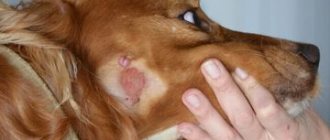
Symptoms
Most often, clinical signs of Perthes syndrome appear between the ages of 4 and 10 months. The puppy suddenly stops using his limb, and the lameness gets worse over time. Depending on the stage of the disease, the gait first changes, and later acute pain appears during manual examination and attempts to move the paw to the side.
At a later stage, a crunching sound is clearly audible when attempting to move. The X-ray image first shows focal changes in the density of the femoral neck.
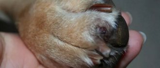
With the progressive stage of Perthes disease, the shape of the head changes. Instead of being smooth and round, it becomes flattened, triangular, with an uneven surface.
The femoral neck is shortened, so the limb looks shorter than others, and noticeably thinner due to atrophied muscles.
Symptoms of the disease
The main indicators of the presence of Perthes disease are a decrease in motor activity and a deterioration in the dog’s mood. As a rule, the first signs of pathology appear in a puppy at the age of 4-6 months. During walks, he may tuck his paw for a short time and refuse to run. Mild pain appears in the animal’s hip joints, which, over time, becomes more intense and prolonged.
With bilateral joint damage, the main symptom of Perthes disease is lameness on both legs. While walking, the animal tries to transfer the load from the diseased limbs to the healthy ones, so the gait visually changes.
Main symptoms of the disease:
- muscle atrophy, noticeable when visually comparing two limbs;
- sudden pressing of the paw during active games;
- lameness in one or two limbs;
- deterioration of mood and physical activity, increased fatigue;
- the animal stops relying on one of its limbs and constantly holds it in an elevated position;
- aggressive reaction to increased attention from the owners and attempts to examine the paw;
- in advanced stages of the disease, a characteristic crunching sound can be heard in the joint.
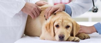
Owners who discover similar symptoms in their pets are advised to immediately seek help from a veterinarian. For this disease in dogs, treatment is long, complex and does not guarantee a positive result.
Other treatments
Conservative treatment of Perthes disease involves pain relief and a course of injections of chondroprotectors into the joint. However, due to the irreversibility of blood supply disruption, this method is ineffective.
Moreover, the condition may worsen, since with temporary relief of the pain syndrome, the dog begins to fully rely on the limb, further injuring the tissue.
Important! Physiotherapy is necessary to quickly restore muscle function. It includes massage, as well as the use of non-steroidal anti-inflammatory drugs.
Diagnostics, breed predisposition
In general, anything can be taken as manifestations of “Perthes.” There have been cases when owners thought that their pet had this pathology, but in fact, veterinarians even discovered fresh fractures! So you need to know how this disease differs from other pathologies and pathological conditions:
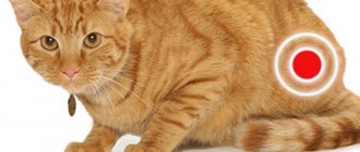
- The most important diagnostic sign is that the disease affects only the hip joints. Simply put, only the hind limbs are always affected ; the front legs never suffer from Perthes.
- Almost always, either puppies or very young animals get sick. If, for example, an old dog started having problems with his paws, then this is almost 100% arthritis or arthrosis, but not Perthes disease.
- Unlike injuries, oncology and similar pathologies, with Perthes disease, pain develops slowly, over many months, or even years.
Prevention
Since Perthes disease is polygenic in nature, it is necessary to exclude sick dogs from breeding. For a growing puppy from breed groups at risk, exercise should be given according to age, as well as adequate nutrition to avoid hypocalcemia.
But since the mechanism of the disease is not fully understood, all these measures will not provide a 100% guarantee of the absence of the disease.
Similar diseases
Diseases of the hip joints of various etiologies often have a similar clinical picture: lameness appears suddenly or gradually, the dog does not step on its paw, and the gait changes.
Arthritis. There are several types of disease, dividing them according to the root cause, among them:
- Traumatic. Due to prolonged or heavy loads, when an injury occurs, the muscle fibers surrounding the joint are stretched. A hematoma forms, but with timely treatment, this type of arthritis goes away. In more severe cases, the integrity of the joint capsule is damaged. Fluid leaks out, forming swelling. Most often, the process is accompanied by the development of infection and traumatic arthritis turns into a purulent form.
- Purulent. The disease is caused by staphylococcal and streptococcal infections. Symptoms include increased body temperature and painful swelling of the joint. In this case, you cannot do without a course of antibiotics.
- Rheumatoid. An autoimmune disease that occurs when the body attempts to eliminate bacteria that are similar in structure to bone and cartilage cells. As a result, your own tissues suffer.
The symptoms are similar everywhere: age group at risk (dogs over 6 years old), rapid fatigue on walks, difficulty walking up stairs, pain during manual examination, decreased activity.
Arthrosis. With age, due to prolonged stress, the cartilage surrounding the head wears out. Blood circulation worsens, calcium content decreases. With advanced arthrosis, bone osteoporosis is observed. Animals with increased weight, untreated arthritis, and trauma have a predisposition.
Hip dysplasia. The disease is polygenic in nature. It consists in the fact that the acetabulum, where the epiphysis of the femur is inserted, ceases to fit tightly and becomes flat.
The head changes shape and can take an uncharacteristic position, injuring the cartilage. Lameness and pain when moving appear.
Outwardly, it is manifested by waddling movements from side to side; when accelerating, the dog uses “hare running”, pushing off with two hind legs.
After sleep, there is stiffness and whining when body position changes. The diagnosis is made after an X-ray examination performed strictly under sedation. And also after the Ortolani test. Depending on the stage, surgical or conservative treatment is possible.
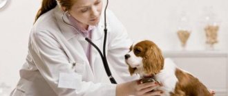
Diagnosis of the disease
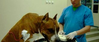
An experienced veterinarian can diagnose Calve-Perthes disease based on examination, palpation of the hip joint and instrumental studies.
If a dog’s paw hurts for a long time, he becomes lethargic and depressed, and upon examination, moving the sore limb back causes him severe pain.
To confirm the diagnosis, the dog is given an x-ray, during which it must lie strictly on its back with its hind legs abducted. Since this position provokes pain in the affected hip and it is difficult for the dog to maintain it, the dog may be put under short-term anesthesia to obtain a high-quality image.
The radiographic image clearly shows both the initial signs of the disease and later ones. In the first case, the image will show compaction of bone tissue and a change in the shape of the femoral head, and in the second, bone growths on the head, osteoarthritis, and in the most severe case, a fracture of the femoral neck or its destruction are visible.
But the best way to get accurate information about Perthes disease is to perform a computed tomography scan. As a result of this study, you can see signs of the disease at a very early stage, which will help you start treatment as quickly as possible. However, this diagnostic method is quite expensive and is not carried out in all clinics.