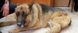
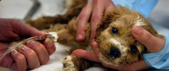
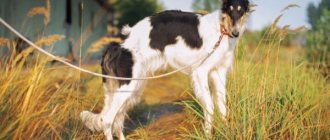
It happens that you suddenly notice a limp in your pet. At first it may go away after an hour or a day, and the dog runs as before. But over time, the dog’s lameness becomes permanent and prevents him from moving freely.
That’s when the owner goes to the veterinarian to find the cause of the disease.
Causes of lameness in dogs
After receiving a description of the problem from the owner, veterinarians first suspect the presence of a medial luxation of the patella in the dog.
This type of dislocation is a genetic disease that is inherited in small breed dogs. Pathology is a displacement of the kneecap relative to its natural position. If the displacement occurs inward, then it is a medial dislocation, if outward, it is lateral. Moreover, veterinarians diagnose medial displacement in almost 80% of cases.
Other causes of medial patella luxation in dogs include trauma and an o-shaped or x-shaped curvature of the hind limbs. Lateral dislocation is most often observed in large animals, since the x-shaped position of the hind legs is more typical for them.
In addition to the above reasons, dislocation is caused by weakness of the ligamentous-muscular system and degenerative changes in the joint that appear with age.
Patella dislocation in a dog
Medial patellar luxation is a displacement of the kneecap from its normal position in the groove of the femoral trochlea.
Etiopathogenesis
The exact cause of medial dislocation has not been established; it is believed to be a hereditary disease. With medial dislocation of the kneecap, coxa vara (decrease in the angle of inclination) and a decrease in anteversion (forward inclination) of the femoral neck are noted.
These changes cause medial displacement of the quadriceps femoris muscle, which ultimately causes a cascade of changes in normal hindlimb biomechanics, namely:
- lateral rotation of the distal femur;
- lateral bending (tilting) of the distal third of the thigh;
- dysplasia of the femoral epiphysis;
- reducing the depth of the groove of the femur block;
- rotational instability of the knee joint;
- tibial deformity.
The extensor mechanism of the knee joint consists of the quadriceps muscle, the patellar tendon, the patella, the patellar ligament, and the tibial asperity. This mechanism is normally located in a straight line from the proximal femur to the knee joint.
Medial deviation of the quadriceps muscle causes an uneven distribution of pressure on the distal femur; increased pressure on the medial portion slows down its growth and development; decreased pressure on the lateral portion, on the contrary, accelerates its growth, which ultimately causes lateral bending (tilting) of the distal third of the femur.
The severity of the lateral bending of the distal third of the femur depends on the severity of patellar dislocation during the development of the animal. A change in pressure on the distal femur can also cause a disruption in the formation of the groove of the distal femoral block - a decrease in its depth (up to the complete absence of the groove).
Also, exposure to abnormal forces on the tibia during animal development can cause medial displacement of the tibial roughness, medial bending (varus) of the proximal tibia, and lateral rotation of the distal tibia.
Clinical signs
Morbidity
There is a pronounced predisposition to the disease in dogs of small and miniature breeds (Yorkshire Terrier, Miniature and Toy Poodle, Pomeranian, Pekingese, Chihuahua), but medial patellar luxation can develop in any breed.
The disease is not typical for cats, but this may be due to the fact that the dislocation remains undiagnosed, because most cats do not exhibit lameness.
Medical history
Dogs with medial patellar luxation can be roughly divided into four groups, depending on age and clinical manifestations.
- Young puppies (from birth) with III and IV degrees of the disease - a manifestation of lameness in the early stages of life.
- Young and mature animals with stages II and III of the disease usually exhibit intermittent lameness.
- Elderly animals with grades I and II of the disease may exhibit a sudden increase in lameness due to secondary degenerative changes in the knee joint or rupture of the anterior cruciate ligament.
- Asymptomatic animals
Physical examination findings
When palpating the knee joint, the presence of a dislocation as such is determined and its degree is also determined.
- I degree. On palpation, the patella moves medially, but immediately returns to its normal position. Spontaneous displacement of the patella during movement is extremely rare.
- II degree. On palpation, the kneecap easily displaces and remains in a dislocated state until the animal straightens the limb. Possible spontaneous displacement of the patella during movement and the manifestation of periodic lameness.
- III degree. The patella remains luxated most of the time, but manual displacement is likely when the limb is extended. Subsequent movement of the joint leads to re-dislocation.
- III degree. The patella is constantly in a dislocated state and cannot be returned to its normal position by palpation.
In addition to determining the instability of the kneecap and the degree of dislocation, when palpating the knee joint, one should pay attention to the following points:
- the presence of crepitus,
- degree of rotation of the tibial roughness,
- bending or twisting of a limb,
- presence of joint contracture,
- presence of a torn anterior cruciate ligament (drawer).
Degrees of the disease
Medial dislocation is divided into four degrees, and the main parameter for this gradation is the possibility or impossibility of returning the patella to its normal position:
- In the first degree of pathology, the kneecap shifts during load on the paw or, conversely, during relaxation and can easily return to its natural position. The dog often gets used to this and “straightens” the knee on its own by extending its paw. The disease of the first degree does not lead to irreversible changes in the joint and can sometimes go away without treatment, since with age the ligaments and muscles of the paw strengthen and hold the kneecap in place.
- In the second degree, the dislocated knee no longer recovers on its own; the dislocation can only be corrected by hand. Due to repeated episodes of kneecap displacement, the cartilage tissue in the knee wears away and inflammation occurs.
- The third degree is characterized by a position in which the patella is constantly displaced and only sometimes is it possible to restore its anatomical position. This stage of the disease forces the dog to keep its paw in a bent position and try not to lean on it.
- In the fourth degree, the dislocation is permanent and the kneecap can no longer be returned to its place without surgery; the animal’s paw is no longer involved in the walking process.
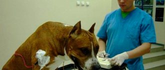
Treatment Methods for Patella Dislocation
The disease is diagnosed during examination by a veterinarian, by palpation and x-rays. For any degree of dislocation, it is necessary to limit physical activity and monitor the pet’s diet and weight. In the first stage of the disease, therapeutic methods of treatment can be used. It is necessary to eliminate the inflammatory process and strengthen the ligaments. Next, you will have to monitor the course of the disease so as not to miss changes.
The remaining stages are treated only with surgery. At the same time, the second and third can be completely cured, and the fourth, even with a successful outcome, does not guarantee a complete restoration of activity.
There are several treatment methods. The choice of technique depends on the cause and stage of the disease. Osteotomy is used for hereditary causes of the disease. It is an artificial fracture, which, when restored, improves the functioning of the joint. Sometimes suturing of the ligaments and plastic surgery of the knee groove is required.
The surgical knee reconstruction procedure involves deepening the groove of the femur. This is trochleoplasty, the most popular procedure in this case. The second method is called trochlear chondroplasty. With this technique, an osteotomy of the tibial tubercle occurs in the direction opposite to the dislocation. Then the tubercle is fixed with a special rod.
After the operation, you need to limit activity and stress for some time, then begin to develop the limb. The recovery period is usually quick and painless. It is important to ensure your pet is eating properly and follow all the veterinarian’s recommendations. It is possible to prescribe special physiotherapeutic procedures.
Symptoms
As we have already noted, the main symptom accompanying medial luxation of the patella in dogs is lameness. In this case, the frequency and conditions for the occurrence of limping may be different:
- on a walk, the dog may suddenly begin to fall on its paw, but this soon passes;
- during sleep or a relaxed state, the kneecap moves out of place, and the animal cannot then lean on its paw;
- if the disease is already in the later stages, then a paw or two is bent, while the dog moves in jumps;
- the range of motion of the joint decreases;
- When moving there is pain and crunching in the knee.
The very first episodes of lameness that appear for no particular reason should force you to take your pet to the veterinarian, since this pathology is easier to treat in the first stages.
General information
By “dislocation” we mean the movement of the kneecap from its natural “guides”, the condyles (the medial condyle especially suffers). The causes of this phenomenon may be congenital, genetic and/or traumatic .
A pronounced breed predisposition is recorded in the following dogs:
- Miniature, “toy” varieties of poodles.
- Bolonki.
- Jack Russell Terriers.
- Yorkshire Terriers.
- Pekingese.
- Chihuahua.
- King Charles spaniels.
- Boston Terriers.
As for large breeds, Labradors, golden retrievers, malamutes, boxers, and St. Bernards suffer. Even if a particular pet does not belong to a predisposed breed, but it has some congenital defects in the structure of the patella, the likelihood of developing the disease is very high.
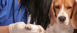
Diagnostic methods
To make a correct diagnosis, you will need a thorough examination by a veterinarian, palpation of the diseased joint and an x-ray.
During the examination, the doctor will evaluate the position of the dog's paws and the nature of the limp. Since lameness occurs periodically in the initial stages, it is important to conduct additional research.
When palpating the joint, the orthopedic veterinarian will determine where the kneecap is displaced relative to its anatomical position (outward or inward) and whether it is possible to return to its normal position.
X-rays are performed in two projections - lateral and direct. In the picture in the lateral position, the kneecap will be in place, but in the straight position, its shift in one direction or another will be visible. Simultaneously with the x-ray of the diseased joint, it is necessary to conduct an examination of the hip joints to detect Legg-Calvé-Perthes disease, since these diseases often accompany each other.
Also, medial dislocation can occur with an injury that results in rupture of the anterior cruciate ligament, and this situation must be taken into account in order to make the correct diagnosis and determine treatment tactics.
As you understand, the diagnosis must be carried out by a competent orthopedic veterinarian so that further treatment is adequate and leads to the dog’s recovery.
Causes, diagnosis and treatment of the disease
The patella is a rounded bony element that protects the knee joint and is located at the top of the knee. This is the largest and most mobile bone, located deep in the tendons.
A luxating kneecap is a traumatic injury in which the articular surfaces of the bones are displaced relative to each other. In this case, not only the anatomical conformity is violated, but also the integrity of the joint capsule, as well as the ligamentous apparatus.
Such damage leads to loss of ability to work and mobility of the joint and requires immediate medical examination.
Types of damage
Patella luxation is classified as follows:
- Medial dislocation of the patella is rare and is accompanied by the cup coming out inward from the area of the knee block.
- It is possible to identify a congenital (genetically determined) or acquired type of injury.
- With lateral dislocation, the cups move outward, from the area of the knee blocks.
- Also, injuries can be unilateral or bilateral.
- With habitual dislocation and instability of the patella, the development of imbalance syndrome is observed. When a luxation occurs, the kneecap may move away from its normal physiological position. Unlike the acute type of injury, the kneecap returns to its place on its own.
- Taking into account the age of the injury, the dislocation can be acute or chronic.
Depending on the type of damage, the doctor selects the appropriate treatment regimen.
Causes
Dislocation of the kneecap is rare; people whose professional activities are related to sports are at risk. There are other possible reasons that can cause such damage:
- Congenital disorders of the development of tendons, ligaments, muscles.
- Injuries resulting from a fall or accident.
- Excessive loads that lead to ligament rupture.
- A strong, sharp turn of the knee.
- Excessive weakness of the vast intrinsic muscles.
- Defects in the shape of the lower limb: detection of patellar dysplasia, hyperextension of the knee joints.
Diagnostics
Diagnosis is carried out taking into account a number of factors:
- Orally question the patient regarding emerging symptoms and medical history.
- X-ray results. In order to get as much information as possible, doctors recommend taking an x-ray of two patellas at once and then comparing the images.
- If there is a suspicion of the development of an old form of damage, the doctor recommends magnetic resonance imaging.
- If there is a need to involve surgical intervention, diagnostic arthroscopy may be required.
Modern surgery makes it possible to provide high-quality and safe surgical treatment of dislocations starting from the age of three months. The earlier treatment is started, the better the prognosis for the patient.
Treatment
If an acute dislocation of the patella is detected, it is recommended to be treated using conservative methods:
- Using local anesthetics, the surgeon reduces the dislocation. To do this, the injured limb is bent at the hip joint. This helps reduce tendon tension. After this, the doctor moves the kneecap with careful movements until it reaches its normal anatomical position.
- After carrying out the necessary manipulations, it is recommended to apply a special plaster cast to the injury site.
- After the doctor reduces the dislocation, it is recommended to immediately perform a repeat X-ray. This study allows you to monitor how successful the reduction procedure was.
- The patient is recommended to be immobilized for 1 to 1.5 months.
- Under the supervision of a physiotherapist, procedures are selected to speed up the recovery of the victim.
- Full loads on the injured leg are allowed no earlier than 30 days from the date of injury.
X-rays also reveal the presence of osteochondral bodies that may form during injury. If they are diagnosed, the patient may be recommended to undergo surgery. The operation is also recommended if there is a risk of developing recurrent patellar dislocations.
Treatment of chronic dislocations involves immediate surgery. After surgery, immobilization is recommended for up to 1.5 months. Loads are allowed no earlier than 9-11 weeks after the operation.
What complications may arise?
If a dislocation is not treated promptly, the likelihood of developing serious complications increases:
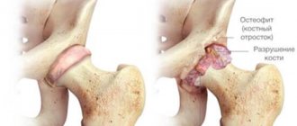
- With each subsequent displacement of the cups from the knee block area, the knee cartilage is damaged more and more. As a result, this can become one of the main causes of the development of osteoarthritis, accompanied by intense pain and inflammation.
- Such an injury can lead to external deformation of the injured limb and rupture of ligaments.
- Prolonged exposure to a dislocation can cause the development of muscle contracture, which leads to loss of motor function of the limb. In severe cases, treatment may not have the desired effect.
In order to prevent the development of such damage, moderate physical activity is recommended in compliance with safety precautions.
Source: https://travmoff.ru/travmy/kolennyj-sustav/vyvihj-chashechki.html
Dislocation of the front and rear paws
A dislocation of a dog's front paw can be determined because the dog is pulling it close to its body to make movement as easy as possible and not strain the affected paw. It is impossible to determine the exact cause without contacting a specialist and undergoing a number of tests in addition to x-rays, since the cause could be, for example, arthritis, muscle weakness or another disease as a side effect. How to treat a dislocated paw will depend on the reason that caused the dislocation of the joint.
It is important to accurately determine the specifics of the disease in order to be sure that the treatment received is correct.
All that is needed from the owner is to take the pet to a veterinary clinic for an examination and until this moment do not allow the dog to lean on its paw, I try to use it and overcome the pain. If you called a doctor to your home, then ensure that your pets are at rest and apply something cold to the damaged area, for example, a frozen item from the freezer
Clinical picture
As a rule, owners of dogs that are initially predisposed to the disease quickly realize that something is wrong with their pets, since the symptoms are quite characteristic. The animal from time to time begins to limp for no apparent reason, its gait becomes unstable, “wobbly” . The dog falls on his sore paw from time to time, tries to sit less, preferring to lie down more often, he gets up with difficulty, very carefully.
Chronic cases can lead to erosion of the cartilage on the femur (due to constant mechanical pressure) and eventually osteoarthritis . It’s easy to find out about this - if the animal simply has some kind of “wrong” with the kneecap, he does not feel pain. If osteoarthritis is involved in the process, everything becomes much worse.
Shoulder dislocation
This is a pathological displacement of the scapula relative to the shoulder joint. The most common cause of dislocation is injury after jumping, fighting, etc., that is, injury to the shoulder girdle occurs due to the activity of mobility of the forelimbs.
Since the shoulder complex works in several planes at once, that is, it produces flexion-extension of the limb, rotation and adduction-abduction forward-backward, it is not surprising that the risk of damaging the joint increases with the number of movements.
Shoulder dislocation in dogs most often occurs at the site of a strong blow to the shoulder girdle, or due to excess load.
Subluxation of the shoulder joint is also highlighted, this is the moment when the sockets and heads of the bone have already moved away from each other, but are still in contact, thus, the head of the articular bone falls out of the socket, but then returns to its place. Such subluxation can lead to the development of dislocation.
First aid will consist of simple manipulations, such as:
- Calm the animal and examine it if it is not showing aggression
- even if there is no aggression, you should still wear a muzzle, since pain symptoms will help temporarily change the character and behavior of even the most obedient pet, and the veterinarian should not get hurt trying to help your dog
- the injured joint should be on top, that is, try to lay the dog on its side with the healthy side
- In no case should you correct a dislocation yourself, as you can only worsen the condition, since you are not aware of the internal condition - bleeding or additional damage
- Do not wrap the wounded area tightly; even an elastic bandage becomes tight when worn. Therefore, it is best to apply a soft splint and fix the limb below and above the joint that is damaged
- Also, if the naked eye can see that the joint is swollen, then the swelling can be relieved by cooling. To do this, wrap the joint with film and several layers of fabric, and put something frozen on top. This will not only help relieve pain, but will help stop bleeding
If you were able to detect a dislocation in the first couple of days after receiving it, then in most cases, without additional internal damage or rupture, the doctor will be able to straighten the joint and fix it with a special bandage. In case of repeated dislocation or delayed treatment, surgical intervention is most often necessary.
Classification of dislocations
Congenital – pathology develops during intrauterine development. The born puppy is quite viable, but it is extremely rare to completely cure the disease.
- Traumatic - is a consequence of injuries that can occur when colliding with an obstacle at high speed, falling from a great height, or getting a limb stuck while moving. Often happens in car accidents.
- Pathological – arising as a complication of various pathologies of the musculoskeletal system, with thinning of cartilage or bone tissue.
- Paralytic - it is caused by atrophy of the muscle group supporting the joint.
- Habitual – this is the name for constantly occurring repeated dislocations, the cause of which is once stretched muscles or ligaments that weakly support the joint.
- Complicated - the bone is displaced, affecting nerve endings and important vessels.
- Unreducible - an old dislocation in which new tissues form between the articular heads.