Owners do not always notice the appearance of neoplasms on the body of pets. The similarity of such seals to bumps diverts the attention of owners of four-legged friends from the problem. Lack of knowledge in this area is fraught with the development of minor pathologies into dangerous diseases. The article will discuss one of the skin pathologies - histiocytoma in dogs, its inherent symptoms and treatment methods.
Histiocytoma in dogs: symptoms, treatment
What is histiocytoma in dogs?
This is a benign tumor of epidermal Langerhans cells (the precursors of which are histiocytes).
The skin consists of three layers: outer (epidermis), middle (dermis) and inner (subcutaneous fat). Each of them has its own structural features. Langerhans cells are located within the epidermis and participate in the protective function of the skin.
What you should know about histiocytoma:
- It mainly occurs in young dogs under 2–4 years of age; it is rarely detected in older animals.
- Develops quickly, over several days or weeks.
- There is no breed predisposition to the disease, but short-haired pets experience it somewhat more often than others.
- The sex of the animal does not affect the development of the tumor.
- According to various studies, histiocytoma in dogs ranks third in frequency of occurrence after mastocytoma and lipoma.
Its types in animals
Based on the type of histiocyte proliferation, pathology is divided into three groups:
- non-tumor - cutaneous histiocytosis (when the process affects all layers of the skin) and systemic (lesions are detected on the skin, mucous membrane and lymph nodes). The systemic form is rare in animals.
- benign neoplasm – cutaneous histiocytoma. In dogs, it can be single and rarely multiple, when neoplasms (more than two) simultaneously develop on different parts of the body. There is no data on the possible subsequent degeneration of this tumor cells into malignant ones.
- malignant tumors – histiocytic sarcoma. It grows rapidly and is difficult to treat. Unlike histiocytoma, sarcoma occurs in older animals (8 years and older).
Symptoms and signs of cutaneous histiocytoma
Dogs of smooth-haired breeds are more prone to skin histiocytoma, regardless of gender. Education rarely makes itself known and leads to inconvenience for the animal. Cutaneous histiocytosis from Langerhans cells is characterized by an abundance of such skin formations (sometimes the mucocutaneous frame of the mouth), which can be of different sizes: from minor nodules to large tumors. Mostly Shar-Peis suffer from it.
Cutaneous histiocytosis from IDC also makes itself felt by multiple approximately 4-centimeter formations in the head, neck, torso, and paws (sometimes lymph nodes are also affected). Moreover, the disease is typical for dogs of any breed.
A distinctive feature of systemic histiocytosis is lymphohistiocytic destruction of the walls of blood vessels, which makes itself felt in the form of dermatitis and panniculitis (inflammation of subcutaneous fatty tissue). This process results in anorexia, weight loss, eye lesions such as swelling of the apple and conjunctiva, and conjunctivitis. Mostly representatives of large breeds are affected.
Histiocytic sarcoma in dogs manifests itself as single and multiple tumors, which can be in one place or spread to other organs. The disease is typical mainly for Bernese Mountain Dogs, Rottweilers, Golden and Straight-Coated Retrievers. Hemophagocytic histiocytic sarcoma is a proliferation of macrophages in the spleen in dogs. It is characterized by regenerative destruction of red blood cells by anemia, thrombocytopenia, and bilirubinemia.
How is histiocytoma diagnosed in dogs?
The generally accepted understanding of this disease is that such types of neoplasms are exclusively benign. There have never been any deaths from histiocytomas in practice.
Skin ailments of this type are easily identified using clinical signs and diagnostic biopsy, the results of which must undergo histological examination. A detailed description of the onset of the disease, as well as the presence of signs provided by the pet owner, will help to correctly identify the disease. As well as conducting biochemical tests of blood, urine, ultrasound, x-rays, etc.
Causes of neoplasms
There are no exact reasons why a dog might develop histiocytoma. Predisposing factors are believed to be:
- genetic factors;
- unfavorable radiation conditions;
- exposure to carcinogens;
- malfunctions of the immune system;
- During inflammatory processes or skin trauma, histiocytes are activated. And this nuance is associated with the predominant location of histiocytes on the most mobile parts of the dog’s body (on the face, paws, tail). Young, active animals can injure their skin while exploring the world around them. Foreign objects (pollen, plant particles, dust, etc.) get there and inflammation develops. Histiocyte cells are drawn into the process, resulting in the development of histiocytoma.
Histiocytic skin diseases in dogs
This group includes rare skin disorders caused by excessive cell proliferation.
Histiocytes are cells of hematopoietic stem origin. During development, they turn into macrophages or dendritic cells, to which IDCs and Langerhans cells belong.
This pathology is classified into several types:
- cutaneous histiocytoma;
- Langerhans cell histiocytoma;
- histiocytoma from IDC;
- systemic and malignant histiocytosis.
This tumor is not always found in a benign form. There are known cases of a neoplasm transforming into a cancerous form.
Histiocytes are connective tissue cells that have a protective function
The disease manifests itself in the form of a tumor that forms in tissue cells during immune failures. Neoplasms form mainly on the head and hind limbs. The favorite place for pathology is the ears.
Histiocytoma in dogs is the most common skin pathology, occupying a leading position in the list of sarcomas.
This type of tumor is characterized by spasmodic growth
Disease susceptibility
Regardless of gender, the disease is most often observed in pets during the juvenile period. In 80% of cases, the disease attacks dogs under 2 years of age. Representatives of smooth-haired breeds are most prone to the disease. Genetic predisposition is observed in Bernese Mountain Dogs , Rottweilers, Golden Retrievers, and Flat-Coated Retrievers.
Rottweiler is at risk
Causes of the disease
The factors that provoke the development of this disease are not fully understood. Scientists have found that mutations occur in the cell genome, leading to rapid tissue growth. Also among the possible reasons are the following:
- Cellular inflammation.
- Exposure to carcinogens.
- Non-cancerous pathologies.
- The influence of ionizing rays.
- Poor immune response.
- Soft tissue injury
Among existing breeds, up to 25% of neoplasms are observed in the Bernese Mountain Dog
Symptoms of histiocytoma in dogs
Histiocytoma is similar to some other skin tumors. Therefore, these symptoms may indicate not only this pathology, but also more dangerous tumors.
Typically, histiocytoma is located on:
- muzzle (near the nose, on the chin, in the infraorbital, occipital and frontal areas, near the lips);
- on the inside of the ear;
- on the tail;
- on the limbs (on the elbows, between the fingers, in the area of the metatarsus and wrist);
- on the body they are less common and mainly in those places where friction occurs (elbow joint area, etc.).
The main symptoms that indicate that your pet has a skin form of pathology:
- it is a clearly circumscribed, rounded neoplasm;
- small sizes - from 0.2 to 2 cm (in rare cases it can reach up to 4 cm);
- appears and grows quickly (usually from 1 to 3 weeks). Having reached a certain size, growth stops;
- there is no hair on the affected area;
- the skin is pink and smooth (if there are no ulcers);
- As a rule, there is no pain;
- does not cause itching;
- there may be ulcerations (which in turn predispose to bacterial infection).
With systemic histiocytosis, in addition to multiple lesions on the skin, the animal experiences a general depressed state and a gradual decrease in body weight. With this pathology, the animal may experience temporary improvements, but they are quickly replaced by further progression of the disease.
Expert opinion
Kuzmenko Olga Olegovna
Information about the expert
Ask a Question
The malignant form develops mainly on internal organs. Symptoms will appear depending on which system is affected.”
Symptoms of the disease
The manifestations of the disease depend on the location of the tumor development:
- If a tumor forms in the tissues, the animal suffers from abdominal pain and fever. The disease is accompanied by increased body temperature, loss of appetite, decreased body weight and severe malaise.
- If a histiocytoma appears on the skin, the pet loses mobility of the joints located in the area of tumor growth. The seal on the dermis is colored red, and when pressed, discomfort occurs. As the formation grows, it puts pressure on neighboring tissues, causing peeling and ulcers.
Symptoms depend on the location of the tumor
If a malignant tumor develops on the extremities, there is a risk of fractures. In pets with cutaneous and systemic histiocytosis, the listed symptoms may be absent.
Cutaneous histiocytoma
Belongs to the category of fast-growing tumors. The surface of the skin often becomes bald, but the new growth does not cause any discomfort.
Main symptoms:
- lesions are concentrated in the dermis and subcutaneous layer;
- the skin is dotted with numerous seals on the trunk, muzzle, limbs and neck.
The pathology is characterized by a chronic nature, in which periodic relief of symptoms is noted. In the advanced form of the disease, there is an overgrowth of secondary microflora, leading to severe itching.
Cutaneous histiocytoma is the most common type of disease
Cutaneous Langerhans cell histiocytosis
Langerhans cells contribute to the development of the disease. Normally, these structural units are part of the mucous membranes and dermis. When the disease occurs, they grow rapidly and provoke multiple histiocytoma of the skin. The size of the tumor ranges from small nodules to large red formations. Compared to the previous variety, the pathology has a worse prognosis. If in normal histiocytosis systemic organs are not involved, then in this form penetration into them is possible. The tumor also affects the lymph nodes.
Shar-Peis are susceptible to Langerhans cell histiocytoma
Cutaneous histiocytosis from IDC
Interstitial dendritic cells contribute to the formation of compactions in the subcutaneous tissue and dermis. The formation of numerous neoplasms, reaching a diameter of up to 4 cm, is characteristic. Localization is the neck, head, limbs and torso. Damage to the lymph nodes is sometimes observed. The average age of pets most susceptible to the disease is 4 years. Unlike the pathology caused by the proliferation of Langerhans cells, this disease has an inherent tendency to penetrate into the deep layers of the epidermis.
Langerhans cells are concentrated exclusively in the epithelium
Systemic histiocytoma
It is not as common as the cutaneous type. The disease is characterized by generalized damage to the mucous membranes, skin and lymph nodes.
Main symptoms:
- multiple formations in the form of nodules, covered with scabs;
- alopecia of the dermis;
- conjunctivitis;
- weight loss;
- noises when listening to the lungs.
Symptoms of conjunctivitis in dogs
The disease most often attacks Bernese mountain herding dogs between 2 and 8 years of age.
The Bernese Mountain Shepherd is susceptible to a systemic type of pathology
Malignant histiocytoma
Histiocytic sarcoma originates predominantly from IDCs. Main symptoms:
- paleness of the skin;
- lethargy;
- dyspnea;
- the presence of noise in the lungs;
- temporary paralysis of the hind legs;
- nervous attacks;
- enlargement of the liver, lymph nodes and spleen.
This type of pathology practically does not affect the skin, progressing mainly on the internal organs. The average age of infected animals is 7 years. The disease develops rapidly and ends in death.
With malignant histiocytoma, compactions form in the liver and spleen
Methods for diagnosing the disease
During the diagnostic process, the veterinarian performs the following procedures:
- general examination of the animal and collection of information about the pet’s condition;
- external examination of the lesion;
- standard tests are carried out at the discretion of the doctor (blood tests, urine tests, x-rays, ultrasound).
To accurately determine the nature of the tumor, laboratory diagnostics of the sample is carried out:
- cytology – for tumors measuring at least 1 cm (small tumors, as a rule, are not examined).
- Punch biopsy (for the skin form), when a piece of the tumor is removed with a special device. This method is convenient if the histiocytoma is located in a location that is difficult to access surgically.
- Fine needle biopsy.
- The resulting samples are prepared and histologically examined under a microscope, after which histiocytoma in a dog can be unequivocally confirmed or refuted.
- Bacteriological culture is rarely performed in the presence of ulcers on the affected area.
Diagnosis of pathology
Since there is a risk of the neoplasm becoming malignant, timely detection of the disease becomes important. Due to the fact that it is not always possible to obtain accurate test results, diagnosing the disease is not easy. During the examination of the pet, the veterinarian performs the following tests and examinations:
- Blood and urine analysis.
- Biopsy of affected tissue.
- Cytology of bone marrow cells.
- Immunohistochemical examination. It is this diagnosis that allows us to identify the pathological origin of cells.
- MRI. Makes it possible to identify hidden tumors and their locations.
- Ultrasound. Shows the size of the tumor located on the internal organs.
Depending on the type of pathology, the specialist uses the appropriate diagnostic method.
Diseases are determined using immunohistochemical studies
Treatment of histiocytoma in animals
How to treat histiocytoma in dogs largely depends on the nature of the tumor and the laboratory diagnostic results obtained.
Reference! They try not to treat a neoplasm less than 1 cm in size, because in most cases, it resolves on its own after 2-3 months (or even several weeks).
If regression does not occur or the tumor causes discomfort in the pet, then medical or surgical treatment methods, cryosurgery, are used.
- Drug treatment includes the use of glucocorticosteroids. Prednisolone is usually prescribed in injection form for 10 to 14 days, after which the dosage is gradually reduced.
- If there is a bacterial infection, the ulcers are treated with Chlorhexidine solution. Next, local medications containing antibiotics are used. After performing a culture culture, the doctor will prescribe a drug to apply to the lesions.
Expert opinion
Kuzmenko Olga Olegovna
Information about the expert
Ask a Question
With the surgical method, the tumor is removed. The animal is given general anesthesia. The tumor is excised, after which sutures are applied (if necessary). Bandage dressings are used if the surface of the wound is in constant contact with any surface (on the limbs, for example).”
What diagnostic methods are needed to make a diagnosis?
Any suspicion of tumor malignancy requires examination in the form of a biopsy . A biopsy is necessary to obtain a sample of tumor tissue. the resulting sample is subjected to histological examination.
The results of histological analysis make it possible to accurately determine what type of sarcoma the patient has. The results are determined by the presence of certain markers in the tissues.
MRI allows you to accurately determine the shape of the formation, its size and exact location parameters. These data are necessary for further surgical intervention.
Ultrasound is necessary to evaluate the tumor itself and damage to nearby lymph nodes.
Computed tomography is used to detect distant metastases. The tumor can metastasize to the liver, brain, and lungs.
Obtaining a complete picture of what type of tumor the patient has and the extent of its prevalence allows oncologists to select treatment that will save the patient’s life.
Pet care
Caring for your pet involves following all therapeutic measures prescribed by the veterinarian.
If the dog has undergone surgery, the pet is fed softened food on the first day after the procedure. You can take the animal for a walk after complete restoration of coordination of movements.
Seams are treated daily. The wound is treated with a solution of Chlorhexidine or hydrogen peroxide, removing all crusts and discharge. Next, Levomekol ointment is applied in a thin layer. The pet is put on a protective collar for the entire recovery period until the stitches are removed.
Important! If the operation was performed on the paw, it is advisable to limit the animal’s mobility. In wet weather, walking is kept to a minimum, and afterward, the wound must be cleaned.
Forecasts
The prognosis for skin histiocytoma is favorable.
As a rule, regression of this tumor can be observed after several weeks or months. The tumor grows rapidly, but once it reaches a certain size, growth stops. After some time, the histiocytoma begins to gradually decrease. After complete resorption, hair grows on the affected area.
Important! If regression does not occur for more than 3–4 months, the tumor is excised.
With systemic histiocytosis, the prognosis is guarded. Pathology requires individual selection of a method of therapy, which, as a rule, includes the use of antitumor drugs.
Histiocytic sarcoma in most cases has a poor prognosis. The tumor is characterized by a high risk of metastasis. Even after removal of the affected organ, the animal can live only a few months.
Treatment
Due to the fact that skin histiocytoma can often resolve on its own, doctors leave it under observation for three months. This is only possible if the tests are good and there is no suspicion of a malignant etiology. To avoid injury to the affected areas, the doctor prescribes antibiotics and cortisone-based ointment. This prevents the development of complications if, as a result of itching, the dog begins to scratch the affected area and an ulcer forms in its place.
If the diagnosis of skin histiocytoma is confirmed and it is truly a benign neoplasm, treatment is carried out using the method of cryodestruction. Surgical intervention requires formations that are not amenable to drug treatment or are localized on vital organs, for example, on the eyelids. Removal of the affected area is carried out with cutting off adjacent tissues in diameter of 2 cm.
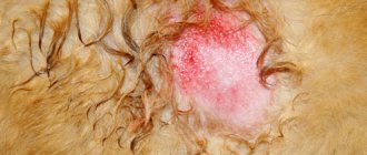
Histiocytoma on a dog's paw
In the case of histiocytic sarcoma, a course of radiation and chemotherapy is carried out along with surgical treatment. If the tumor is inoperable, medications are prescribed. Most often these are hormones that are used for blockade. Injections with hormonal drugs are given directly into the tumor, which allows the tumor to be preserved, and in some cases, to reduce it. A good therapeutic effect is achieved by the use of dimethyl sulfoxide and corticosteroids. Antibiotics include the anthracycline series, and the same drugs are used for systemic histiocytosis.
Prevention methods
There are no specific methods for preventing this disease. In case of injury, the damaged area of skin must be treated until complete healing. Inflammatory processes must be fully cured.
If a neoplasm is detected in a dog, the owner must take the pet to the veterinarian. At the discretion of the doctor, the necessary tests are carried out, after which further treatment is prescribed, or observation of the animal at home with periodic follow-up examinations by a specialist.
General methods of prevention include high-quality and balanced feeding, walking the animal in environmentally friendly and safe conditions, timely treatment for parasites, and routine vaccinations.
Cutaneous histiocytoma does not pose a health risk to your dog. To know for sure that your pet has a benign neoplasm, you need to go to a veterinary clinic, where the animal will undergo a biopsy followed by histological analysis. As a rule, the tumor goes away on its own without additional intervention within several months.
Veterinary clinic of Dr. Shubin
Description and reasons
Canine cutaneous histiocytoma is a benign tumor (neoplasm) of mononuclear cells that develops from cells in the superficial layer of the skin called Langerhans cells. This is a fairly common disease at the veterinary clinic; young dogs under the age of 4 years are predisposed to cutaneous histiocytoma; in cats, this type of tumor is observed quite rarely. The exact reasons for the development of cutaneous histiocytoma have not been determined; there is an opinion that it is not a true tumor, but just excessive cell growth.
Clinical signs
Cutaneous histiocytoma is more often observed in dog breeds such as boxer, cocker spaniel, Great Dane, dachshund, sheltie and bull terrier. Cutaneous histiocytoma appears externally as single (rarely multiple) rapidly growing round and hard raised skin nodes with a diameter of 0.5 cm to 4 cm. The surface of the lesions loses hair and ulceration may occur. The typical localization of lesions in cutaneous histiocytoma is the head, ears and limbs. Cutaneous histiocytoma is characterized by rapid growth of lesions, the nodes reach their maximum size within 1-4 weeks, after this period there is a spontaneous decrease in size and complete recovery within one to three months.
Diagnostics
Cutaneous histiocytoma is easily diagnosed by fine-needle biopsy followed by cytological examination of tissue under a microscope. Examination of tissue samples reveals layers of characteristic round cells, and making an accurate diagnosis in a veterinary clinic is usually not difficult.
Treatment and prognosis
Cutaneous histiocytoma is characterized by spontaneous recovery, and in most cases does not require special treatment (observation without treatment). Resolution of the tumor process is usually observed within 3 months from the date of diagnosis. In rare cases, the formation of multiple cutaneous histiocytomas of dogs is noted, a slight increase in recovery time is likely, and the formation of new tumors may be observed as old ones decrease, but even in this case, the disease goes away on its own.
If there is significant inflammation and itching with canine cutaneous histiocytoma, surgical treatment can be performed in the form of complete excision of the tumor followed by suturing. In areas where, for one reason or another, it is impossible to excise the tumor, spontaneous regression of the tumor can be accelerated by local application of corticosteroids or dimethyl sulfoxide (DMSO).
In general, the prognosis for cutaneous histiocytoma is favorable.
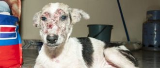
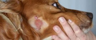
Photo 1 . Characteristic appearance of histiocytoma in a young dog
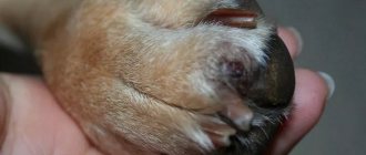
Photo 2-3 . Spontaneous regression of histiocytoma in a young Yorkshire terrier. Photo 2 - view of the histiocytoma at the time of contacting the veterinary clinic.
Photo 3 . Type of histiocytoma in the same Yorkie during a re-examination at the veterinary clinic a week later.
Photo 4. View of a cutaneous histiocytoma involving the chin in a one-year-old dog at an appointment at a veterinary clinic.
Veterinary clinic of Dr. Shubin, Balakovo.