Among dog diseases, liver pathologies occupy an “honorable” place. Their problem is that these ailments remain undetected for a long time due to the specific characteristics of the organ. It is for this reason that even relatively “harmless” cholangitis in dogs can end very badly.
This is the name for inflammation of the bile ducts in the liver. It should be noted that “pure” cholangitis is quite rare in dogs; more often it involves inflammation of the liver tissue and ducts. For this reason, this disease in veterinary medicine is often called simply “hepatitis” , without making any special adjustments to the specifics of the disease. Divided into three types:
- Neutrophilic.
- Lymphocytic.
- Parasitic origin. In this case, the liver is “inhabited” by parasitic species of trematodes. In our country, opisthorchid is the most common.
Neutrophilic cholangiohepatitis is characterized by the infiltration of large numbers of neutrophils into the portal areas of the liver and directly into the bile ducts. The disease occurs when pathogenic microflora penetrates from the small intestine. E. coli, various types of staphylococci, streptococci, clostridia, pathogenic fungi and salmonella - all of them can easily lead to this outcome.
“Parallel” diseases that form a “triad” (pancreatitis, inflammation of the small intestine + cholangitis) in dogs, unlike in cats, practically do not occur. Predisposition factors include: congenital or acquired pathologies of the biliary system, various anomalies in the development of the gallbladder, adhesions resulting from an infectious disease, obstruction of the bile duct, parasitic infestations . The lymphocytic type of the disease develops in a similar way.
Cholecystitis in dogs
Cholecystitis is an inflammation of the gallbladder.
Cholecystitis in dogs usually occurs with inflammation of the biliary tract - cholangitis. Anatomical data of the gallbladder in a dog.
The gallbladder is a reservoir for bile, in which bile thickens 3-5 times, since it is produced more than is required for the digestion process. The color of gallbladder bile in dogs is red-yellow.
The bladder lies on the quadrate lobe of the liver high from its ventral edge and is visible from both the visceral and diaphragmatic surfaces. The bubble has a bottom, a body and a neck. The wall of the bladder is formed by the mucous membrane, a layer of smooth muscle tissue and is covered on the outside by peritoneum, and the part of the bladder adjacent to the liver is made up of loose connective tissue. The cystic duct, in which the spiral fold is located, originates from the bladder.
The fusion of the cystic duct and the common hepatic duct forms the common bile duct, which opens into the S-shaped gyrus of the duodenum next to the pancreatic duct at the apex of the major duodenal papilla. At the point where it enters the intestine, the duct has a sphincter of the bile duct (sphincter of Oddi).
Thanks to the presence of the sphincter, bile can flow directly into the intestines (if the sphincter is open) or into the gallbladder (if the sphincter is closed).
Etiology . The causes of cholecystitis in dogs are:
- Parasitic diseases - isosporosis, echinoccosis (worms in dogs), alveococcosis, dicroceliosis, helminthiasis, opisthorchiasis (worms in dogs). Protozoa and helminths attack the internal organs of the dog. Parasitizing organs and tissues, helminths in the process of their vital activity secrete strong toxins that cause irritation of the mucous membrane of the bile ducts, surrounding tissues and the development of inflammation of the gallbladder.
- Infectious diseases affecting the intestines of dogs. In case of infectious diseases affecting the dog's intestines (canine parvovirus enteritis, infectious hepatitis in dogs, canine distemper, salmonellosis in dogs), pathogenic microorganisms enter the gallbladder through the bile ducts and lead to inflammation of its inner lining.
- Diseases of the digestive system . Fatty liver, pancreatitis, colitis of various etiologies, and duodenal ulcers often become the triggering mechanism for cholecystitis. Especially often, cholelithiasis leads to inflammation of the mucous membrane of the gallbladder.
- Errors in feeding . Sometimes the cause of cholecystitis in a dog is feeding dry food of dubious quality, especially if there is a lack of free access to water. With natural feeding, cholecystitis can cause the dog to feed leftover food from the common table. Periodically giving your dog sweets, sausages, and flour products leads to diseases of the digestive system and gall bladder. Failure to comply with the feeding regime, prolonged fasting or overfeeding the dog. A diet low in carotene and vitamin A, which are responsible for the condition of the epithelium and mucous membrane, promotes the appearance of cholecystitis.
- Obesity , overweight, sedentary lifestyle (hypodynamia), lack of constant physical activity causes a decrease in the contractility of the muscular layer of the gallbladder (dyskinia).
- Heredity – Large dog breeds are more susceptible to hereditary factors.
- Injuries in the abdominal area.
Clinical picture. Cholecystitis is characterized by indigestion. After feeding, a sick dog burps and frequently vomits (vomiting in a dog). The vomit is liquid in nature, with undigested food, and a small amount of mucus. Sometimes the presence of bile can be detected in vomit. As a result of the irritating effect of bile acids on the intestinal mucosa, the dog experiences flatulence (the dog's stomach is seething), bloating and diarrhea (the dog's diarrhea). As a result of advancing dehydration, the skin becomes dry, the skin becomes dull, and the dog has an unkempt appearance. When the bile ducts are blocked, the stool becomes pale in color. Some dogs become constipated (dog constipation). The dog becomes lethargic, apathetic, and reluctant to move. The body temperature may rise for a short time, and sometimes we experience a fever.
As a result of pain, a dog develops a characteristic posture - the animal lies on its stomach and arches its back upward. Palpation in the abdominal area is painful.
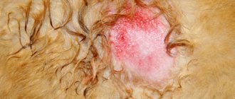
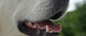
On clinical examination, the dog's gums and sclera are pale and icteric (jaundice). Due to the large amount of bilirubin, urine has a bright carrot tint.
Chronic cholecystitis is usually asymptomatic in dogs and is detected only during an exacerbation of the disease. In the dog, owners note lethargy after eating, nausea, vomiting, bowel irregularities accompanied by diarrhea or constipation.
Diagnosis . The diagnosis of cholecystitis is made by the veterinary specialists of the clinic based on a clinical examination, collection of anamnesis of the disease and additional research methods:
- General blood test - we find an increased number of leukocytes, with a shift in the leukocyte formula towards immature cells. Increased levels of bilirubin and bile acids. Increased alkaline phosphatase activity. High levels of transaminases.
- Urine and feces analysis - increased levels of bile acids and bilirubin.
- X-ray examination - we detect the presence of stones in the gall bladder.
- Ultrasound - decrease in the lumen of the bile ducts, thickening of the bile itself.
Differential diagnosis . Cholecystitis is differentiated from liver diseases (liver disease in dogs), gastroenteritis (gastroenteritis in dogs), and peritonitis.
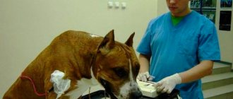
Treatment. Veterinary specialists at the clinic treat cholecystitis based on the form of the disease and the general condition of the sick dog. In severe cases of the disease, when experts believe that there is a threat of rupture of the gallbladder and the development of peritonitis, they resort to emergency surgery to remove the inflamed gallbladder.
If the disease is in an acute phase, then to begin with the dog may be prescribed therapeutic fasting for 2-3 days or a strict diet following a certain diet.
To eliminate pain, the dog is prescribed painkillers and antispasmodics - baralgin, no-shpu, papaverine, spasgan, atropine sulfate.
To normalize the outflow of bile and at the same time to disinfect the biliary tract, choleretic drugs are used - allohol, magnesium sulfate, cholenzyme, ursosan, ursofalk.
Herbal medicines such as immortelle flowers and corn silk are excellent choleretic agents. These drugs are used in the form of infusion and decoction.
If the cause of cholecystitis is an intestinal infection, then the sick dog is prescribed a course of antibiotic therapy after titrating the isolated microorganisms for sensitivity to antibiotics. Typically, veterinary specialists use cephalosporin antibiotics when treating cholecystitis.
Based on the fact that the disease affects the liver, the dog is prescribed hepatoprotectors – Essentiale Forte, Heptral.
To eliminate dehydration and at the same time to detoxify the dog’s body, infusion therapy is carried out by intravenous administration of 5-10% glucose solution, saline solution, polyglucin, hemodez, rheopolyglucin, calcium chloride, borglucanate.
If a dog has a parasitic disease, deworming is prescribed.
Prevention .
Prevention of cholecystitis in dogs should be based on compliance with rational, complete feeding (basics of feeding dogs, feeding pregnant females, feeding lactating dogs, feeding aging dogs, feeding puppies, golden rules of rational feeding of dogs). Do not feed your dog cheap food or table food. Spicy, fried, smoked, sweet and flour products are strictly prohibited. Dry food should only be of high quality. When feeding a dog, owners should pay attention to the presence of vitamins in the food, especially vitamin A (vitamins for dogs).
Why is he dangerous?
When released into the blood, a large amount of the enzyme provokes weakness, high fever, yellowness of the eyeballs and gums, and itchy skin. If cholecystitis occurs in a dog, bile enters the peritoneum through the perforated walls and the animal may die from peritonitis.
Veterinarians distinguish between acute and chronic forms of this disease. Chronic cholecystitis in dogs is usually almost asymptomatic. This is where its danger lies. Often, when the dog is brought to the doctor, the disease is already quite advanced. An attentive owner may notice nausea, lethargy after eating, and signs of vomiting in the animal. In some cases, problems with stool begin: constipation alternates with diarrhea.
Acute cholecystitis in a dog is much easier to notice. The animal may experience fever, the sclera and gums turn yellow. The most severe situation occurs due to rupture of the gallbladder. Here the dog can only be saved by immediate assistance from a veterinary surgeon. No less dangerous is the formation of stones and other neoplasms.
Based on many years of medical practice, veterinary experts recommend that owners adhere to the following tips and rules to prevent the disease:
Liver
– a vital organ that performs many functions in the body necessary to maintain life. It is the main metabolic organ, performing the functions of producing necessary substances, accumulating, processing and neutralizing waste.
The liver has significant regenerative abilities and enormous functionality; as a result, many liver injuries do not lead to disruption of its function and clinical manifestations until more than 70% of functional liver cells are lost. That is, liver failure and liver disease are not the same thing.
The result of the diversity of liver functions is that the clinical manifestations of diseases of this organ are very diverse and nonspecific, which can complicate diagnosis.
Liver diseases (hepatopathy)
is a general term that combines a fairly extensive list of damage to the liver parenchyma or biliary system.
Liver diseases in dogs can generally be divided into diseases of the hepatobiliary system and diseases of the liver parenchyma, acute and chronic, inflammatory and non-inflammatory, neoplasia, vascular anomalies. Hepatopathies can be primary (for example, with infectious hepatitis, exposure to hepatotoxic substances) and secondary (for example, with gastroenterocolitis
,
pancreatitis
,
sepsis
).
Signs that may occur with liver disease in dogs are due to the diversity of its functions. The most common symptoms are loss of appetite (anorexia), lethargy, vomiting, and sometimes diarrhea
, weight loss.
Polydipsia or polyuria, jaundice, bleeding disorders, ascites, edema, neurological symptoms (convulsions, blindness, behavioral changes, loss of coordination) due to hepatoencephalopathy or hypoglycemia
, signs of abdominal pain (forced postures, pain on palpation), anemia may occur.
The causes leading to liver diseases may be infectious agents, invasions, exposure to hepatotoxic compounds (phenolic compounds, aflatoxins, paracetamol, halothane, phenobarbital, iron, arsenic, copper, zinc), toxic substances coming from food (smoked meats), toxins, arising from diseases of the gastrointestinal tract and diseases of other organs (pyometra, sepsis).
Diseases of the hepatobiliary system
Cholangitis/cholangiohepatitis/cholecystitis
– inflammation of the mucous membrane of the gallbladder (cholecystitis), bile ducts (
cholangitis
), covering to one degree or another the liver parenchyma (cholangiohepatitis). Inflammation can be acute or chronic. Acute inflammation of the gallbladder and bile ducts in dogs is usually caused by an ascending infection from the intestines with gastroenterocolitis and pancreatitis. Errors in nutrition contribute to the manifestation of the disease. True chronic inflammation has not been noted in dogs; as a rule, this is a consequence of untreated acute inflammation or its repeated repetitions.
Cholelithiasis
(cholelithiasis) is very rare in dogs, and even rarer to cause clinical signs.
Diseases of the biliary system are usually manifested by signs of pain in the abdominal cavity, anorexia, vomiting, and increased gas formation. If the outflow of bile is significantly impaired, jaundice may develop.
Acute inflammatory and non-inflammatory diseases of the liver parenchyma
Acute hepatitis
can be either primary (with infectious hepatitis, for example) or secondary (with gastroenterocolitis or pancreatitis).
The causes of acute hepatitis can be various infections and invasions: leptospirosis, adenoviral hepatitis, herpes virus (in small puppies), salmonellosis, piroplasmosis, systemic mycoses, septicemia, pyogenic infections (can lead to the formation of liver abscesses), parasitosis (Capillaria hepatica, Opisthorchis felineus). Also, acute hepatitis can be caused by exposure to bacterial toxins during inflammatory processes, infection of other organs (most often the pancreas, gastrointestinal tract, uterus, peritonitis).
Acute non-inflammatory hepatopathies
may be caused by exposure to toxic substances and medications; inadequate reaction to drugs that generally do not have a hepatotoxic effect; ischemic damage (for example, due to trauma, hemolytic anemia, heart failure); metabolic disorders (deficiency of certain amino acids, for example, methionine, choline deficiency).
Signs largely depend on the degree of dysfunction. In milder cases, the disease is accompanied by anorexia, vomiting, varying degrees of depression, diarrhea, jaundice, pain on palpation of the liver area and enlarged liver, hyperthermia. In more severe cases, symptoms of shock and bleeding due to coagulopathy
(coagulation disorders), hypoglycemia, neurological disorders due to hepatoencephalopathy.
Chronic hepatopathy
We can talk about chronic hepatitis when hepatopathy exists for more than 3 months, when there are no external causes. Chronic hepatitis in dogs can develop after an acute illness, but in most cases the cause remains unknown. Infections (leptospirosis, adenoviral hepatitis), hereditary predisposition, impaired immune response, and ongoing exposure to allergens, exotoxins and medications are likely to play a role.
Chronic hepatitis
is mostly asymptomatic until liver function is significantly impaired. As the disease progresses and liver failure develops, apathy, weakness appear, mild jaundice, polyuria/polydipsia, vomiting, blood coagulation disorders, signs of portal hypertension (ascites), and hepatoencephalopathy may occur.
Chronic copper hepatitis
Bedlington Terriers are associated with the accumulation of copper in the lysosomes of liver cells, and the development of chronic hepatitis as a result. This is a genetically determined disease, common among Bedlington Terriers, but also occurs in other breeds (Doberman Pinschers, West Highland White Terriers, Skye Terriers). More often, the disease is chronic, with a gradual development of symptoms (apathy, anorexia, vomiting, weight loss, ascites, possible hemolytic anemia), but acute development of symptoms with rapid death of the dog is also possible.
Other storage diseases
- these are specific congenital anomalies that lead to a lack of enzymes involved in metabolism. As a result, the corresponding metabolic products accumulate in cells, leading to disruption of their function. Most of these diseases manifest themselves as neurological diseases, but internal organs, including the liver, can also be affected. Liver dysfunction can accompany Gm1 gangliosidosis in hounds, glucocerebrosidosis in Silkie terriers, and impaired glycogen storage in German shepherds. All of these diseases are rare.
Cirrhosis
and
liver fibrosis
is characterized by a combination of nodular, disordered regeneration and degenerative changes accompanied by fibrosis. Ultimately, chronic liver diseases lead to this, regardless of their cause. In addition, chronic congestive heart failure can lead to similar changes due to hypoxia.
Symptoms are mainly limited to symptoms of liver failure and progress gradually.
Liver amyloidosis
– a disease associated with the deposition of pathological amyloid protein in tissues. Liver amyloidosis occurs as part of a systemic disease accompanied by damage to the kidneys, intestines and liver. Immune reactions play a role in its occurrence. Symptoms of this liver disease in dogs may include jaundice and ascites, but symptoms of renal failure and intestinal malabsorption usually appear earlier and are more severe. There is a genetic predisposition to amyloidosis, which is quite common among Shar Peis.
Lipidosis (obesity) of the liver
– in dogs it is more common due to diabetes mellitus or hyperadrenocorticism, and can be observed in obese animals. Unlike cats, liver lipidosis does not cause serious clinical symptoms in dogs, and is quite susceptible to reverse development. As a rule, only enlargement of the liver is observed; there is an increased risk of rupture of such a liver due to injury. Symptoms predominate the underlying disease (diabetes, hyperadrenocorticism).
Neoplasia (neoplasms, tumors) of the liver can be primary or metastatic.
Primary liver tumors are rare in dogs. Hepatomas, hepatocellular carcinomas, cholangiocarcinomas, and hemangiosarcomas were noted. Most often occur in dogs 6 years of age and older. In the case of benign neoplasms, there may be no symptoms for a very long time until the tumor reaches such a large size that it begins to put pressure on surrounding tissues and organs (for example, due to compression of the bile duct, jaundice may begin to develop and progress). Some tumors can produce insulin, leading to symptoms of hypoglycemia in dogs. Malignant neoplasms cause more symptoms due to significant impairment of liver function (may be severe jaundice, coagulopathy, ascites, anorexia, vomiting, hepatoencephalopathy).
Secondary, or metastatic, neoplasia develops as a result of the spread of cells from primary malignant tumors (for example, carcinoma
mammary glands, splenic hemangiosarcoma). The liver is also affected in lymphosarcoma. Symptoms may be associated with the development of liver failure or with intra-abdominal bleeding due to tumor rupture (often this is the first symptom, for example, with hemangiosarcoma).
Vascular anomalies - in this case we are talking about porto-systemic anastomosis, when vessels are formed that allow blood to pass from the portal vein into the caudal vena cava, bypassing the liver. Most often, this is a congenital anomaly, to which mainly small breeds of dogs are predisposed (the pathology is most often found in Yorkshire terriers, but it can also occur in dwarf Spitz dogs, Jack Russell terriers, etc.). Adult dogs may develop secondary, usually multiple intrahepatic, shunts due to chronic hepatopathy and the development of portal hypertension. Congenital portosystemic shunts are characterized by growth retardation, a tendency to hypoglycemia, and neurological disorders (their manifestation in connection with feeding can be noted). In the case of secondary shunts in adult dogs, signs of the underlying disease will predominate, and symptoms of hepatoencephalopathy may also develop.
Diagnosis of liver diseases
It is worth noting that with various liver diseases in dogs, the signs are generally similar, depending more on the degree of damage than on the cause. To choose effective treatment, you need to know a specific diagnosis. A number of laboratory and instrumental studies may be required to make a diagnosis.
First of all, based on the history and results of examination of the animal, the doctor may suspect a possible liver pathology. Primary studies will require a biochemical and clinical blood test, as well as an ultrasound of the abdominal cavity. If an infectious or parasitic cause of the disease is suspected, tests for relevant diseases (leptospirosis, adenoviral hepatitis, toxoplasmosis) may be necessary.
An important diagnostic measure to confirm hepatoencephalopathy in the presence of neurological symptoms is a blood test for bile acids. To make an accurate diagnosis if a portosystemic shunt is suspected, diagnostic laparotomy and angiography may be required. If ascites is present, cytological examination of ascites fluid will be required. In chronic hepatopathy, definitive diagnosis is only possible on the basis of a liver biopsy.
Treatment of liver diseases in dogs comes down, first of all, to influencing their cause (prescribing appropriate antibiotic therapy for infection, stopping further intake of toxic substances, discontinuing hepatotoxic drugs, treating the primary disease if hepatopathy is secondary), as well as symptomatic therapy (infusions, antibiotics , vitamins, hepatoprotectors, antispasmodics, antiemetics, enterosorbents, etc.) in order to maintain the life of the animal until regeneration of liver cells occurs and restoration of its function (it is believed that after eliminating the pathogenic factor, regeneration of hepatocytes occurs on average in 10 days) . In the case of chronic hepatopathies, the main goal of treatment is to slow down the development of the disease, if possible eliminating the identified pathogenic factors, as well as maintaining the quality of life of the animal (diet, antibiotics, enterosorbents, hepatoprotectors, vitamins, etc.). Some diseases (congenital portosystemic shunt) require surgical treatment.
Liver diseases (hepatopathy)
is a general term that combines a fairly extensive list of damage to the liver parenchyma or biliary system.
Liver diseases in dogs can generally be divided into diseases of the hepatobiliary system and diseases of the liver parenchyma, acute and chronic, inflammatory and non-inflammatory, neoplasia, vascular anomalies. Hepatopathies can be primary (for example, with infectious hepatitis, exposure to hepatotoxic substances) and secondary (for example, with gastroenterocolitis
,
pancreatitis
,
sepsis
).
What it is?
This is the name for inflammation of the bile ducts in the liver. It should be noted that in dogs, “pure” cholangitis is quite rare; much more often we are talking about inflammation of the liver tissue and ducts. For this reason, this disease in veterinary medicine is often called simply “hepatitis” , without making any special adjustments to the specifics of the disease. Divided into three types:
- Neutrophilic.
- Lymphocytic.
- Parasitic origin. In this case, the liver is “inhabited” by parasitic species of trematodes. In our country, opisthorchid is the most common.
Neutrophilic cholangiohepatitis is characterized by the infiltration of large numbers of neutrophils into the portal areas of the liver and directly into the bile ducts. The disease occurs when pathogenic microflora penetrates from the small intestine. E. coli, various types of staphylococci, streptococci, clostridia, pathogenic fungi and salmonella - all of them can easily lead to this outcome.
“Parallel” diseases that form a “triad” (pancreatitis, inflammation of the small intestine + cholangitis) in dogs, unlike in cats, practically do not occur. Predisposition factors include: congenital or acquired pathologies of the biliary system, various anomalies in the development of the gallbladder, adhesions resulting from an infectious disease, obstruction of the bile duct, parasitic infestations . The lymphocytic type of the disease develops in a similar way.
Symptoms
Diseases of all types described above appear rather blurred. So, symptoms of cholangitis in dogs include:
- Refusal of food.
- Frequent vomiting.
- Pain when palpating the right hypochondrium.
- Gradual development of jaundice: everything turns yellow - from the skin to all visible mucous membranes.
- The dog becomes lethargic, apathetic, and in severe cases (with the development of cholemia) it can fall into a coma.
Therapy depends on the original cause of the disease (if it can be determined). Antibiotics are most often prescribed , since in any case there is a high risk of secondary bacterial infection. When choosing a drug, you should take into account that it must be at least 40% excreted from the body with bile, since otherwise the treatment of cholangitis in dogs will be ineffective.
Amoxiclav , which is potentized with clavulanic acid, is a good choice for cholangitis. Metronidazole is used if contamination of the bile ducts with anaerobic microflora is suspected. In cases where ultrasound or radiography revealed stones, tumors or adhesions that are compressing the bile ducts, surgery must be resorted to. When the disease is severely advanced, the gallbladder is involved in the pathological process (especially if it is completely clogged with stones ), the latter must be removed.
Ursodeoxycholic acid is prescribed in almost all cases, since it is an effective hepatoprotector. It has anti-inflammatory, immunomodulatory and antifibrotic properties, and has a beneficial effect on the overall health of the organ. In addition, this substance enhances bile secretion and makes bile more fluid.
Intensive replacement therapy is required when the condition of the sick animal is of serious concern. Intravenous infusions of buffer compounds are prescribed to relieve intoxication; administration of vitamin K is useful, which prevents the development of internal bleeding. Interestingly, it is useful to feed dogs with cholangitis food rich in easily digestible protein (cats, which is typical, the opposite) . This will prevent the development of exhaustion in a sick pet.
Remember that a pet with cholangitis must receive an unlimited amount of clean drinking water!
The course of the disease and its symptoms
Hepatoencephalopathy can progress rapidly or manifest itself in the form of periodic exacerbations. The insidiousness of the disease lies in the complexity of diagnosis. It is almost impossible to detect pathology at an early stage.
The following symptoms may indicate the presence of the disease:
- lack of appetite, refusal to eat;
- frequent bouts of vomiting;
- decreased activity, apathy;
- severe thirst, frequent urination;
- diarrhea;
- eating strangers, one's own excrement, and inedible objects.
If left untreated, the disease develops and becomes severe. At this stage:
- there is copious secretion of saliva;
- complete loss of strength;
- visual acuity decreases;
- severe epileptic seizures occur;
- active consciousness decreases, a coma may occur.
There are signs that may raise suspicions about the presence of the disease. The dog's behavior changes. She refuses to play, tries to run away from her owner, wanders around the house aimlessly, and looks depressed. Typically, such symptoms appear after eating protein foods.
Cholecystitis - inflammation of the gallbladder in dogs
It would seem, what could people and dogs have in common? But it turns out that our smaller brothers suffer from the same diseases as people.
One of these ailments is cholecystitis. This disease behaves very secretly and is diagnosed at fairly late stages, so owners need to know what cholecystitis is in a dog, how it manifests itself and what causes it.
How can you tell if your pet is sick?
As you already understand, the danger will be much less if you start treatment immediately. Symptoms of cholecystitis in a dog are not always immediately noticeable. In humans, this disease is accompanied by a feeling of disgusting bitterness in the mouth, as well as pain in the right hypochondrium. In a dog, in principle, everything is exactly the same. Only she can't tell you about it.
Owners should be wary if:
- the animal has lost its appetite and refuses to eat;
- the dog often lies on its stomach and arches its back;
- the dog is lethargic and gets tired very quickly;
- vomiting often occurs with insufficiently digested food particles, and sometimes with bile;
- there are digestive disorders (constipation, diarrhea, flatulence, belching, bad breath).
Have you noticed these symptoms of cholecystitis in your dog? Treatment should be started immediately.
Cholecystitis and its classification
Normally, bile, consisting of bile acids, water, bilirubin, cholesterol and electrolytes, is located in the gallbladder and passes from here to the duodenum, where it plays an important role in the digestive process:
- promotes the digestion of fats by breaking them down into small particles;
- improves the absorption of processed fats;
- enhances the elimination of cholesterol.
Any disruption in this well-established process is the cause of disease of the biliary system. Cholecystitis is a pathological condition caused by inflammation of the mucous membrane of the gallbladder in dogs and damage to its ducts.
Depending on the cause, cholecystitis is divided into:
- Obstructive, occurring against the background of compression of the bile ducts by an enlarged pancreas, neoplasms of the liver, intestines, as well as due to the development of mucocele (dropsy) of the gallbladder or the formation of stones in it.
- Non-obstructive, which is associated with the presence of infection in the dog’s body. In this case, the infection may be of bacterial or parasitic origin.
- Rupture of the gallbladder due to long-term chronic processes or mechanical trauma to the organ.
Acute and chronic cholecystitis are also distinguished.
What causes cholecystitis?
Veterinarians believe that dogs suffer from cholecystitis for the following reasons:
- The chronic form of the disease can be inherited.
- Congenital anomalies of the gallbladder.
- Infectious diseases (enteritis, plague, etc.), as a result of which the causative agent of the disease enters the gallbladder from the intestines.
- The presence of endoparasites (giardia, liver fluke, etc.) affecting the liver.
- Unbalanced diet, lack of vitamin A.
- Mechanical injuries of the gallbladder.
Most often, problems with the biliary system occur in middle-aged or elderly animals, and in German shepherds the risk of cholecystitis is especially high.
Diagnosis of the disease and treatment of cholecystitis
To make an accurate diagnosis, your veterinarian will prescribe your pet a comprehensive examination, which includes various types of diagnostics:
- General and biochemical blood tests.
- General urine analysis.
- Ultrasound of the abdominal organs.
- X-ray.
According to the results of a blood test, changes in liver parameters are visible, which, first of all, indicate problems with the gallbladder. An ultrasound examination will show congenital abnormalities of the gallbladder, excess bile, suspension or sediment.
If the doctor suspects cholelithiasis, an x-ray may be required, and to exclude an infectious or parasitic cause of cholecystitis, a laboratory test of bile is performed.
After diagnosis, the animal is prescribed comprehensive treatment.
If the disease is in an acute phase, then to begin with the dog may be prescribed therapeutic fasting for 2-3 days or a strict diet following a certain diet.
Medical treatment, which involves cholecystitis in dogs, is aimed at reducing pain, relieving spasms, eliminating the source of infection and parasites in the body.
To begin with, the veterinarian will prescribe ascorbic, salicylic acid and calcium, which will help relieve inflammation. Then, with the help of antispasmodics, spasm of the bile ducts and the gallbladder itself is eliminated.
Allochol and magnesium sulfate have a disinfectant effect and help normalize the flow of bile, and antibiotics and anthelmintic drugs are used to eliminate infections and parasites in the dog’s body.
If the disease is detected in the later stages, therapy does not give the desired result and symptoms of ascites appear in the dog, then a surgical operation can be performed to remove the gallbladder, after which, if the dog follows a diet and maintains a healthy lifestyle, the animal can live for a long time.
In order for your dog to be healthy and not have problems with the gall bladder, you need to carefully monitor its diet and use high-quality food, enriched with vitamins and having a balanced composition. Also of great importance are timely vaccinations, regular use of medications against parasites, and timely contact with a veterinarian at the first signs of illness.
Feline cholangitis syndrome
Feline cholangitis syndrome
Cholangitis is an inflammation of the bile ducts, manifested by indigestion and jaundice.
The main cause of inflammation of the biliary tract is microflora that penetrates from the duodenum through the bile duct, portal vein and hepatic artery. Often the disease occurs as a complication of infectious (colibacillosis, salmonellosis) and parasitic (fascioliasis, dicroceliosis, eimeriosis) diseases. Inadequate diets, especially in vitamin A, as well as feed toxicoses contribute to the development of the disease.
Cholangitis in cats is relatively common and is very different from liver disease in dogs. It can be quite difficult to treat, especially if the cause of the disease is not precisely known.
There are three main types of cholangitis in cats:
Neutrophilic cholangitis is caused by a bacterial infection in the liver, leading to inflammation. Usually develops as a result of migration of bacteria to the bile ducts from the small intestine. The disease is sometimes observed simultaneously with inflammation of the pancreas and intestines.
Lymphocytic cholangitis is non-infectious in nature, although it also leads to inflammation. The exact cause is unknown, but the disease may be related to disturbances in the cat's immune system (immune-mediated disease).
Lymphocytic cholangitis presents with typical chronic tissue changes progressing over months or years and may initially be asymptomatic. The disease is more common in younger and Persian cats. The pathophysiological mechanisms of its development are unknown, but they are assumed to be immune-mediated.
In the third case, the disease develops against the background of opisthorchiasis, a parasitic disease.
Endocrine glands
Clinically, the disease can manifest itself as drowsiness, loss of appetite, vomiting, weight loss, and bloating due to effusion into the abdominal cavity. On palpation and percussion there is severe pain in the liver area. In most cases, signs of obstructive jaundice appear due to obstruction in the outflow of bile. However, sometimes the cat’s condition does not correspond to the diagnosis at all, and the appetite even increases.
Characteristic diagnostic signs of cholangitis are increased levels of alkaline phosphatase; hyperbilirubinemia; increased levels of bile acids in the blood serum. In some cases, neutrophilia with a shift to the left is possible.
Ultrasound examination can reveal thickening of the gallbladder wall and bile stagnation. However, sometimes the ultrasound picture is completely normal. In some cases, the liver tissue is hyperechoic. If bile stagnation occurs, the echogenicity or size of the common bile duct may increase.
Differential diagnosis
Ultrasound of the gallbladder
- Liver lipidosis,
- Acute toxic hepatopathy,
- Feline infectious peritonitis,
- Hyperthyroidism,
- Toxoplasmosis,
- Bartonellosis,
- Portosystemic shunting,
- Extrahepatic biliary obstruction,
- Neoplasms (bile duct cancer, hepatocellular carcinoma, lymphoma), tumor metastases.
The basis for the treatment of cholangitis in cats is properly selected antibacterial therapy. Since infectious agents most often come from the gastrointestinal tract, the optimal choice would be a powerful broad-spectrum antibiotic that is bactericidal and active against anaerobes. It is desirable that this antibiotic is excreted primarily in bile.
Traditionally, treatment begins with the prescription of corticosteroids , both to suppress inflammation and, thereby, immune-mediated liver damage, and to protect against the development of fibrosis.
In addition to primary treatment for cholangitis in cats, supportive or additional treatments may be effective:
- Intravenous hydration and electrolyte administration
Fluids and electrolytes should be selected according to the electrolyte profile. Crystalloids should be given initially for rehydration and maintenance. If the cat's liver dysfunction is more severe, or cirrhosis has developed, hypoproteinemia may be present. In these cases (usually accompanied by ascites), treatment with colloids is recommended.
Cholangiohepatitis in cats
The wide prevalence of cholangiohepatitis in cats is associated with the peculiarities of their anatomy: the pancreatic duct and the gallbladder ducts connect before emptying into the duodenum. Therefore, inflammation of the small intestine or pancreatitis, inflammation of the pancreas also leads to inflammation of the bile ducts, cholangitis. The acute form occurs more often in young cats. It begins with a sudden refusal to feed and lethargy. Vomiting appears, body temperature often rises, and the abdominal area is painful. With acute hepatitis, dehydration quickly occurs.
Catalog.
Diagnosis of diseases of the hepatobiliary system should be based on medical history, the results of a clinical examination and laboratory tests, and the results of instrumental research methods.
Dog, Labrador, not neutered male, 3.5 years old. Reason for treatment: abdominal enlargement, loss of appetite, lethargy. History of piroplasmosis - 5 months before the onset of these symptoms. Vaccination with a complex vaccine 8 months before the onset of these symptoms. VIDEO ON THE TOPIC: Allergy symptoms in dogs.