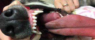
Signs and symptoms
Hydrocephalus in a puppy most often manifests itself at 1.5 months, but this is not a dogma. It all depends on the owner: if he is attentive to his pet and can see the first signs of illness, the dog can be saved.
The exact cause of the disease is still unknown. The following factors can provoke its acquired form, which is rare:
- brain tumors;
- chronic inflammation;
- genetic inheritance.
Therefore, take a closer look at your favorites. The first thing that should alert you is the pet wandering around the apartment for no reason, as well as walking in circles. At the same time, he usually does not notice anything around him and remains indifferent even when bumping into some objects. This sign is very characteristic of brain damage. If your dog begins to behave this way, immediately take him to the veterinarian in order to identify this disease at an early stage.
ATTENTION! Detected and initiated treatment at an early stage of the disease will yield positive results, provided that the owners closely monitor the pet’s healing and take its health seriously. Their dog will live longer.
Among the main signs by which hydrocephalus can be recognized:
- unusual tilting or tilting of the head;
- the appearance of strabismus or even moving the eyes in different directions, deterioration of vision;
- the occurrence of seizures resembling epilepsy;
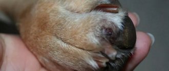
- enlargement and convexity of the skull;
- impaired coordination of movement (especially in puppies);
- delayed psychological development.
As the illness progresses, the dog will be noticeably different in that it has a large head compared, for example, with unusually thin limbs. In addition, she can show aggression or be completely indifferent to everything.
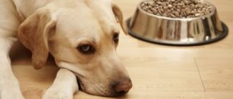
These symptoms only occur in dogs and small breeds. In large representatives of four-legged animals with this disease, the size of the head does not change. Hydrocephalus in them is usually accompanied by loss of appetite, weight, depression, etc.
Clinical manifestations
How does this disease manifest itself? You should rush to the vets immediately if you notice the following symptoms:
- The dog “plays carousel”, spinning in one place for a long time or simply walking in a circle. Often, he bumps into objects and other dogs, not noticing anything around him. These are very characteristic signs, 100% indicating brain damage.
- The dog falls, throwing its head back. She can lie like this for several minutes, squealing and whining, but unable to change her body position.
- For no apparent reason, the dog begins to have convulsions. Depending on the degree of brain damage, they can involve the entire body or individual limbs.
- Often the puppy constantly walks with his head thrown back to one side.
In general, such dogs have a lot of deviations in behavior: their eyes can look in different directions, sometimes they have seizures, the animal can be extremely aggressive or, conversely, completely indifferent to everything. If the disease has progressed greatly, in appearance such a dog stands out among its peers with a large head, skinny and skinny limbs (especially clearly noticeable in the photo).
How is hydrocephalus treated in dogs?
If your pet exhibits the above signs and symptoms, it should be taken to an experienced veterinarian immediately. The disease is diagnosed using various research methods: from ultrasound to computed tomography of the head. Treatment of hydrocephalus in dogs involves drug and surgical therapy. It all depends on the severity of the disease and the reasons for its occurrence. The disease is similar in appearance to dropsy.
ATTENTION! Drug therapy can be beneficial in some cases. But, unfortunately, the relief will be short-lived. It is during this period of improvement that it is advisable to undergo surgery. You can't do without it.
When the symptoms of illness are still mild and the diagnosis was made on time, drug therapy is used. First of all, drugs are prescribed that can reduce intracranial pressure. Dogs are also prescribed diuretic drugs, antibiotics, and glucocorticoids. These are mainly Prednisolone, Furosemide, Acetazolamide, Omeprazole, etc. All these drugs are recommended to be administered with a gradual reduction in therapeutic portions to the minimum required to maintain therapeutic results.
In general, the use of such medications requires strict adherence to the dosage time. This will help, first of all, to obtain the desired impact, as well as protect the animal from their side effects. It should also be taken into account that therapy with these medications can cause a lack of various microelements. Therefore, in addition, it is important to take vitamins.
ATTENTION! It is strictly forbidden to take drugs that are classified as diuretics constantly and uncontrollably. When carrying out therapy with them, you should adhere to the prescribed healing gap. If convulsive activity occurs, the animal is also prescribed anticonvulsant medications.
Such treatment, although it provides a certain level of relief, cannot completely get rid of this disease. In addition, it is not suitable for long-term therapy. The solution is surgery in the form of ventriculoperitoneal shunting. The essence of the operation is that with the help of a shunt (tube) the brain and abdominal cavities are connected and thus it is possible to drain excess cerebrospinal fluid from the brain of the head.
It goes without saying that the dog will have to be with this tube constantly for the rest of its life. In this regard, owners of such animals must comply with the following rules:
- do not allow your pet to be excessively active, playing with other dogs (it is better not to allow this at all);
- carefully pick up your pet so as not to harm him;
- Regularly visit the veterinary clinic with him to monitor the condition of the shunt, which tends to become clogged from time to time.
ATTENTION! Surgical installation of a shunt designed to drain cerebrospinal fluid in case of hydrocephalus is the most effective measure that can alleviate the dog’s condition. Your pet will feel 90% better. But the disadvantage of such a device is that it can move, get damaged or cause rotting at the place of its installation. Therefore, constant monitoring of the dog by a veterinarian is necessary.
Life of dogs after surgery
After installing the shunt, owners will have to somewhat limit the activity of their pet so as not to damage the structure. In particular, you should not play outdoor games with your dog or allow it to frolic with other animals. During games, the drainage tube may be damaged and the operation will have to be repeated.
Dogs with hydrocephalus have to live a very careful lifestyle.
You should lift your pet very carefully so that the shunt does not become dislodged. You will also need regular visits to the veterinary clinic, examination of drainage and the general condition of the animal.
Hydrocephalus requires constant monitoring by a doctor
Over time, the tube may become clogged and stop draining cerebrospinal fluid. The drainage may also be damaged. There is a danger of the development of inflammatory processes at the site of shunt installation, which without timely diagnosis can lead to tissue necrosis.
Forecast
By the way, such an operation cannot be performed on all animals without exception. It is contraindicated if the animal is exhausted, or has an infectious disease, in an advanced form of the disease, when the skull has acquired an unusual shape. The best solution in this case is euthanasia.
From what has been said, it is clear that this ailment cannot be classified as one that should not be paid serious attention to. If it is detected early and therapy is started on time, then there is still hope for overcoming the disease. In a different scenario, the prognosis is unfavorable.
If you don’t forget about this, then your four-legged friend, although he will be limited in some ways, can live a long time and almost fully. But if time is lost, then, unfortunately, nothing can be done, even to alleviate his condition.
Hydrocephalus in puppies is not a death sentence if the pathology is detected in a timely manner and appropriate measures are taken. Then the treatment will definitely be successful, and your pet will delight you for a long time.
A healthy brain is the basis not only for the normal functioning of the body, but also for the socialization of animals, their upbringing and training. It is the parts of the brain that are responsible for the perception and processing of information. This is why brain diseases such as hydrocephalus are so dangerous, because a healthy and intelligent animal can turn into a disabled creature due to pathology.
Main symptoms
It is very easy to determine hydrocephalus, since the disease has a pronounced clinical picture. The first signs appear in a puppy at 1-1.5 months; the most characteristic ones include:
- visual impairment visually resembling strabismus;
- increase in the shape and size of the head;
- seizures, convulsions;
- change in behavior (aggression or lethargy, apathy);
- unusual movements (rolling on the floor, throwing back the head, spinning around oneself).
The sick puppy stands out among the other babies in the litter: its head is much larger, its limbs are thin and thin.
Treatment method and prognosis
Once the diagnosis is made, the doctor prescribes treatment for hydrocephalus. It is carried out in a complex and includes taking diuretics, the action of which is aimed at removing excess fluid from the body. Prescribed:
- Prednisolone: 0.25-0.5 MK/1 kg of weight 2 times a day every 12 hours.
- Furosemide: 0.4-4 MK/1 kg 1 time per day.
- Acetazolamide: 10 MK/1 kg 4 times a day every 6 hours.
- Omeprazole: 10-20 MK/1 kg 1 time per day.
The dosage and course of taking medications are determined by the doctor depending on the weight of the animal, the condition and individual characteristics of the body. The owner must strictly comply with the treatment conditions, since uncontrolled use of medications can have a detrimental effect on the dog’s health and lead to numerous side effects.
Since convulsions are common in sick puppies, anticonvulsant medications are given. To strengthen the immune system, vitamin complexes are prescribed.
But conservative therapy is often ineffective, so there is a need for surgical intervention. The operation involves the introduction of a shunt for the outflow of cerebrospinal fluid.
After surgery, your veterinarian may prescribe corticosteroid hormone therapy.
Contraindications to surgical intervention include exhaustion of the body, advanced hydrocephalus, severe changes and deformation of the skull.
In most cases, the prognosis for hydrocephalus is unfavorable, since even with timely shunting it is impossible to completely get rid of the pathology. It is not always possible to restore vision and motor function. In addition, the risk of death during surgery is very high.
Help and treatment
Treatment is prescribed in a comprehensive manner to cover the entire spectrum of symptoms and influence them.
- Prednisolone is used to reduce fluid production
. Dose – 0.5 mg per kilogram of body weight. Give twice a day, exactly on time. - Furosemide is prescribed at the rate of 4 mg per kilogram of weight, every day
. It is important to give the medicine at the same time every day. Acetazolamide should be taken every six hours, omeprazole every other day. Both drugs are given in a dose of 10 mg per kilogram of animal body weight. - Diuretics and diuretic medications remove excess fluid from the body
. Taking two drugs at the same time is prohibited due to severe toxicity. With each dose, the dose should be reduced so as not to provoke side effects. But you also need to take a break for a few days. - It is advisable to prescribe anticonvulsant medications to eliminate convulsive conditions
. It is important to understand that taking pills can provoke a decrease in electrolyte levels and hypotension. Therefore, treatment should be carried out only under medical supervision. It is forbidden to make decisions about therapy on your own.
The drug Prednisolone is used for treatment.
Surgical intervention
If medication is ineffective, surgical intervention is necessary, during which a shunt is placed to ensure constant drainage of brain fluid.
Surgery is prescribed if medications do not help.
The postoperative period requires special monitoring
, since complications are possible, during which the shunt may become displaced, damaged, or become infected at the installation site, resulting in rotting.
There are contraindications for the operation. If significant exhaustion is recorded, it means that the body is not able to heal wounds. Among other things, there is a possible risk of having a concomitant disease or infectious pathology. These facts are also contraindications for surgical intervention. And you also cannot perform surgery if hydrocephalus is advanced, the skull has undergone significant changes, and has become deformed.
Post-operative care
If, nevertheless, the operation was performed, there are a number of recommendations for caring for the operated animal.
- Limit active games with other dogs and avoid sudden movements.
- Lift the operated puppy very carefully to prevent the shunt from dislodging.
- Be systematically examined in the clinic, undergo routine examinations, and clean possible clogged shunts.
- After the onset of this disease, only a cautious prognosis is given, and then only if treatment is started in a timely manner; in other cases, the prognosis is unfavorable.
After surgery, you cannot play active games with the dog.
Symptoms of hydrocephalus in dogs
The most striking symptoms accompany the congenital form of hydrocephalus in dogs. Such puppies have a disproportionately large head, an “unclosed fontanel,” and are developmentally delayed. Often these dogs have divergent strabismus. In behavior, puppies differ from littermates in aggression, lethargy, disorientation, and may experience seizures and loss of consciousness. Also, puppies with hydrocephalus are almost impossible to train and cannot control their behavior at home.
Congenital hydrocephalus without clinical signs is common among toy breeds. Puppies look almost no different from their peers, but they can exhibit behavioral disorders and be aggressive.
Developing from birth, hydrocephalus does not occur with a sharp deterioration in the dog’s well-being, but the acquired disease in an adult animal progresses quickly and symptoms of damage to the nervous system appear sharply. One of the main complaints of owners is the appearance of seizures in an adult dog or loss of consciousness.
The acquired form of the disease develops in dogs of different breeds and at any age. At an early age, infectious causes of hydrocephalus are more common; in middle and old age – brain tumors and hemorrhages.
With the rapid progression of hydrocephalus in a dog, neurological disorders may progress to the onset of coma and/or death.
Signs of hydrocephalus
The age at which symptoms first appear can vary, on average from 2 months to 5 years, but owners of animals under the age of 1 year most often seek help.
The most characteristic clinical signs of hydrocephalus include seizures and/or unusual behavior of the animal, for example:
- the dog runs in circles (as if chasing its tail);
- throws his head back;
- or tilts it to one side;
In addition, hydrocephalus is indicated by the characteristic appearance of the dog, which distinguishes it from its littermates:
- a very large skull on a thin neck;
- strabismus of the eyeballs;
- behavioral disorders (aggression, bulimia, increased libido, difficulties in training).
Chihuahua with signs of hydrocephalus
If you notice such deviations, you urgently need to take the animal to a veterinary clinic for a comprehensive examination.
Diagnosis of hydrocephalus in dogs
Hydrocephalus in dogs is easily detected when clear clinical signs of the congenital form of the disease develop (impaired growth rates, increased head volume, strabismus, etc.), but in case of seizures or an asymptomatic course of the disease, it is diagnosed only by magnetic resonance imaging (MRI).
The advantages of MRI in making a diagnosis and determining the cause of the disease: detection of neoplasms, signs of infection and inflammation of the meninges, detection of primary pathology (Chiari-like malformation).
An MRI is performed after a preliminary examination: blood tests and ultrasound of the heart.
If the sutures of the skull are open, an ultrasound of the brain is performed; this study does not determine the cause of hydrocephalus, but confirms the presence of an excess amount of cerebrospinal fluid. More often it is performed when it is impossible to conduct an MRI at the time of examination, when there are high anesthetic risks, or when the puppy is too young. A prerequisite for performing an ultrasound of the brain is an “open fontanel”.
Dogs with no clinical signs of hydrocephalus, admitted in a state of epistatus or experiencing short-term seizures, undergo a complex of diagnostics: a neurological examination, tests for hidden infections, an electroencephalogram and MRI.
Who gets sick most often?
You should know that hydrocephalus in large breed dogs is a very rare case, as well as in medium breeds. But the “pocket” varieties (,) are initially predisposed to this pathology. And this is not about some genetic diseases, no. It’s just that in these dogs the size of the brain is quite large, but the volume of the cranium is too small, which leads to problems. However, owners of “pickpockets” aged more than a year can calm down, since in their case the development of the disease is already unlikely. This is due to the fact that the reasons here lie in physiology...
Puppies have every chance of getting sick. The fact is that at a young age such dogs grow very quickly, and the increase in the skull bones may not keep pace with the growth of the brain. If the lag becomes critical, hydrocephalus of the dog’s brain occurs.
Treatment of hydrocephalus in dogs
Hydrocephalus in dogs is treatable, which is based on removing excess cerebrospinal fluid and addressing the cause of the disease.
Congenital hydrocephalus with a severe course of the disease can only be corrected surgically using ventriculoperitoneal shunting. The essence of the operation is to create an artificial vessel (shunt) through which excess cerebrospinal fluid is drained into the abdominal cavity.
For secondary hydrocephalus, the following are indicated: diuretics under the control of dehydration of the dog's body, steroidal anti-inflammatory drugs, anticonvulsants, proton pump inhibitors, etc. Antibiotics are used in case of established bacterial infection. Symptomatic therapy is most often lifelong; the choice of this type of treatment is possible if the drugs are well tolerated, if there is no progression of hydrocephalus and in cases where surgical intervention is contraindicated.
Acute attacks of hydrocephalus in dogs are treated in an inpatient department under the control of vital organ systems, and the issue of surgical intervention is resolved in a short time. The sooner a diagnosis is made and the correct treatment is prescribed, the greater the chance in such a situation that the dog will not suffer irreversible changes in the central nervous system.
High blood pressure in dogs and cats: causes, symptoms, how to measure
Hypertension can be primary due to the pathology of the vessels themselves (idiopathic or essential) and secondary, arising from problems with some organ or system (for example, the kidneys or hormonal system), and sometimes can arise due to the use of certain drugs. Secondary hypertension in cats and dogs is more common than primary hypertension. AH develops more often in animals of older age groups (After 6-7 years).
How is blood pressure regulated in dogs and cats and why does hypertension occur?
Blood pressure (BP) depends on two quantities: the volume of blood that the heart pumps per unit of time (varies depending on the heart rate and cardiac output) and general vascular resistance (elasticity of the vascular walls).
Simply put, the pressure of a liquid in any pipe system is regulated by the pumped volume of this very liquid and the diameter of the pipes through which it flows. An increase in the volume of liquid and/or a decrease in the lumen of the pipe (vessel) leads to an increase in pressure.
The mechanisms of blood pressure regulation are complex. Normally, the relative constancy of blood pressure is maintained due to the coordinated work of the nervous (central and peripheral) and hormonal systems.
One of the main organs that influence blood pressure is the kidneys. The kidneys perform several functions that help regulate blood pressure: they filter salts and water, and also take part in the functioning of the renin-angiotensin-aldosterone system (RAAS).
One of the endocrine glands that influences blood pressure is the adrenal glands (due to catecholamines and aldosterone).
Scheme of operation of the renin-angiotensin-aldosterone system (RAAS)
An example of the participation of the nervous system in the process of blood pressure regulation is shown in the figure below: impulses from baroreceptors (which respond to changes in pressure and are located in the vessels) along the afferent nerve fiber go to the central nervous system to the centers that process these impulses (vasomotor) and return along the efferent nerve fiber to receptors/tissues/organs responsible for changes in pressure.
Scheme of the participation of the nervous system in the process of blood pressure regulation
The main mechanisms leading to the development of hypertension
- impaired filtration of sodium salts by the kidneys and their retention in the body (i.e., an increase in the amount of sodium salts leads to an influx of water into the bloodstream, which is necessary to maintain a stable concentration of the latter, which increases the total blood volume and pressure);
- disruption of the sympathetic nervous system;
- disruption of the RAAS;
- disruption of the functioning of endothelial cells (cells that line the vessels from the inside, take part in the expansion and contraction of blood vessels, which directly affect elasticity);
- vascular hypertrophy (thickened walls, unable to react mobilely and expand the lumen of the vessel to changes, for example, blood volume or increased cardiac output).
Diseases that can cause secondary hypertension
- kidney disease (comes first in both cats and dogs);
- hyperthyroidism (more common in cats);
- hyperadenocorticism;
- diabetes;
- hypothyroidism;
- acromegaly;
- pheochromacytoma;
- hyperaldesteronism;
- cardiac hyperkinesis and arrhythmias;
- intracranial problems (for example, increased intracranial pressure);
- hyperestrogenism.
The mechanisms of development of primary hypertension in cats and dogs (as, in general, in humans) are currently not fully known and understood. That is, what causes the thickening of the walls of blood vessels or leads to disruption of the endothelium (provided there are no other causes of hypertension) has not been fully studied.
Why is AG dangerous?
Any disease has its target organs (those that suffer the most during the development of pathology). In hypertension these are: kidneys, heart, brain, eyes.
- Kidneys : with constantly high blood pressure in the vessels of the nephron, a gradual change in the structure of the tissue of this kidney unit occurs (an increase in the number of fibrous fibers), leading first to a disruption and then to a complete loss of the ability to filter urine. When such nephrons become more than 75%, irreversible renal failure occurs.
- Heart : constantly increased pressure forces the heart muscle to work with greater force, this leads over time to its thickening, complicating the nutrition of the heart muscle and increasing the risk of arrhythmias.
- Brain : the trophism (nutrition) of certain parts of the brain is disrupted as a result of tissue edema (due to high pressure, part of the liquid component of the blood “sweats” into the surrounding tissues) or hemorrhages (as a result of vascular ruptures). In some cases, these changes can lead to irreversible changes in the functioning of the central nervous system. Hydrocephalus (stagnation of fluid in the ventricles of the brain) also sometimes develops.
- Eyes : due to increased pressure in the vessels of the eye, hemorrhages may occur in different parts of the eyeball, retinal detachment, and glaucoma may develop. These changes often lead to partial or complete loss of vision.
As a rule, in the presence of hypertension, not one, but all of the listed organs suffer. And it doesn’t matter for what reason the hypertension arose. It is important how long and how strongly the pressure rises.
What are the symptoms of hypertension in cats and dogs?
Symptoms of hypertension vary in manifestation and strength. They depend, of course, on what, and how badly, the target organ was damaged. Symptoms that owners of animals with hypertension usually pay attention to:
- vision impairment/loss (more often in cats);
- pendulum eye movements;
- hemorrhages (redness) in the anterior chamber of the eye;
- causeless vocalization (in cats);
- dyspnea;
- fainting;
- epileptiform seizures;
- lethargy, apathy;
- disturbance of appetite and water consumption;
- Manege movements (movement in a circle).
Symptoms of high blood pressure in cats and dogs (changes) that can only be identified using specific methods in a veterinary clinic
- proteinuria and hematuria (urinalysis);
- concentric hypertrophy of the left ventricle of the heart (only by echo kg);
- arrhythmia (using ECG);
- systolic murmur (during auscultation);
- retinal detachment or hemorrhage in the fundus of the eye (ophthalmoscopic);
- changes in the structure of the central nervous system (according to CT or MRI).
The listed symptoms are nonspecific and often occur with other diseases. And this significantly complicates the early diagnosis of hypertension.
How to detect high blood pressure?
There are not many options for measuring pressure: these are direct methods when placing sensors directly into the central vessels (a traumatic method), but they are more accurate than indirect ones. In humane medicine and veterinary medicine, it is used in the intensive care unit and during complex operations.
The indirect method is to measure pressure with the tonometers we all know. However, in veterinary medicine, conventional medical tonometers often give too great an error or it is not possible to use them at all, for example, in cats and toy breeds of dogs.
In small animal veterinary medicine, it is recommended to use devices operating on the Doppler principle - one of them is PetMAP. The pressure is measured by placing a cuff on the paw or tail. It is recommended to make up to 3-5 changes in one place and display the average.
At the veterinary, you can measure the blood pressure of cats and dogs using such a device. The price for measuring blood pressure in dogs and cats is indicated in the corresponding section.
For dogs, blood pressure norms range from 100/65mmHg - 160/100mmHg (systole/diastole). For cats - 110/70 - 180/110mmHg. Systolic pressure close to 200 always requires drug correction, and above 280 may require emergency measures.
However, in some pets, a blood pressure of 185/110 may already require therapeutic intervention. Unfortunately, finding out the presence of high blood pressure is not enough; it is important to understand primary or secondary hypertension. And this always requires additional studies, which are prescribed by the doctor based on the clinical symptoms noticed by the owner and abnormalities identified during examination.
This is important, because in the presence of secondary hypertension, therapeutic correction (if possible) of the primary disease removes the symptom of hypertension. If this is not possible, the doctor selects a drug to lower blood pressure. Dose adjustment of the drug often occurs during the first 1-2 weeks of treatment and is then applied for life.
Symptoms of the disease
The manifestation of the congenital disease, visible to the naked eye, can be detected within 2-3 weeks after the birth of the puppies. However, acquired hydrocephalus in dogs can manifest itself much later. The most striking signs of pathology, according to experts, include:
- inappropriate behavior, expressed in the fact that the dog can start circling in one place for no reason, unnaturally throw back its head and twist it in different directions;
- vision is impaired, the dog begins to clearly cross his eyes;
- seizures occur that are in many ways reminiscent of epileptic seizures;
- the shape and size of the skull changes, it becomes larger, acquires a spherical shape, cranial sutures are clearly visible under the skin;
- hydrocephalus causes the dog to become aggressive. When the owner tries to play with him, he may growl at him and even bite him.
The owner must be aware that skull transformation is only possible in representatives of “pocket” breeds; these symptoms will not affect a large dog. In dogs of medium and giant breeds, the disease manifests itself in that they lose their appetite, become apathetic and lethargic, sleep a lot, and when awake wander aimlessly around the house, bumping into pieces of furniture.
Briefly about hydrocephalus
What is this disease? This is an accumulation of fluid in the meninges. The pathology leads to serious consequences, because the compressed nerve cells degrade and die en masse. This is manifested in dogs by seizures and severe behavioral disorders.
You should know that representatives of small breeds of dogs are more susceptible to this dangerous disease. Large and medium-sized ones, as veterinary practice shows, are practically not threatened by the disease. “Pocket” varieties of dogs (for example, Yorkies, Chihuahuas) are initially predisposed to pathology. But the point is not a genetic predisposition of small dogs to hydrocephalus. No. In such animals, the brain size is large, but the volume of the cranium is small. This is what leads to problems. It is worth noting that in representatives of small breeds, pathology practically does not occur after a year, because the growth period ends. Puppies have a high chance of getting sick. When young they grow quickly. And often the increase in the bones of the skull does not keep pace with the growth of the brain. When such a lag becomes critical, the animal develops hydrocephalus.
Diagnosis of the disease
Hydrocephalus in a dog is expressed quite clearly, so it will be quite easy for a specialist to differentiate it from other ailments. The same applies to the detection of the first signs of pathology by the owner himself. Veterinarians strongly discourage owners of sick animals from trying to self-medicate at home.
The brain is an important and complex organ in dogs, which is very difficult to treat with medications, let alone homeopathic remedies or those options offered by traditional medicine. Such treatment will certainly kill the dog, so if you suspect dropsy of the brain, immediately take him for examination to a veterinarian.
As for the diagnosis of the disease itself, it is carried out comprehensively and includes procedures such as:
- general and biochemical analysis of blood fluid;
- general urine analysis;
- cerebrospinal fluid analysis;
- Ultrasound (especially important for puppies whose fontanel is open);
- For those dogs that have reached maturity, X-ray and electroencephalography studies are ideal. Thus, the functioning of the brain is assessed, as well as the degree of its damage;
- in some cases, the method of contrast craniography or computed tomography is acceptable, however, unfortunately, not all veterinary hospitals can boast of such equipment.
Diagnosis of the disease
The symptoms of the disease are quite bright and pronounced. An attentive owner will naturally pay attention to changes in his pet’s condition and take appropriate measures. First of all, this means a visit to the veterinarian. Self-medication, as well as putting off visiting a doctor, is dangerous, since an increase in fluid volumes in the brain area compresses it, which can lead to death.
Before starting treatment, the doctor must make an accurate diagnosis.
In order to prescribe adequate therapy for your pet, the doctor must make an accurate diagnosis. Diagnosis of an animal includes a set of measures. First of all, this is a visual inspection of the animal. Laboratory research methods will also be required:
- Blood and urine analysis
- Cerebrospinal fluid analysis
- If a dog has an open fontanel (relevant for puppies), an ultrasound examination can help in making a diagnosis.
- Adult dogs undergo radiography and electroencephalography. These methods make it possible to assess the functioning of the brain and determine the degree of its damage.
- In some cases, when other diagnostic methods for one reason or another do not allow establishing the picture of the disease, the CT method is used.
Drug treatment for this disease does not last long.
For hydrocephalus, the pet is prescribed drug therapy. First of all, the animal is advised to take diuretics to remove excess fluid from the body. It is important to observe the dosage of the drug, because uncontrolled use of these drugs can lead to very unpleasant side effects. The doctor prescribes the dosage and duration of treatment individually for each specific case.
Most often prescribed:
- Prednisolone. Dosage: 0.25-0.5 MK per kilogram of animal weight. Take the product 2 times a day with an interval of 12 hours.
- Furosemide. Dosage 0.4-4 MK per kg. weight. Take 1 time per day at a strictly defined time.
- Acetazolamide. Dosage: 10 MK per kg. weight. Take 4 times a day with an interval of 6 hours.
- Omeprazole. Dosage: 10-20MK per kg. weight. Take the drug at the same time with an interval of 24 hours.
It is very important to strictly adhere to the time of administration, as this will help to achieve the desired effect faster and reduce the risk of side effects. In addition, it is important to take into account that taking these drugs can provoke a lack of microelements important for the animal. Therefore, taking vitamin supplements is necessary.
Hydrocephalus can cause seizures and convulsions in the animal. Therefore, the animal is advised to take anticonvulsant medications.
Important! Drug treatment can lead to positive results. Unfortunately, this result is short-lived, and, alas, it cannot be done without surgical intervention.
Hydrocephalus of the brain in dogs is a dangerous disease that is more susceptible to small breeds. It is characterized by excessive accumulation of cerebrospinal fluid in the brain cavities of the animal's head. Statistical data have shown that such a pathology is predominantly congenital, although the exact reasons for its occurrence have not yet been identified by medicine. Like any other disease affecting the brain, it is extremely unsafe for a four-legged pet, and without surgical treatment it can lead to serious consequences. The article will discuss the causes and symptoms of this disease, as well as methods of its treatment.
If cerebrospinal fluid accumulates in the brain, the pressure inside the skull increases critically, eventually leading to damage to this organ. First of all, the ventricles of the brain suffer, since the accumulated cerebrospinal fluid in them leads to a reduction in the specific volume of nerve cells, which becomes the cause of various neurological manifestations of a pathological nature.
It is important to understand that the true factors of congenital disease have not yet been identified by veterinarians. However, the causes of congenital hydrocephalus in dogs are quite well known. These include:
- Pathologies of the brain caused by the appearance of various neoplasms in it.
- Pathogenic processes with inflammatory etiology occurring in the dog’s body.
- Vulnerability to dropsy is especially common in small breed dogs (Chihuahua, Yorkshire Terrier). Experts associate this with the physiology of animals in which, during rapid development and growth, the bones of the skull do not have time to grow behind the brain.
Treatment of the disease
Treatment of hydrocephalus in dogs involves two therapeutic options: medication and surgery. The doctor determines the method based on the factors that provoked the pathological process and the severity of the symptoms. In the first case, the specialist prescribes antibiotics, glucocorticoids, and drugs that reduce the production of cerebrospinal fluid to the dog. The most effective among them are Prednisolone, Furosemide and Omeprazole. The dosage is prescribed by the doctor, depending on the weight and general condition of the dog.
The essence of the surgical intervention is that the surgeon performs a bypass of the ventricles of the brain. A tube connecting the abdominal and brain cavities is implanted into the pet's brain. Thanks to this method, excess cerebrospinal fluid will not accumulate in the dog's head. Owners should be prepared for the fact that the prognosis will be disappointing, since the chances of cure depend entirely on how quickly hydrocephalus increases, as well as how much the intensity of its symptoms decreases.
Owners are often interested in the question: is it possible to reduce the risk of side effects from treatment? Doctors answer this in the affirmative. Vitamin complexes, careful care and care for a sick animal significantly reduce the chance of relapse of the disease, and also increase the body’s natural resistance to infections and bacteria that can attack a weakened pet. If bypass surgery has been performed, then the dog will need to be carefully picked up, active games and possible conflicts with fellow dogs should be excluded from his life, and also taken to the veterinarian for examination at least once every two weeks.
Hydrocephalus is a formidable pathology that should never be ignored. It is the owners’ quick response to the first symptoms that determines how effective subsequent treatment will be. After therapeutic intervention, the dog’s quality of life will decrease, albeit slightly, but it will still live quite fully and delight its caring owner.
Treatment
If hydrocephalus is confirmed, treatment is prescribed, which can be carried out using both medical and surgical methods. The specific type of therapy is selected depending on the clinical picture.
During the period of acute neurological disorders, a number of drugs are used to reduce the production of cerebrospinal fluid and lower pressure - prednisolone, furosemide, acetazolamide. The exact dosage and duration of therapy depends on the condition of the animal.
If drug treatment is ineffective and acute symptoms persist even after completing the course, ventriculoperitoneal shunting is used - a surgical intervention that provides a significant improvement in the dog’s condition.
This operation consists of inserting a special drainage tube that connects the cavity of the cerebral ventricles and the abdominal cavity. Thus, excess cerebrospinal fluid is removed from the cerebral ventricles, and pressure in the brain is reduced.
X-ray of a dog after installation of a ventriculoperitoneal shunt
Causes of hydrocephalus
Hydrocephalus can develop in both open (compensated) and closed forms (occlusive). The closed form is usually congenital. In such cases, the puppy is born with abnormally narrowed ducts through which the cerebrospinal fluid moves. The reasons for the development of pathology are not fully understood. Basically, the development of congenital occlusive hydrocephalus is associated with genetic failures or intrauterine infection. The congenital form of hydrocephalus most often appears between the ages of 1 month and 1 year. Moreover, against the background of stress or complex diseases, the disease can develop in adulthood.
Most often, the disease manifests itself in dogs at a very early age.
Acquired hydrocephalus appears against the background of other diseases, the course of which leads to damage to the ducts. The main reasons for the development of acquired hydrocephalus are:
- head injuries;
- oncology;
- infectious diseases.
Compression of the brain leads to the gradual death of nerve cells
Injuries received by a dog can provoke the appearance of hematomas in the brain. Thus, the brain tissue is displaced under the pressure of the blood clot, and the duct narrows. Oncological diseases act in the same way as hematomas. Tumors, both malignant and non-malignant, mechanically disrupt the circulation of the cerebrospinal fluid. As a result of infections, foci of inflammation appear, which subsequently transform into scars and thickening of brain tissue.
Certain breeds with specific skull structures are predisposed to hydrocephalus
Video about the dog's brain
Small breeds of dogs are susceptible to such an unpleasant disease as hydrocephalus, a disease characterized by abnormal fluid content in the brain. The main cause of the disease has not yet been established, however, there are a number of factors that can trigger the development of pathology. Read about these factors, as well as the symptoms of the disease, methods of diagnosis and treatment in the article.
Hydrocephalus, a congenital disease.
In some cases, a phenomenon such as excessive accumulation of cerebrospinal fluid in the brain area can be observed in a pet. This leads to increased pressure inside the skull, and, as a result, to various types of brain damage.
Hydrocephalus is classified as a congenital disease (although acquired cases are also known, but they are much smaller). The disease manifests itself due to impaired functionality of the ventricles of the brain. A large amount of fluid accumulates in them, as a result of which the volume of nerve cells is reduced. This causes various neurological disorders.
Signs of disease development
It is easy for an attentive owner to notice warning signs. The first signs may appear when the puppy reaches one month of age. If you can identify the disease at an early stage, you can successfully resist its development and save the life of your pet.
The earlier the puppy is diagnosed, the higher the chance of stopping the development of the disease.
Symptoms that should alert the owner:
- unnatural head position;
- convulsions;
- disturbance of consciousness and perception of space;
- divergence of eyes;
- behavior uncharacteristic for an animal;
- impaired coordination of movements;
- deformation of the skull.
One of the most common symptoms that may indicate hydrocephalus is the dog's confused movement in a circle. The animal wanders or runs as if chasing its tail. At the same time, it does not notice anything in its path and, having encountered any obstacle, stubbornly tries to continue moving along the established trajectory.
An ordinary action in the life of a healthy dog becomes a constant ritual for a puppy with hydrocephalus.
No less often, hydrocephalus is accompanied by convulsions that affect the entire body or only the limbs. It happens that the cramp is repeated only on one paw. In addition, pets with developing hydrocephalus may suddenly fall to the floor with their head thrown back. The animal remains in this position for a long time.
Seizures in dogs with hydrocephalus resemble epileptic seizures
In this state, the dog may squeal, whine and twitch, but is unable to rise or change body position. In between attacks, you can notice that the animal's head is constantly leaning to one side.
Symptoms of hydrocephalus include not only an enlarged skull, but also behavioral abnormalities
In addition to external manifestations, the dog develops behavior that was not typical for it before. The dog suddenly becomes aggressive and rushes around the apartment, biting and tearing everything he can reach. While running, he may bump into pieces of furniture without noticing them. Such states are replaced by complete apathy, when the animal lies, indifferently staring at one point, and does not react to external stimuli. The dog does not respond to the owner’s voice and refuses water and food.
Strabismus with hydrocephalus
With further development of the disease, an increase in intracranial pressure provokes a disproportionate increase in the animal's head. The skull becomes large and convex, while the rest of the body is emaciated. The animal's limbs become thin and weak. During this period, the development of strabismus is often observed. The pupils begin to diverge in different directions, or one of them is directed upward and the other downward.
If any of the listed symptoms appear, the animal must be immediately shown to a veterinarian and an appropriate examination must be carried out.
About the clinical picture of the disease
The owner of the animal needs to contact a veterinarian as soon as possible if he notices the following symptoms in the dog:
- Carousel game.
The dog spins in one place for a long time or simply walks in a circle. At the same time, she does not notice foreign objects around her and stumbles upon them, as if blind. This is the main sign of brain damage, almost 100% indicative of this disease. - Falling on your back.
The animal can lie like this for several minutes, without moving its head, whining, squealing. Because of the pain, the dog is simply unable to change his body position. - Cramps.
They can start suddenly, for example, while eating, playing, or sleeping. Depending on how advanced the inflammation of the meninges is, seizures can affect only the limbs or the entire body. - Walking with your head thrown back.
Pain causes the puppy to walk this way. In this position, the pain is slightly relieved. - Seizures and sudden changes in behavior.
The dog's eyes may look in different directions, and it becomes aggressive. And sometimes the dog is completely indifferent to everything that is happening, as if he were in a coma.
Externally, such a pet stands out for its too thin limbs and large head.
Treatment of hydrocephalus
Hydrocephalus is a disease that cannot be completely cured. However, the means available to doctors make it possible to contain the disease for many years. Based on the indications, the veterinarian may prescribe drug treatment aimed at reducing intracranial pressure. For this, diuretics, glucocorticoids and antibiotics are used.
Table. Drugs against hydrocephalus
| Name | Action | Reception frequency | Dosage |
| "Prednisol" | Relieves swelling, reduces fluid production in the body | 2 times a day | 0.5 mg per kilogram of weight |
| "Furosemide" | Diuretic | Every 6 hours | 10 mg per kilogram of weight |
The dose of drugs is gradually reduced, reducing it to nothing. Long-term treatment with diuretics can lead to the development of serious pathologies in the animal’s body.
The more accurately the timing of taking medications is observed, the better the effect of treatment will be.
Medicines against hydrocephalus work with symptoms, but do not relieve the animal from the disease
It is impossible to cure hydrocephalus with medication alone. In rare cases, in this way it is possible to achieve a relatively stable remission, but with the slightest stress the disease goes into the acute phase.
Surgery
Drug treatment usually precedes surgery. By reducing blood pressure, the chances of a successful operation increase significantly. Ventriculoperitoneal shunting is used for the surgical treatment of hydrocephalus. The essence of the method is simple. A thin, flexible tube with a pump is placed into the dog's brain where cerebrospinal fluid accumulates. The drainage tube is placed under the animal's skin, passes through the chest and is discharged into the abdominal cavity.
Installation of a drainage tube in the brain
In this way, excess fluid is removed from the skull and absorbed into the surface of the internal organs. As a result, intracranial pressure decreases, the unpleasant symptoms that accompanied the disease disappear, and the animal’s quality of life improves.
Bypass process
Video - Shunting for hydrocephalus
When should surgery not be performed?
The operation cannot be performed if the animal is severely emaciated or has acute infectious diseases. In this case, surgery is postponed until the infection is cured or the required weight has been gained. If the disease was detected late and the dog’s skull was already severely deformed at the time of examination, surgical intervention no longer makes sense. In this case, the animal is euthanized to save it from suffering.
If there is a severe deformation of the dog’s skull, surgery is powerless
Hydrocephalus, despite the fact that the disease is a serious pathology, can be quite successfully controlled in the early stages. Timely detection of disease symptoms and timely diagnosis are not a death sentence. Carrying out therapeutic measures will extend the life of your pet for many years. The main thing is to be an attentive owner and notice any changes in behavior that were previously unusual for the dog.
Diagnostics
To make a diagnosis and prescribe treatment, the veterinarian carries out certain diagnostic measures. In addition to a visual examination of the animal, the specialist will need to take tests of urine, blood, and cerebrospinal fluid. If the fontanelle is open, an ultrasound examination is performed.
If we are talking about acquired hydrocephalus, encephalography and radiography are mandatory to establish the degree of brain damage and assess its functioning. In some cases, the computed tomography method is used. It is used in cases where it is not possible to accurately establish the picture of the disease.