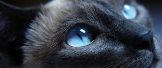
The term hyphema describes the presence of blood in the anterior chamber of the eye - what to do if you find blood in your cat's eye. Bleeding usually comes from the iris blood vessels, but can also come from the ciliary body (tissue behind the iris), from the choroidal vasculature (the layer of tissue under the retina), or from the blood vessels of the retina.
Many cats suffering from hyphema experience decreased visual function or eye damage. However, if only one eye is involved, the cat's behavior is usually normal. Eyes affected by hyphema may be painful, appear half-closed, and be characterized by increased tearing and persistent blinking of the eyelids. Hyphema caused by trauma is often accompanied by bleeding or bruising of the conjunctiva and tissues surrounding the eye.
Causes of hyphema
Ophthalmologists identify 4 main factors that provoke hyphema: 1. Mechanical eye injuries.
They are the most common cause of pathology. With a through puncture (injuring all eye membranes), the vessels rupture, the blood of which fills the front wall. Hemorrhage due to blunt trauma is explained by a sudden drop in pressure in the eyeball, causing micro-tears in the blood vessels of the eye membranes. A similar condition often occurs with wounds and concussions.
Preventive actions
To reduce the risk of styes occurring on a cat’s eye, owners will need to monitor the animal and prevent foreign objects from entering the eye organs. After walking your pet, it is recommended to wash and treat the eyes with tea leaves. To prevent a decrease in immunity, which can trigger an inflammatory process, veterinarians recommend normalizing the animal’s diet.
The menu should include foods that contain sufficient amounts of microelements and vitamins. The animal should be given fermented milk products, boiled potatoes, legumes, and fish. However, it is important to do this in moderation. A cat can also get useful components from vitamin-mineral complexes, which are presented in a wide range in pharmacy chains. It is better to give them in the autumn-spring period.
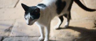
Not only people are susceptible to eye diseases. Problems with the visual organs also occur in cats, although they do not watch TV or play computer games. But they fight, climb trees and fences, sleep in the basement or attic, and this is fraught with injuries, colds and infections. Knowledge of the main symptoms and signs of eye diseases will help the owner to promptly identify the pathology and cure it. Without human help, an animal can lose its sight, which will significantly worsen its future life.
Painful tearing
Experienced breeders know that if a cat has one eye watering for a long time, this is a sign of a serious pathology:
- Cold. Animals often have watery eyes if they are hypothermic and have contracted the virus. In addition to eye discharge, body temperature rises. The pet refuses food and constantly sleeps.
- Conjunctivitis is an infectious disease caused by pathogenic microflora, for example, staphylococci or chlamydia. As a rule, with such an ophthalmological pathology, the cat is afraid of bright light, hides under the sofa or bed, and has watery eyes in one or both eyes. The veterinarian will determine what to do.
- Worm infestation. Intestinal parasites very often cause excessive tearing in pets. They affect not only the digestive tract, but also settle in the visual organs, causing severe suppuration and inflammation. The owner immediately notices that the cat’s eye is swollen and watery. After antihelminthic therapy, these symptoms disappear.
- Trichiasis is a rare pathology characterized by abnormal eyelash growth in cats. They appear inside the eyelid, irritate the tear ducts, and cause infection. The animal constantly rubs its eyes and squints. The problem can only be solved surgically. Simple rinsing with medicinal or folk remedies does not help in such a situation.
- Entropion of the eyelids is a problem that often appears in Sphynxes and other hairless breeds. It can only be solved surgically. Antibacterial therapy is carried out after the operation. After a few days, the animal’s vision is completely restored, and all unpleasant symptoms disappear.
- An allergic reaction often causes swollen eyelids in pets and excessive tear production. It is triggered by various allergens: pollen, household chemicals, cosmetics, especially aerosols, new food with additives, tobacco smoke, poplar fluff or household dust. The pet's eyes become red, frequent sneezing and scabies appear. To save him from suffering, they are treated with antihistamines and, if possible, protect the pet from contact with irritants.
- Chemical or thermal burns of the eyelids and cornea. Pets get such injuries if household chemicals, all kinds of solvents, dyes, and varnishes come into contact with their faces. Before sending to the veterinarian, you need to find out what solution the pet was burned with - alkaline or acidic. If alkali gets into a cat's eyes, at home they are washed with an aqueous solution of boric acid, and if the eyelids are burned by acid, a soda solution is suitable. When you don’t have such products at hand, it’s enough to rinse the face with plain tap water.
- Foreign objects. If your cat's eye is red and watery, it could be a piece of hard food, a splinter of wood, a speck of sand, or sand. They rupture the membranes of the eyelid, cause inflammation, swelling, and activate the production of tear fluid. To remove foreign objects, the pet's eyes are washed with running water and then wiped with an antibiotic solution. If the cornea is seriously damaged, independent actions can only cause harm and cause complete blindness.
- Injuries. Very often, if several mature cats live in a house, clashes and fights occur, as a result of which the pets injure each other’s eyelids with their claws. It often happens that a cat's red eye becomes watery after a walk. He could have been injured by a dry branch on a tree or a stem in the grass.
Important! In any case, if pathological symptoms appear in a pet, a veterinarian examination is necessary. He will examine the cornea and determine the extent of damage to the eye. If conjunctivitis is suspected, an analysis of the eye fluid will be required to determine the causative agent - fungi, bacteria or viruses. In special cases, an ultrasound of the eyes, tests for the patency of the lacrimal ducts, and ophthalmoscopy are performed.
Symptoms of hyphema
Symptoms of hyphema depend on the amount of blood visible in the eye. In most cases, there is a red stripe with clear edges. Depending on its size, the hyphema is divided into 3 stages, which are characterized by their own clinical manifestations:
- Stage 1
– the anterior chamber of the eye is filled with blood to less than 1/3. This stage is characterized by a feeling of slight fog before the eyes; pain will appear if the hyphema occurs after an injury. - Stage 2
– blood is from 40 to 50%. The clarity of vision decreases slightly, a veil forms before the eyes, a sensation of flying “flies”, headaches and dizziness appear. - Stage 3
– the eye is more than half filled with blood. The symptoms are the same as in the second stage, only more pronounced. - Stage 4
(“black eye”) – the anterior chamber is 100% filled with blood. Typically, this condition occurs after penetrating wounds to the eyeball. In this case, the eye becomes completely filled with blood, the person loses the ability to see, only light perception remains (the difference between shadow and light).
If such symptoms appear, you should urgently seek medical help to avoid serious consequences, including complete loss of vision.
Determining the severity of the pathology
It is important to understand that an eye swollen with blood and bleeding from the conjunctival cavity are completely different phenomena, of varying degrees of severity. If a small amount of bloody exudate simply flows out of the eye, then there is no particular reason to panic. But in situations where the cat's eye resembles a blood globule, it is necessary to show the pet to the veterinarian as soon as possible.
As a rule, eye diseases accompanied by the appearance of bloody exudate are quite easy to treat. Pathologies that turn the eyeball into a bloodshot sphere almost always lead to blindness of the animal. In addition, it is necessary to contact the clinic as soon as possible if the following signs appear:
- Small red dots appeared on the surface of the eye.
- Some pets develop thin reddish stripes on their corneas.
All these signs may indicate the initial stages of a pathology called “hyphema.” If you do nothing, the dots and/or stripes will gradually merge, the eye will fill with blood, as a result of which the cat will go completely blind.
The cat's eye is bloodshot photo
If blood appears in the eyes, many ophthalmologists also recommend excluding other viral infections (immunodeficiency, panleukopenia and rhinotracheitis). Very often their course or complications lead to different types of conjunctivitis.
Treatment of hyphema
There is no universal method for treating hyphema. Therapy is carried out for each patient individually and depends on:
- Factors that caused hyphema;
- The volume of blood filling the anterior wall of the eye;
- Developed complications;
- The duration of the observed hemorrhage.
In most cases, patients with hyphema are hospitalized. In case of microscopic hemorrhage, treatment is carried out at home, strictly following the doctor’s prescription. Treatment can be carried out either by surgical intervention or by conservative methods.
Conservative therapy includes:
- Bed rest with head raised;
- A bandage is applied to the injured eye;
- Rinsing the affected area with glucocorticosteroids;
- Taking medications.
Your doctor may prescribe the following medications:
- Painkillers of analgesics;
- Hemostatic and vascular strengthening;
- Eye drops – Lidaza or Emoxipin;
- M-anticholinergics.
Typically, correctly selected therapy leads to complete recovery already on the 5th day of treatment.
A radical method, such as surgery, is resorted to if drug therapy does not produce positive results. Also indications for operations are:
- Significant decrease in visual function;
- Leakage of blood into the cornea;
- Filling of the entire lower eye chamber with blood.
- Increased intraocular pressure that cannot be normalized with medications within 5 days.
What not to do with hyphema
In case of hemorrhage in the anterior wall of the eye, doctors categorically prohibit:
- Use contact lenses;
- Rub your eyes as this stimulates bleeding;
- Self-medicate – use eye drops without being prescribed by a specialist.
In most cases, with timely treatment of hyphema, therapy gives the desired results, and serious complications can be avoided.
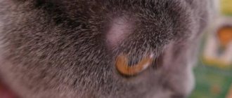
Surgical methods for the treatment of lymphoextrovase in cats and dogs
There are several surgical techniques for solving the problem of lymphoextrovase and hemolymphoextrovase in cats and dogs. All of them involve evacuating the hematoma exudate and placing one or more sutures to create an adhesion - a cohesive fusion between the skin of the ear and the cartilage of the auricle. To do this, through stitches are made in the damaged part of the auricle, the purpose of which is, first of all, to ligate the damaged lymphatic or blood vessels and prevent further leakage. The sutures are left in place for approximately three weeks to create favorable conditions conducive to the development of scar tissue, which exerts additional pressure and compresses long-term bleeding vessels, preventing the hematoma cavity from filling with lymph or blood.
All surgical methods for solving this problem, unfortunately, can lead to some degree of scarring of the auricle. In this case, scarring is not only inevitable, but also a desirable process, since the main cartilage of the animal’s auricle is damaged and thinned, and the resulting collagen scars, although not able to completely restore the functions of the cartilage, can still partially support it. The more scars are formed, the more irregular, corrugated in shape and hard to the touch the auricle
after healing. If the owner of the animal does not seek veterinary help from specialists at all, then within several weeks or even months, provided that the hematoma does not rupture earlier, its contents gradually resolve, and the surrounding tissues grow into powerful dense scar tissue. This is an inevitable process. Not only will the animal experience severe discomfort and pain throughout this period, but complete drying out and severe deformation of the auricle as a whole are possible.
Causes of bleeding from the eyes
Most often in veterinary practice, specialists encounter the following causes of “bloody eyes”:
- Eye injuries. Blood will definitely ooze from deep scratches and abrasions.
- Contact with foreign bodies in the eyes. Large particles of sand, glass or other debris will certainly scratch and injure the delicate mucous membranes of the organs of vision.
- Advanced forms of conjunctivitis (especially purulent). In this case, the capillaries of the conjunctival membrane may be destroyed, resulting in bloody or even bloody discharge from the eyes.
- Blepharitis, inflammation of the eyelids. If the process has affected the inner surface of the organ, then bloody discharge will also appear from the eyes.
- Ulcerative keratitis , i.e. inflammation of the cornea, accompanied by ulcerations of the surface of the organ. In this case, the cornea may bleed.
- Turning of the eyelids. In this case, the cornea is scratched and the eyelashes are damaged, as a result of which the eye begins to bleed.
As mentioned above, all of these are the main and most “harmless” pathologies, which in most cases are treatable. But there is a disease that, without immediate veterinary care, in almost 100% of cases leads to complete blindness of the animal.
Causes of lymphoextrovasation in cats and dogs
Shaking your head can cause these tiny blood or lymph vessels to rupture, bursting and spilling into the surrounding area, gradually peeling the skin away from the cartilage tissue. The accumulated exudate causes excessive irritation, and the animal begins to shake its head even more, thereby aggravating the process. If the situation is not resolved, then blood or lymph continues to accumulate under the skin in greater and greater quantities, increasingly peeling the skin away from the cartilage and forming a large pocket, which in some cases can block the ear canal.
In some cases, constant shaking of the head can eventually lead to spontaneous rupture of the hematoma and splashing of its contents outward. However, a responsible pet owner should never allow such a development to occur.
In what cases is it necessary to contact a veterinarian?
If there is no appetite and lethargy, you should show the animal to the veterinarian. If the cat, in addition to the desire to hide in a dark place, has the following deviations, it is necessary to show the animal to the veterinarian:
- Lack of appetite;
- Increased thirst;
- Refusal of water;
- Heavy discharge from the mouth, nose, or eyes;
- Purulent discharge;
- Changes in the consistency and smell of excrement;
- Change in the color and odor of urine;
- Dull coat, baldness;
- Skin rashes, suppuration;
- Frequent scratching, licking;
- Cough, sneezing, vomiting;
- Lethargy, drowsiness;
- Loss of coordination;
A cat’s desire to hide in a dark place, especially when it is not eating or drinking, is caused by many reasons. An attentive and caring owner will respond in a timely manner to changes in the pet’s behavior and will do everything possible to restore a comfortable environment.
Consequences and complications of hyphema
The complication and consequence that hyphema can cause will depend on the degree of filling of the anterior chamber of the eye with blood. When completely filled, the cornea becomes stained with blood, which negatively affects vision. Even if the hyphema goes away soon, it takes a long period of time for the recolored cornea to recover.
Hyphema, occupying less than half of the anterior chamber, is complicated in 4% of cases by increased intraocular pressure (IOP) and restoration of vision in 78% of cases. Hyphema, occupying more than 50% of the chamber space, provokes an increase in pressure in the eye in 85% of patients, among whom vision is completely restored in only 28%. Increased intraocular pressure leads to the fact that bleeding resumes with greater intensity after 3-5 days.
The condition is also dangerous because along the periphery of the eyeball the iris and cornea can grow together, which, in combination with increased IOP, leads to the death of the optic nerve.
Patients with damaged drainage systems of the eye chambers are required to be examined by an ophthalmologist once a year for the prevention and early diagnosis of glaucoma.
Main symptoms and treatment methods
For timely and adequate treatment, it is necessary to know the characteristic signs of eye diseases.
One of the common diseases in pets is conjunctivitis. Inflammation of the mucous membrane occurs for the following reasons:
- injuries;
- infectious diseases;
- allergic reactions;
- infection with worms and ectoparasites;
- lack of vitamins in the body.
There are several forms of conjunctivitis: catarrhal, follicular, ulcerative and purulent. The main symptoms indicating the development of conjunctivitis are:
- redness of the mucous membrane;
- swelling of the eyelids;
- lacrimation;
- the appearance of pus;
- eyelid gluing;
- photophobia;
- cloudiness.
Purulent conjunctivitis is indicated by bilateral inflammation. In addition, the owner notes the following symptoms of the disease:
- general depression of the pet. He becomes lethargic, does not want to play and frolic;
- high body temperature. Normally, the body temperature of an adult animal is 38-39°C; in children, the figure may be half a degree higher (38.5-39.5);
- poor appetite or its complete absence also indicates a painful condition;
- the cat tries to avoid inspection and palpation, this process causes him pain in the area of the eyeball;
- in advanced cases, inflammation spreads to the cornea.
Treatment steps are as follows:
- A visit to the veterinary clinic to remove a foreign object from the eye.
- Washing can be done at home every 3-4 hours. For the procedure, use a warm solution of manganese, furatsilin, chamomile or calendula decoction. Herbs help reduce inflammation, eliminate swelling and itching. In addition, natural decoctions have anti-inflammatory properties. Manganese copes well with infection, but at the same time dries out the mucous membrane. Therefore, a pale pink solution is used. It is recommended not only to thoroughly dissolve the manganese crystals, but also to filter the resulting composition through several layers of gauze. This guarantees the absence of crystals in the water, the accidental entry of which can cause a burn to the mucous membrane. Furacilin is diluted in hot water (1 g per 5 l). Used for washing while warm.
- Applying ointment. At a pharmacy or pet store you need to buy any eye ointment, for example, Tetracycline, Syntomycin or Erythromycin. The composition is placed behind the eyelid. The procedure is carried out wearing gloves. The ointment should be applied carefully with a special glass rod. Applying with fingers or cotton swabs is unacceptable. After each application of the ointment, the stick is disinfected in boiling water. This is necessary in order not to spread the infection in the future. Some people use an ointment tube to apply the ointment. This must be done extremely carefully, otherwise bacteria will settle on the spout of the tube and, during the next treatment, will again get into the eyes.
- Instillation is carried out only in washed eyes. It is necessary to instill 2-3 drops every 4 hours.
- If the disease is caused by an infection, a course of antibiotic therapy is necessary.
- A solution of novocaine and hydrocortisone (ratio 1 ml/0.2 ml) helps relieve swelling. The composition is instilled into the conjunctival sac.
- Chronic pathology is recommended to be treated with silver-based drugs.
- In a veterinary hospital, a novocaine blockade is carried out. It reduces pain and improves tissue nutrition, which leads to a speedy recovery.
- For follicular conjunctivitis, surgical intervention is performed. After the operation, ointments and antibiotics are prescribed.
We suggest you read: Muscovy ducklings and their cultivation
Keratitis and its types
Inflammation and clouding of the cornea in cats indicate the development of keratitis. The disease is acquired and requires proper treatment. The reasons for the development of keratitis are:
- mechanical injuries of the cornea. The ingress of solid dust particles, grains of sand, blades of grass, and fibers leads to mechanical damage;
- conjunctivitis. The combination of diseases leads to the formation of keratoconjunctivitis;
- thermal or chemical burns;
- infections and viruses. Typically, keratoconjunctivitis develops against the background of a general infection of the body (herpes, adenovirus). In this case, complex treatment is required, otherwise it will not be possible to get rid of keratitis;
- allergic reactions to food, dust, pollen, household chemicals;
- inflammatory processes in the lacrimal glands, leading to drying out of the cornea;
- lack of vitamins in food;
- genetic predisposition to the disease (in British, Sphynx, Persian and Siamese cats).
The appearance of the following symptoms should alert the cat owner:
- clouding of the cornea, its dullness and roughness;
- accumulation of blood vessels on the cornea;
- the discharge causes the fur around the eye to become wet;
- accumulation of pus;
- fear of bright light;
- the appearance of scars indicates the irreversibility of the process. The animal may go blind.
Treatment is prescribed after a thorough examination and confirmation of the diagnosis. The initial stage of the disease can be treated with drugs. In advanced cases, laser correction of the cornea is used.
The following groups of drugs are used as complex therapy:
- antibacterial agents (Tetracycline, Erythromycin, Gentamicin eye ointment, Tobrex);
- antiviral and immunomodulatory drugs (Cycloferon, Gamavit).
Keratitis requires long-term treatment and special care for your pet. If the veterinarian's instructions are not followed, the animal may go blind.
The nictitating membrane in cats is a kind of protection provided by nature. The third eyelid is a whitish film located in the inner corner of the eye. The prolapse of this membrane can be noticed by the following signs:
- uncontrollable trembling, closing eyelids;
- increased lacrimation;
- redness around the eye;
- copious discharge of mucus or pus.
Prolapse of the nictitating membrane is associated with the following diseases:
- conjunctivitis;
- eye damage;
- gastrointestinal diseases;
- fungal pathologies;
- allergic reactions;
- helminthic infestations.
If you suspect third eyelid prolapse, you should wash your pet's eyes with a warm decoction of chamomile or calendula. If after a few hours the nictitating membrane has not returned to its place, a visit to the veterinarian is necessary. Surgery may be required.
There are several types of blepharitis in cats:
- simple;
- with the formation of ulcers;
- scaly;
- meibomian;
- demodectic;
- allergic.
If left untreated, simple blepharitis develops into ulcerative blepharitis. The plaque that appears along the growth of the eyelashes disappears, and ulcers appear in its place. The progression of the disease is accompanied by loss of eyelashes, further damage to the conjunctiva and eyes, and panophthalmitis develops.
If a vessel bursts in the eye for the first time - reasons
There is probably no person who has not experienced an eye capillary rupture at least once. If this happened for the first time, then you shouldn’t be scared and come up with incredibly scary diagnoses for yourself.
This pathology, as a rule, does not cause any painful sensations. You should calm down and wait a few days. The bloody spot or vascular network disappears quite quickly.
In most cases, after 5-7 days, and sometimes even earlier, not a trace will remain of it.
If after a few days the bruise has not changed much, then you should consult an ophthalmologist. This is an alarming symptom.
The vessels need a lot of time to recover, but this does not apply to the hemorrhage itself. Normally, its self-removal occurs in a short time.
Among the possible reasons for the appearance of a bloody mark on the white of the eye are the following circumstances.
- Accumulation of fatigue associated with lack of sleep, possibly chronic.
- Eye strain. The eye muscles, like any other, get tired and need proper rest. Unregulated watching of TV shows, continuous use of a computer (smartphone, tablet), reading at night in poor lighting or in road conditions during shaking - all this leads to overwork of the oculomotor muscles. Bursted capillaries are the first alarming signal of the development of pathology. Failure to comply with the rules of visual hygiene and lack of necessary therapy will soon lead to various ophthalmological problems.
- Physical overload can be of various types. Heavy physical labor, severe overexertion during sports, contractions and labor in women, and a strained cry can easily lead to rupture of the ocular vessel. In this case, the described phenomenon is not systematic. By the way, strong, prolonged crying can cause a vascular rupture in the baby’s eye. There is nothing particularly terrible about this, but such phenomena should be prevented. Next time the muscles may not be able to withstand it and a hernia may form.
- Intense heat. A significant increase in body temperature during illness can cause deformation of the blood vessels in the eyes and lead to their rupture.
- Manifestation of allergies. The body can react differently to all kinds of allergens, including rupture of capillaries.
- A fair amount of alcohol. Alcoholic drinks cause a sharp expansion of blood vessels, including the eye ones. Therefore, if you drink too much, you may find in the morning that a capillary has burst in your eye. But this phenomenon can be not only random. People who abuse alcohol can often see bloody spots on their eyes. In chronic alcoholism, this is a characteristic symptom.
Possible causes of a ruptured vessel in the eye
A bloody spot, indicating a ruptured vessel, can appear for various reasons. It could be some minor trifle that is random in nature, such as a speck that has gotten under the eyelid, a child’s prolonged crying, or a heavy burden.
The factor that provoked damage to the capillary may be more significant - a cold, minor injury, vitamin deficiency, etc. But this lesion can also signal the presence of serious pathologies or systemic diseases (diabetes, tumor, leukemia, etc.).
So you shouldn’t take a burst vessel lightly, especially if it happens repeatedly.
External factors that can lead to rupture of a vessel in the eye
A capillary can also burst due to external factors. It can be:
- weather conditions - windiness, heat, intense sunlight, frost, as well as sudden climate changes or changes in atmospheric pressure can cause a bloody spot to appear on the eye;
— dry air , high concentration of dust particles or smoke in the room;
- going to the steam room or visiting the bathhouse - rupture of the vessel is associated with high temperature;
— contact with the mucous surface of the eye by a foreign particle (dirt, dust, eyelashes) – microtrauma of the surface occurs, which is accompanied by minor hemorrhage;
- prolonged wearing of lenses - rupture of capillaries can be associated with rubbing, drying out of the mucous membrane, vasodilation due to insufficient oxygen permeability of this device;
- prolonged exposure to excessively bright lighting.
As a rule, in these cases, hemorrhages in the eye are also one-time in nature.
But there are a number of pathological conditions and serious diseases in which the eye capillaries burst more than once.
Internal causes associated with various pathologies
When a vessel in the eye bursts not for the first time, there is no need to put off visiting an ophthalmologist.
The reasons for this phenomenon can be very serious.
- Pressure surge. Arterial or eye pressure may suddenly rise. In this case, the vessels become overfilled and rupture may occur. Particularly dramatic changes occur during a hypertensive crisis. Nosebleeds may occur. This condition requires immediate normalization of pressure. Preventive measures should be taken - pressure changes can cause ischemia, stroke, heart attack, aneurysm, coma, and death.
- Diabetes. This condition is characterized by damage to the capillaries, which leads to a narrowing of their lumen, inelasticity, thinning, and necrosis. Blood flow is disrupted, tissues lack nutrients, and blood vessels can burst. Such damage occurs both spontaneously and during trivial loads (bending, squatting, etc.). There is no point in delaying visiting an endocrinologist. Progression of the condition can lead to a gradual decrease in vision up to its complete loss.
- Inflammation of the cornea (keratitis). Symptoms: itching, burning, excessive tearing, intolerance to light, the appearance of red spots on the whites of the eyes, spontaneous drooping of the eyelids. The disease develops as a result of injury, the action of pathogenic organisms (fungi, colds, herpes, etc.), exposure to foreign particles, an allergic reaction, burns, and the use of hallucinogens. It is necessary to promptly identify and eliminate the cause of the disease. The advanced form of the disease threatens tissue ulceration and loss of vision.
- Damage to the conjunctiva (conjunctivitis). Irritation and inflammation of this membrane of the eye has symptoms similar to keratitis. Pain, burning, itching, redness, tearing, purulent discharge, high photosensitivity, and a feeling of the presence of foreign inclusions are observed. The reasons for the development of conjunctivitis are also similar - the action of an infectious pathogen (influenza, coccus bacteria, etc.), allergens, foreign objects (dust particles, smoke, litter). The disease can also develop due to subsidence of immune defense and significant vitamin deficiency. An untreated disease can become chronic.
- Other ophthalmological diseases. Inflammation of the eyelids, lacrimal glands, eye membranes, increased intraocular pressure and other ailments cannot be ignored. Specific treatment must be carried out to prevent the development of complications.
- Vitamin deficiency. A deficiency of vitamin compounds, primarily rutin (Vit. P) and ascorbic acid (Vit. C), leads to a deterioration in the condition of blood vessels and an increase in their fragility. Therefore, hypovitaminosis can lead to repeated appearance of bruises on the proteins. Increasing your daily diet with vegetable and fruit supplements or taking additional vitamin complexes will help cope with the problem and avoid further damage to blood vessels.
- Taking certain medications can cause weakening of the vascular walls, which in turn increases the likelihood of rupture of the eye capillaries.
- Nicotine addiction. In smokers, blood vessels lose their elasticity and become prone to atony. Therefore, this addiction can cause periodic damage to the capillaries in the eyes.
- Pathological changes in the blood , malfunctions of the lymphatic system. Leukemia, decreased coagulability, platelet, lymphocyte disorders and other similar abnormalities cause increased bleeding. It can be observed in various areas of the body, including the orbital area. Such diseases are extremely dangerous due to sudden bleeding. In addition, even minor injuries can lead to serious blood loss.
- Injuries. Traumatic eye injuries of varying degrees of complexity cause the appearance of red spots. Head injuries can also lead to bleeding in the eye area. In this case, dizziness, migraines, sleep disturbances, nausea, and high fatigue are observed. Damage should be identified promptly and repaired.
Types of conjunctivitis
All types of conjunctivitis have similar signs and symptoms. But each type has a certain degree of severity of the disease.
1. Allergic conjunctivitis in cats
There are many irritants that cause the disease. But at the same time, each cat has only one source that causes allergies. Irritants include:
- small pollen particles;
- dust particles, particles of woolen products;
- sunlight, ultraviolet;
- chemicals (washing powders, paint, varnishes, perfume)
This type of disease appears suddenly and develops rapidly. It's difficult to define. If the cat does not have a fever, there are no serious reasons for inflammatory processes, and at the same time the eye swells, tearing appears. This is already a reason to contact the veterinarian.
2. Purulent conjunctivitis in cats
The main source of inflammation of the mucous membrane of the eye is the animal’s weak immune system.
Stages of disease development
Stage 1 – development of catarrhal inflammation: photophobia in the pet.
Stage 2 – the appearance of a tumor on the eyelid: the eyeball becomes red.
Stage 3 – the conjunctiva begins to bleed: pus in the corners of the eyes.
Stage 4 – presence of weeping eczema.
3. Catarrhal conjunctivitis in cats
This type is a harbinger of the onset of the development of other viral eye diseases. Symptoms:
- profuse lacrimation;
- presence of viscous liquid;
- pain accompanies swelling of the eyelids.
4. Chronic type of conjunctivitis : an untreated form of the disease becomes chronic. This type is dangerous because the animal looks healthy, but the disease progresses in the animal’s body. If it lasts for a long time, the eyelid may turn in.
5. Follicular conjunctivitis in cats: the presence of increased pain is the main sign of this type. The cat's eyes narrow, muscle spasms occur, and copious amounts of liquid of a cloudy color and thick consistency occur.
6. Fibrinous type of disease : in practice, this type is very rare. Conjunctivitis occurs after chemical burns. When affected by this type, the animal’s eye is covered with a cloudy film, under which the retina separates.
Two forms of the disease:
- lobar - the mucous membrane is covered with viral necrosis, the removal of which is accompanied by heavy bleeding;
- diphtheroid - necrosis affects the deep layer of the mucous membrane.
7. Viral conjunctivitis in cats : the inflammatory process occurs due to various viruses and bacteria. Sign of this type: cloudy serous fluid.
8. Chlamydial type of conjunctivitis : the causative agent of the disease is chlamydia. This type of conjunctivitis mainly affects newborn kittens due to weak immunity. Kittens experience swelling of the eyelids and excessive lacrimation. Purulent accumulations have blood streaks.
9. Phlegmonous conjunctivitis : this type is characterized by a complex period of the disease and has a long treatment period. The animal has hemorrhage at the affected area. The eye becomes covered with abscesses, and during the development of the disease there is a high temperature.
10. Bacterial species : the cause of the disease is the following infections: streptococcal, hemophilic, staphylococcal. A swollen eye periodically fills with pus, the disease is accompanied by painful sensations in the pet.
11. Eosinophilic conjunctivitis : a characteristic feature of this type is the formation of specific formations of light pink color. The main factor is a malfunction of the immune system. There are no pronounced symptoms. Long-term progression of the disease affects the third eyelid and cornea.
Symptoms of eye lacrimation
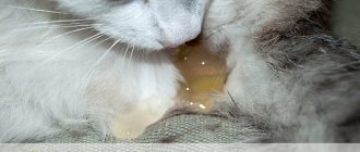
If a cat has watery eyes for a long time in one or both eyes, there is brown discharge or with a whitish tint, this is a manifestation of several serious diseases at once. Along with them, the animal also exhibits other signs of pathology:
- severe itching (the cat constantly rubs its eyes with its paws);
- lethargy and poor appetite;
- redness of the whites;
- increased body temperature;
- photophobia and swelling of the eyelids;
- dried crusts and hair loss in the corners of the eyes.
Pathological discharge may change its character. At first they are liquid and transparent, and after a day they are already thick and with pus, then they pass, then they appear with renewed vigor. If one of your cat's eyes is watery, swollen and squinting, you should not delay your visit to the veterinary clinic. Timely measures will completely relieve your pet from suffering and restore his vision.
Prevention
To prevent panophthalmitis, it is recommended to vaccinate your cat annually against blood-sucking insects, treat it with special means, and treat diseases in a timely manner. It is better not to walk your pet and avoid contact with other animals or aggressive cats. Even with a minor injury to your pet, you should immediately take it to the veterinarian.
Panophthalmitis is rare in domestic cats. However, if the first signs of the disease appear, you should immediately contact a veterinarian. This will help not only preserve the eyes and vision, but also the life of the pet.