Pets, like people, sometimes get sick. To make a correct diagnosis, your veterinarian will often order laboratory tests, one of which is a urine test in cats and dogs.
Urinalysis in cats and dogs
The composition of urine is determined by the metabolic processes occurring in the animal’s body. It can vary depending on the composition of food and liquid consumed, seasonal and climatic factors, and the physiological state of the animal (sleep, stress, pregnancy, illness, etc.). Over 160 substances formed during the metabolic process are excreted in the urine of animals.
Tests are the starting point for diagnosing a pet’s disease.
The physicochemical characteristics of urine can tell us about the condition of the kidneys and urinary tract, the presence of infection, toxins, and the order of metabolism. Based on the results of the analysis, the doctor can diagnose and predict diseases, monitor complications, monitor the effectiveness of therapy, judge the functional state of organs, and identify metabolic disorders.
Indications for urine analysis:
- diagnosis of diseases of the kidneys, bladder, ureters, urethra;
- diagnosis of diabetes mellitus;
- assessment of the condition of internal organs in case of poisoning with toxins;
- control of therapy, assessment of effectiveness, prevention of complications.
Caring owners can independently collect biomaterial and request analysis if they notice unnatural behavior of the pet: frequent visits to the litter box, strained urination, plaintive meowing or whining, uncharacteristic color or smell of discharge.
A cat urinating too often or too rarely is an important reason to immediately contact a specialist.
With some kidney diseases, the temperature rises and the animal may stop urinating or do so in unusual places. Delay in such cases can cost the animal’s life; owners must immediately take samples of discharge and come to the clinic for an appointment.
The chemical structure of urine changes quickly, so it must be delivered to the clinical laboratory within the first two hours. The required minimum volume of liquid is 20 ml.
How to collect samples for testing from domestic cats and dogs?
In order for laboratory test results to be reliable, you must correctly collect a urine sample from your pet.
Collecting urine from cats
Biomaterial is collected from feline representatives at any time of the day. There are several simple and proven collection methods. The choice depends on the habits of the pet itself.
- a tray with a grid insert is the most convenient method for the owner if the animal is accustomed to a tray with a grid without filler. You just need to rinse it first and dry it with a clean, moisture-wicking cloth. Do not use foaming detergents to clean the tray. It is better to treat the surface with hydrogen peroxide or laundry soap. After the pet has emptied, the material is poured into a special sterile container, closed and delivered to the laboratory for analysis;
Example of a tray with grid
- box with filler. If your cat is used to emptying into a litter tray, you can use white office paper, new aquarium soil, or foam balls. They do not absorb moisture and do not have dyes. Urine is collected from the corner of the tray with a syringe without a needle;
- toilet, bathtub, washbasin. Some pets are trained to urinate directly into the toilet or bathtub. Then the animal owner must first thoroughly rinse the surface of the toilet bowl and lay cling film on top so that it completely covers the drain. Then, after visiting the animal, collect biomaterial with a syringe;
To collect urine from the toilet you will need improvised means
- specialized urine collector for cats.
Cat urine collection agent
Collecting urine from dogs
Urine collection from dogs is done in the morning. The container must be prepared in advance: washed and disinfected.
- collection during free urination. Discharge samples are collected during a morning walk. For male dogs, you need to prepare a plastic bottle (suitable for mineral water or oil). Cut off the lower half, and rinse and disinfect the upper half with the lid well.
Collecting urine while walking depends on the gender of the pet
For females, take a tray with low sides or a cup. Don't forget to take a sterile urine container and disposable gloves. The dog is held on a short leash, located slightly behind it. At the right moment, a container is placed under the stream. It is better to take a medium portion of urine. To pour into a container, simply unscrew the cap of the bottle;
- collecting from a tray. It will not be difficult to take the material from litter box trained dogs. You just need to disinfect the container in advance.
A children's urine bag, which can be purchased at any pharmacy, is quite suitable for dogs.
Below are the simplest ways to do this:
- If your dog urinates in the same place every time, you can put a clean film in advance and then collect the result with a syringe;
- You can use a urine bag for children. To secure it to the body, use diapers or accessories for dogs (overalls, pants, bodysuits)
Below are additional tips on how to collect urine from your pet on the street without causing resistance.
Collection of tests on the street
If you have difficulty taking samples at home, you can seek help from specialists. In veterinary laboratories, urine collection may be done using a catheter. However, this method has a number of disadvantages: pain, the need for fixation, trauma and contamination in males. Therefore, this method is used for emergency purposes.
A catheter is used only in extreme cases, as it can threaten the health of the dog
The most sterile and informative method is cystocentesis - puncture of the bladder with a syringe. This manipulation is performed by a doctor. The procedure is painless and is done in a position that is comfortable for the animal. Sometimes cystocentesis is done under ultrasound guidance.
Cystocentesis procedure
Video - Collecting tests from cats and dogs
How to collect urine for research
Incorrect sampling may result in false negative or positive results. For the accuracy of the study, the owner will also have to take care of storing and transporting the resulting sample to the veterinary clinic.
In bitches
Wash your pet's genitals before a walk and prepare a flat container or bottle with the neck cut off at an angle. Do not use detergents to clean the containers, but simply pour boiling water over them.
When you go outside, wait until the dog sits down to urinate and place the prepared instrument under the stream. Pour the resulting specimen into a special container for collecting urine.
Alternative methods include using a baby urine bag, absorbent diaper, or catheter. In addition to catheterization, the clinic recommends bladder puncture.
In males
Males are more suspicious, so do not reveal the presence of the container before urination begins. It is better to collect biomaterial with a partner to keep the dog on a leash until 20-100 ml of liquid gets into the container.
Remember that male dogs urinate in small amounts. Collecting everything in one go is very problematic. For convenience, you can use a condom:
- Make 2 holes in its base and thread an elastic rope through them.
- Place the resulting product on the dog's penis and tie the ties on his back.
- Wait until the required portion is collected and carefully cut off the tip of the condom.
- Pour the contents into a container.
If there is frequent and light urination, it is better to seek help from a veterinarian. Collecting it yourself using a pipette and syringe will take a lot of time, and any dirt on the floor can greatly distort the results.
Storage and transportation
The collected biomaterial must be taken to the veterinary clinic within half an hour. If this is not possible, then the jar can be placed in the refrigerator for 3 hours. Transportation at a later date will distort the results and complicate the diagnosis.
If your pet often asks to go for a walk, and suspicious inclusions appear in its urine, take the sample to a veterinary clinic for examination. Changes in smell and color are alarming symptoms that require the owner’s attention.
When you receive results, try to understand their meaning, but avoid self-medication. Remember that only a doctor can make an accurate diagnosis.
The article is for informational purposes only. Contact your veterinarian!
How is urine testing performed on pets?
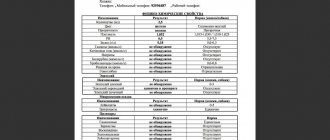
The simplest and most informative diagnostic method is a general (clinical) urinalysis (OAM), which consists of three interrelated studies:
- Analysis of physical properties.
- Study of chemical indicators.
- Microscopic examination of sediment.
Analysis results can be ready in as little as 30 minutes.
The speed of obtaining test results depends on their type and degree of detail.
To determine pathological microflora, bacterial culture of urine is performed. The results will be ready in 10 – 14 days.
conclusions
In order for therapy to be successful, you need to have the correct research results on hand. With the help of urine analysis, not only the disease is detected, but also differential diagnosis is carried out. There should be no inaccuracies here, otherwise the doctor will prescribe the wrong treatment.
Indicators that are considered “norm” are averaged. You can’t discount the dog’s gender, age, individual characteristics, diet and medications used.
Currently reading:
- 4 types of disease development due to dark urine in dogs
- Causes of kidney stones in dogs and methods of treatment
- Is it worth it or not to include natural food in your dog’s diet?
- Seven Signs and Remedies for Getting Rid of Fleas in Dogs
Physical indicators of urine analysis in cats and dogs
The physical characteristics of urine are determined by visual examination. These include:
- daily amount;
- specific gravity or density;
- color gradation;
- transparency, presence of sediment;
- consistency;
- reaction;
- smell.
Daily amount
70% of the fluid entering the body is excreted with urine. The daily amount depends on many factors: the volume of liquid drunk, the composition of the feed, the functioning of the sweat and sebaceous glands, the heart, lungs, digestive tract organs, kidneys. The quantitative indicator of urine excreted per day helps the doctor characterize the condition of the body as a whole and recognize pathological processes.
Depending on where the pet urinates, the count is carried out either by the owner himself or by a specialist.
If the animal uses a tray without filler, then the owners can calculate the daily amount of urine at home. In other cases, counting may cause difficulties, then this procedure is done in a hospital setting.
Normally, the daily amount of urine should be proportional to the liquid drunk, per 1 kilogram of body weight: 20-50 ml for dogs, 20-30 ml for cats.
Polyuria is dangerous because your pet is dehydrated
An increase in the volume of daily urine is called polyuria. The reasons may be:
- diabetes (sugar and insipidus);
- subsidence of edema;
- kidney infections;
- tumor neoplasms,
- metabolic disorders;
- hypercalcemia;
- liver dysfunction;
- inflammatory processes.
Oliguria is fraught with the accumulation of harmful toxins in the pet’s body
A decrease in daily urine output is called oliguria. Oliguria is caused by:
- gastrointestinal disorders (vomiting, diarrhea);
- the appearance of edema;
- small amount of fluid consumed.
Lack of urine (urinary retention) – anuria. A serious pathology, the cause of which may be shock, acute nephritis and advanced chronic kidney disease, blockage of canals with stones or tumors.
Specific gravity
Specific gravity (USG) or relative density shows the average amount of solid compounds dissolved in urine and characterizes the ability of the kidneys to thicken and dilute the fluid contents.
Example of a urinometer
This indicator changes throughout the day and is affected by food and water intake, environmental temperature, medications, and the functional state of internal organs. When dehydrated, the discharge will be concentrated; with a high degree of hydration, it will be diluted. The density of urine is determined by special devices: a urometer, a hydrometer, a refractometer.
Normal specific gravity of urine: in dogs is 1.015 – 1.030 g/l, in cats – 1.020 – 1.035 g/l.
An increase in the density of urine is called hypersthenuria. May indicate dehydration, which may be caused by:
- large loss of fluid (fever, diarrhea, vomiting, profuse sweating);
- low water consumption;
- liver diseases.
The density of urine also increases with oliguria, kidney disease (acute nephritis), heart and kidney failure, accompanied by swelling of the legs and arms, and bacterial infections. At the same time, the level of protein in the urine often increases.
Hypersthenuria is an indicator of kidney or heart problems
If the increased density is accompanied by an increase in the daily amount (polyuria), this is a pronounced symptom of diabetes mellitus. Every 1 percent of sugar in urine increases the specific gravity by 0.004 g/l.
The readings may be affected by medications, for example, radiocontrast agents or diuretics (mannitol, dextran).
A decrease in urine density is called hyposthenuria. Accompanies many kidney diseases (acute and chronic nephritis - “wrinkled kidney”, nephrosclerosis, chronic renal failure). For example, in severe nephrosclerosis, USG approaches a value of 0.010 and is complemented by oliguria.
Hyposthenuria indicates renal failure or diabetes mellitus
A very low specific gravity, similar to that of water (1.002 - 1.001), occurs in diabetes insipidus. A decrease in density is also observed when taking diuretics, ketosis, and dystrophy.
Color
The color of urine (COL) is also determined by various factors: type of food, medications taken, amount of fluid taken, condition of internal organs.
The normal color of urine of cats and dogs is considered to be a uniform yellow color of various shades.
The table shows possible pathologies and natural causes of changes in urine color.
Table 1. Relationship between the color of urine and the state of the pet’s body
| Color | Pathology | Norm |
| Colorless | Diabetes mellitus, polyuria, nephrosclerosis | |
| Light yellow | Increasing the amount of fluid consumed | |
Yellow | Natural color | |
| Bright yellow | Fever, increased sweating | Dyes in food or medicines: riboflavin, furagin |
Dark yellow | Oliguria | Reducing the amount of fluid |
Pink | Urine acquires shades of red when blood penetrates it (hematuria), fresh blood turns a rich scarlet color, brown tones indicate chronic and infectious diseases. Reasons: 1. Injuries and hemorrhages. 2. Helminths and endoparasites in the urinary tract. 3. Chronicle cystitis, urethritis, glomerulonephritis. 4. Acute infectious diseases of the excretory organs. 5. Chemical, feed poisoning. 6. Metabolic disorders, manifestation of porphyrin and myoglobin pigments in urine. 7. Presence of tumor formations (lymphosarcoma), urinary tract cancer | Presence of food colors: carrots, beets, medications: aspirin and amidopyrine |
Scarlet | Alkaline reaction to santonin, taking medications - antipyrine, phenazol, pyramidon | |
Orange-red | ||
Meat wash color | — | |
Reddish | — | |
Yellow-green | Green-brown shades: diseases of the liver and biliary tract, release of bilirubin into the urine | Acid reaction to the administration of santonin |
Beer color | — | |
Brown | Taking sulfonamides, activated carbon | |
Tea color | — | |
Black | Hemoglobinuria, upon settling, separation occurs into a transparent and sedimentary dark part | Introduction of carbolic acid preparations |
Lactic | Pyuria - leukocytes in the urine, pus, due to inflammatory processes (lipoid nephrosis, cystitis, polycystic disease, renal tuberculosis, phosphaturia, etc.) | — |
Greenish-brown | — | |
Grey | — | |
Gray pink | — | |
Blue-green | Intravenous administration of methylene blue (for poisoning or diagnostic procedures) |
Changes in the color of urine are short-term in case of exposure to medications
It should be remembered that a sharp change in the color of urine due to food or medications is normally short-term. If the unnatural color persists for more than two days, this is a sign of disease.
Transparency, precipitation
The transparency of the urinary secretions of cats and dogs depends on the amount of dissolved salts, the reaction medium, and the presence of pathological phenomena in the body. The urine of healthy domestic cats and dogs is completely clear. To determine the level of transparency, the secretion is poured into a narrow glass vessel. Urine is considered transparent if printed text can be read through it.
Turbidity of urine depends both on inflammation in the pet’s body and on violation of its storage conditions
If turbidity, flakes, or visible sediment are observed, this indicates inflammatory processes, the presence of bacteria, leukocytes, mucoid (mucus from the urinary canals), epithelial cells, salts, and red blood cells. Further analysis of the sediment will clarify the cause of the turbidity. In addition, the transparency and turbidity of the urine of cats and dogs depends on environmental and transportation conditions: with a decrease in temperature and long-term storage, a salt precipitate may form.
Consistency
This parameter is determined by slowly pouring the liquid into another container. In domestic breeds of cats and dogs, urine should flow in drops, i.e. have a thin, watery consistency.
Normally, the consistency of urine from cats and dogs is liquid.
A change in urine consistency indicates bladder problems in your pet.
In case of illness, the composition of urine changes; it can become thicker, even jelly-like and porridge-like. With cystitis, inflammation of the urinary tract, decreased diuresis, the consistency may become mucous.
Reaction
The reaction of urine (pH environment) determines the type of nutrition. In domestic cats and dogs it is slightly acidic, because... they eat mainly meat. When eating plant foods, urine becomes alkaline. In the morning on an empty stomach the levels will be the lowest, and the highest after eating.
Changes in urine acidity are monitored if urolithiasis is suspected in order to identify the nature of stone formation: at pH < 5, urates are formed, at values from 5.5 to 6 - oxalates, above 7.0 - phosphates.
Acidity is checked if diseases associated with the kidneys, endocrine glands and nervous system are suspected
Also, the pH of urine is checked for endocrine disorders, dieting, taking diuretics, and neurological pathologies.
Acidity is checked with special litmus test strips. This is done immediately after collecting the material, before submitting it to the laboratory, because urine tends to become alkaline over time.
Normal pH values for domestic cats and dogs are 5.5 - 7.
An increase in pH value means alkalization of the medium (pH >7). May indicate bacterial urinary tract infections, hyperkalemia, increased protein levels in the urine, metabolic disorders (alkalosis, hyperthyroidism), renal acidosis, chronic renal failure, and oncological processes in the genitourinary system.
Increased acidity can be a dangerous harbinger of malignant tumors
A decrease in pH value means acidification of the urine (pH < 5). This occurs with an increase in meat in the diet, hypokalemia, diabetes, dehydration, and fasting.
Smell
The smell of urine is caused by ongoing metabolic processes, the state of internal organs, the nature of the food, and the use of medications.
The normal smell of urine in domestic cats and dogs is specific and not strong.
Sometimes a change in the smell of your pet's urine is caused by food additives.
The occurrence of an uncharacteristic odor in urine discharge may be due to a number of reasons listed below.
Table 2. The smell of urine and the reasons that caused it
| Smell | Causes |
| Ammonia | Bacterial infections, bladder inflammation |
| Acetone | Diabetes mellitus, ketosis |
| Ether | Roundworm infection |
| Rotten | Cystitis, rotting processes in the urinary tract |
| Feces | Paraproctitis |
| Specific | Food supplements, medicines (onion, garlic, enzyme-containing medicines (mezim), menthol, essential oils, valerian, etc.) |
Indications for urine testing
Timely contacting a veterinarian and taking a urine test allows you to identify latent sources of inflammation and prevent the progression of diseases. The study is especially relevant in dogs after 5-6 years. At this age, the body's resource is already significantly worn out. Therefore, regular examinations and tests at least once a year are very important.
A urine test should be taken in the following cases:
- suspicion of diseases of the urinary system, diabetes mellitus;
- assessment of treatment effectiveness;
- characteristics of the toxic state of the body;
- dynamics of the disease.
For what symptoms is it prescribed?
Owners must submit a urine test if the dog exhibits the following disorders:
- frequent urge to urinate;
- urinary incontinence, the pet can leave puddles behind in inappropriate places;
- uncharacteristic color of urine (cloudy, dark), unpleasant odor;
- weight loss with normal appetite;
- weakness, lethargy.
Why does a dog drool and in what cases is hypersalivation normal? Read useful information.
The signs of intestinal obstruction in dogs, treatment and the danger of the pathological condition for the pet are written on this page.
Chemical indicators of urine analysis in domestic cats and dogs
Analysis of chemical elements allows us to identify organic and inorganic compounds in urine. It is performed using special reagent test strips or an analyzer. Chemical components of urine:
- protein level;
- glucose (sugar);
- bile pigments (bilirubin and urobilinogen);
- ketone bodies (acetone and acetoacetic acid);
- nitrites;
- red blood cells;
- hemoglobin.
Protein
Protein (PRO) is a product of cellular breakdown, so finding it in the urine is an alarming symptom. He states the presence of destructive inflammatory processes and disruption of organ systems. In normal urine it can be present only in the form of traces.
In normal urine of domestic cats and dogs, the protein level should not exceed 0.3 g/l
The loss of protein compounds into the urine is called proteinuria. This may be a temporary phenomenon (physiological proteinuria), which occurs after stress loads or hypothermia.
In some cases, the cause of proteinuria may be food with a high protein content.
Protein fluctuations can also occur in the last days of pregnancy and in newborns in the first 72 hours. With physiological proteinuria, protein is found within the normal range of 0.2 - 0.3 g/l.
Pathological proteinuria is characterized by a significant excess of protein molecules in the urine. It can occur for three reasons:
- inflammation in the kidneys themselves (renal proteinuria): pyelonephritis, glomerulonephritis, renal tuberculosis, nephrosis, malignant tumors, etc.;
- inflammation in the genitourinary area (extrarenal proteinuria): cystitis, urethritis, pyometra, amyloidosis, prostatitis, vulvovaginitis, etc.;
- inflammation in other organs and systems (symptomatic proteinuria): heart failure, diabetes mellitus, poisoning, burns, parasitic diseases, etc.
Pathological proteinuria is characterized by persistent loss of protein compounds into the urine
Glucose
Glucose (GLU) should not be present in the urine of healthy animals. Stressful conditions, ingestion of carbohydrate foods, childbirth, injury, and uncontrolled use of medications can provoke a physiological increase in sugar in the urine. However, this phenomenon is short-lived and disappears when the forming factor is removed.
Glucose in the urine of healthy domestic cats and dogs should not exceed 0.2 mmol/l.
An increase in glucose levels in urine is called glucosuria. At the same time, other characteristics also change: the urine becomes light, almost colorless, has an acidic environment, and quickly becomes cloudy. Pathological glycosuria can be provoked by a number of diseases:
- Diabetes. At the same time, urine density increases and blood sugar levels rise.
- Impaired renal tubular function (secretion, absorption, etc.)
Certain dog breeds, such as the Scottish Terrier, are predisposed to glucosuria.
Some dog breeds are predisposed to this type of disease: Scottish terrier, besenge, Scottish shepherd, Norwegian Elkhound, etc. In the case of dogs, diseases that cause increased blood glucose are:
- Diseases of the nervous system, damage to the brain and spinal cord, distemper, rabies.
- Toxic poisoning.
Testing for sugar in urine is often not enough because it can give inaccurate results.
Sometimes test strips are not informative and may show incorrect results: in cats with cystitis a false positive response is possible, in dogs when taking ascorbic acid a false negative response is possible.
Bile pigments
Bile pigments include bilirubin (BIL) and its derivative urobilinogen (UROBIL). They are indicators of the functionality of the liver and bile ducts. In a healthy body, they should not be detected in the urine. May be present in dogs in the form of traces, especially in males.
The normal level of bilirubin in domestic cats is 0.0, in dogs - 0.0-1.0, and the level of urobilinogen in domestic cats is 0.0-6.0, in dogs - 0.0-12.0.
Bile in a pet's urine indicates possible diseases of the digestive tract
An increase in indicators may be a consequence of damage to the liver and bile ducts, jaundice, toxin poisoning, disorders in the digestive tract (enterocolitis, peptic ulcers, intestinal obstruction).
Ketone bodies
Ketone bodies (KET) are acetone, acetoacetic acid and beta-hydroxybutyric acid. They are synthesized in the liver during fasting, low-carbohydrate nutrition, stress, and fatty foods. Their function is to break down fats and maintain the body’s energy balance when there is a lack of glucose.
Ketosis is more common in cats
If ketone bodies appear in the urine, it acquires a pungent odor of acetone. This phenomenon is called ketonuria. There are no ketone bodies in a healthy body.
Normally, the urine of cats and dogs does not contain ketone bodies.
If glucose is detected simultaneously with ketonuria, then this is a criterion for diabetes mellitus. An increase in ketone bodies can also occur with oncological degeneration of the pituitary gland, comatose states, and severe intoxication.
Nitrites
Nitrites (NIT) are a waste product of pathogenic bacteria. Their presence in the urine indicates an infectious infection of the urinary tract.
The urine of healthy cats and dogs does not contain nitrites.
The appearance of nitrites in the blood is caused by the action of bacteria in the genitourinary system
Analysis for nitrites is also done for diagnostic purposes in animals after operations on the genitourinary organs.
Red blood cells
The appearance of blood cells - red blood cells - in urine gives it shades of red. This is a serious symptom indicating injury and infection of the excretory system. In medicine this is called hematuria.
The urine of healthy cats and dogs does not contain red blood cells.
The most common cause of blood in urine is kidney stones, which, when moving, injure the urinary canals. Also, the source can be injuries and ruptures, parasites, infectious diseases.
Red blood cells in dog urine may appear during distemper
If blood appears in the first drops of urine during urination, then the urethra is injured; if in the last drops, the bladder is injured. In the presence of kidney stones, the blood increases when they move, combined with pain when palpated. If detected in an animal’s urine, you should immediately contact a veterinary clinic.
Hemoglobin
Hemoglobin (HGB) is a blood protein that enters the urine during the breakdown of red blood cells from exposure to hemolytic poisons. These are dangerous toxins such as arsenic, lead, insect and snake venom. Urine becomes dark brown, sometimes black. When settling, it separates into a transparent upper part and a dark sediment. The appearance of hemoglobin in the urine is called hemoglobinuria.
Normally, the urine of cats and dogs does not contain hemoglobin.
The cause of increased hemoglobin in urine may be the effect of various poisons
Reasons for the appearance of hemoglobin in urine:
- sun and heat stroke;
- extensive burns;
- poisoning by chemical and plant poisons (sublimate, phenol, eating lupine, bitterling, etc.);
- sepsis;
- blood parasitic diseases;
- hemolytic anemia.
What can affect the result of the analysis?
As you can see, there are quite a lot of urine indicators by which one can judge the animal’s condition, and therefore it is important to conduct the most accurate study possible.
When following the standards of proper preparation for analysis, you need to pay attention to the following factors:
- Taking corticosteroids, nicotinic acid and diuretics increases the level of glucose in the urine, and ascorbic acid and tetracycline lower it.
- Taking methionine and ascorbic acid increases the number of ketone bodies in the analysis.
- Penicillin, anticoagulants, and indomethacin increase the number of red blood cells in the urine.
- Iron salts, acetylsalicylic acid, ampicillin increase the level of leukocytes in the analysis.
- Storing the analysis at room temperature leads to the destruction of up to half of the urine cells in 2-3 hours.
In an emergency, urine testing in cats is carried out without complying with the specified standards. The veterinarian at the appointment can take a test using cystocentesis, puncture the bladder with a syringe, or use a catheter.
These methods can be useful when you need to take a cat urine test for cystitis. Severe pain and frequent minor urination make it impossible to obtain the test material naturally.
Microscopic analysis of urinary sediment of domestic cats and dogs
The final part of the laboratory analysis of urine from cats and dogs is a microscopic examination of the sediment. It helps to differentiate genitourinary diseases. The objects of research are:
- crystalline sediments (salts);
- epithelial cells;
- leukocytes (white blood cells);
- erythrocytes (red blood cells);
- urinary cylinders;
- bacteria;
- mushrooms;
- fat;
- slime.
Crystalline precipitation
Salt crystals precipitate when the urine reaction changes to the acidic or alkaline side. They are also observed in healthy animals and may appear when drugs are removed from the body. Some crystalline precipitates can diagnose diseases.
Table 3. Types of crystalline precipitation and associated diseases
| Crystalline precipitate | Norm | Concomitant diseases |
Trippelphosphate | No | Cystitis, pyelitis, dehydration, vomiting |
Calcium carbonate | No | In large quantities - urolithiasis |
Amorphous calcium phosphate | No | Alkalinization of urine, gastric lavage, vomiting, arthritis, rheumatism |
Ammonium urate | No Exceptions are Dalmatians | Cystitis, pyelitis, pyelonephritis |
| Calcium oxalate | Single | Can form oxalate kidney stones, pyelonephritis, calcium metabolism disorders, diabetes mellitus |
Calcium sulfate | No | Inflammation of the small intestine |
Uric acid | No Occasionally found in Dalmatians and English bulldogs | Acidified urine, high fever, pneumonia, leukemia, high protein diet |
Urats | Single | Form urate stones, chronic kidney failure, glomerulonephritis |
| Leucine | No | Liver damage, leukemia, poisoning |
Tyrosine | No | Damage to the nervous system, liver disease, intoxication |
Bilirubin | No | Diseases of the liver and bile ducts, jaundice |
Cholesterol | No | Pyelitis, echinococcus, fatty kidney degeneration |
Cystine | No | Cytinosis, liver cirrhosis, hepatic coma, viral hepatitis |
Indigo | No | Hepatitis, cystitis |
Epithelial cells
Epithelial cells are usually divided into three types according to the place of their formation:
- genitals – flat;
- urinary tract (ureter, bladder, pelvis) – transitional;
- renal epithelium.
Normally, only single cells (0 – 2) of squamous epithelium can be present in the urine of cats and dogs; there should be no other epithelial cells.
To avoid inaccuracies in test results, carefully follow the veterinarian’s instructions and monitor your pet’s hygiene.
If the amount of squamous epithelium in the urine is increased, it may be:
- poor preparation for analysis, poor hygiene when collecting urine;
- inflammation of the vaginal mucosa (in females);
- squamous metaplasia.
If transitional epithelial cells are found in the urine, the cause may be:
- inflammation of the urinary tract: cystitis, urethritis, urolithiasis;
- intoxication;
- postoperative period;
- urinary tract tumors.
When renal epithelium appears in urine, kidney damage is indicated:
- pyelonephritis;
- nephritis;
- necrotic nephrosis;
- lipoid nephrosis;
- Kidney amyloidosis.
Leukocytes
Leukocytes are white blood cells that protect the body from foreign invasions. There should be very little of them in the urine of a healthy animal.
Normally, in the urine of cats and dogs, leukocytes should be 0 - 3 cells in the microscope field at 400x magnification.
An increase in the number of leukocytes more than 3 is called leukocyturia, more than 50 is called pyuria. The urine becomes cloudy and purulent.
An excess of leukocytes in the urine can be identified by the characteristic loose sediment
An increased number of leukocytes is a sign of inflammation in the genitourinary area: cystitis, pyelonephritis, glomerulonephritis, pyometra, endometritis.
Red blood cells
Under a microscope, you can see more than just the presence or absence of red blood cells. Red blood cells may appear changed (without hemoglobin) and intact. The first ones diagnose kidney damage (bleeding, nephritis, kidney tumors). The latter appear when the urinary tract is damaged (urolithiasis, cystitis, etc.).
The type of red blood cells found in urine determines the group of diseases to which the pet is susceptible
Normally, in the urine of domestic cats and dogs, there should be no more than 3 red blood cells in the field of view of a microscope.
Urinary cylinders
Urinary casts are protein formations that clog the lumens of the urinary canals. They are washed out with urine, while maintaining the shape of the canal. Depending on the cells that formed them, the cylinders are divided into different subtypes (epithelial, leukocyte, fat, etc.). The loss of casts of any kind in urine is a sign of pathological changes in the renal structures.
There should be no cylinders in the urine of healthy cats and dogs in the field of view of the microscope.
The loss of casts in the urine is called cylindruria. The shape and origin of the cylinders are used to judge the nature and area of damage.
- Hyaline cylinders are barely visible under a microscope, translucent, light gray in color. They can take on the color of a coloring pigment - red if there is blood in the urine or yellow if bilirubin is lost. They are formed by kidney protein, so their appearance in the urine is a sign of degenerative phenomena in the kidneys (nephrosis, pyelonephritis, etc.).
- The waxy cylinders are dense, sometimes with cracks. They are formed from the surface cells of the renal tubules, which indicates their inflammation and degenerative decay.
- Red blood cell casts are formed from blood cells - red blood cells. Formed during bleeding in the kidneys.
- Leukocyte casts form white blood cells – leukocytes – using a similar principle. A sign of purulent inflammation in the genitourinary tract.
- Bacterial casts are a collection of bacteria that clog the kidney ducts.
- Granular cylinders are similar in appearance to grains - this is what disintegrating epithelium and coagulated protein look like. This is a sign of deep pathological changes in the structures of the kidney.
Types of cylinders in urine
Cylinders are a sign of acidified urine, because When exposed to alkalis, they disintegrate.
Bacteria
In healthy animals, the secretions are sterile. If bacteria are found in the urine sediment under a microscope, this indicates either a violation of hygiene during the collection of the analysis, or an infectious infection of the genitourinary tract.
The quantity is of diagnostic importance: less than 1000 microbial bodies per ml of urine means contamination (in females it is normal), from 1000 - 10,000 - infection of the urinary tract (cystitis, urethritis), over 10,000 - damage to the bladder and kidneys (pyelonephritis).
There should be no bacteria in the urine of healthy domestic cats and dogs within the field of view of a microscope.
If an infection is suspected, a bacteriological analysis of urine (tank culture) is performed. Cultures of urine bacteria are grown on a special medium, their type and sensitivity to drugs are determined.
Mushrooms
Microscopic examination of urine sediment may reveal yeast fungi of the genus Candida. The cause may be high sugar, antitumor drugs.
Yeast can also be detected with frequent use of antibiotics
There should be no fungi in the urine of healthy domestic cats and dogs within the field of view of a microscope.
A urine test for fungi differentiates a mycotic infection, which is performed in a similar way to a bacterial test.
Fat
Fat (lipids) is found in urine in microdoses. Associated with the quality of feed and the level of metabolism in the animal.
Normally, fat is found in single drops in the urine of cats; in dogs, only traces.
On average, lipids in urine are more common in cats than in dogs.
An increase in the rate is called lipuria. This phenomenon is rare, indicates a pathology in the functioning of the kidneys, and may be a consequence of urolithiasis.
Slime
Mucus in urine is found in microdoses. It is synthesized by epithelial cells and increases during inflammation and infections.
In the urine of healthy domestic cats and dogs, mucus appears in small quantities.
Vitamin C
Ascorbic acid (VTC) does not accumulate in the body and is excreted in the urine, so its volume in urine can be used to judge the transport of vitamin C in the body, vitamin deficiency or overdose.
The urine of healthy domestic cats and dogs can contain up to 50 mg of vitamin C.
Bacteriological analysis
If from 1000 to 10,000 microbial bodies are detected in 1 ml of a female’s urine, this is normal. For males, these figures indicate inflammation of the genitourinary organs. This analysis is necessary to titrate the effect of antibiotics that will subsequently be used for treatment.
Neoplasms and crystals. If crystals of tyrosine or leucine are detected, one can safely indicate pathologies caused by leukemia. The presence of cholesterol indicates neoplasms in the kidneys or degenerative processes occurring there.
Analysis for the presence of fungi. At a certain analysis temperature, microscopic fungi grow. They should not be present in a normal analysis. But with prolonged use of antibiotics and in the presence of a disease such as diabetes, the painful microflora is activated.
Urine analysis for fungi can be carried out using test systems - these are special strips adapted for veterinary diagnostics and in laboratory conditions.
In some cases, the initial analysis done using the test system may deviate from the norm in one direction or another. In this case, there is no reason to panic. It is necessary to carry out repeated tests from a veterinarian who is licensed to perform the work. Any veterinarian can interpret a urine test.
Almost any dog's disease can be cured. It is important to monitor the dog’s health and, at the slightest suspicion, contact a specialized medical facility.
Diagnosis and treatment
If at the time of detection of the problem there is no direct threat to life, observe the pet for 6-12 hours before going to the veterinary clinic. To make a diagnosis, the doctor will need the following indicators:
- frequency of urination;
- presence and severity of pain;
- associated symptoms;
- amount and shade of urine excreted;
- features of posture when relieving yourself.
The stronger the pain, the more exotic the position of the unfortunate animal. A sick male does not lift his paw, but sits down as close to the ground as possible.
After a clinical examination and history taking, urine and blood are taken from the animal for general and biochemical analysis. If prostatitis is suspected, a semen sample is examined, and if pyometra is suspected, a vaginal smear is examined. An X-ray of the abdominal cavity and an ultrasound of the bladder and kidneys are required. Additional diagnostic methods include endoscopy and biopsy.
A treatment regimen is prescribed only after diagnosis. When constructing it, the doctor relies on the cause of the ailment. Most situations can be resolved with drug therapy. Surgeries are recommended only as a last resort when other treatment methods remain powerless.
von Willebrand disease
Animals diagnosed with von Willebrand's undergo blood transfusions. In the future, it will be necessary before each surgical operation. Dogs with spontaneous bleeding are discarded from breeding.
Urolithiasis and infections
If there is excessive accumulation of salts or the formation of stones, the dog is given painkillers and prescribed a course of antibiotics, anti-inflammatory and diuretic drugs. In advanced cases, the bladder is washed. During treatment, it is recommended to change the food and switch to a veterinary line. Urinary tract infection is treated in a similar way.
Helminthiasis
Diseases caused by parasites are treated with anthelmintic and acaricidal drugs. When secondary infections occur, antibiotics are prescribed. To restore internal microflora, it is recommended to take a course of probiotics, vitamin and mineral complexes.
Diseases of the genitourinary system
Gynecological diseases are treated only surgically. The likelihood of relapse is minimal with complete sterilization. In this case, both the ovaries and the uterus are removed.
Males have a chance to do without surgery - a course of antibiotics is enough. But in this case, there is a risk of developing a chronic disease, which will require re-treatment in case of relapse.
Severe poisoning
In severe intoxication, the animal loses a large amount of fluid. Infusions of saline solution can help prevent dehydration. The effect of the poison is suppressed by the introduction of an antidote. Further treatment depends on the symptoms.
Malignant tumors
Oncology is treated surgically followed by a course of chemotherapy. The exceptions are elderly dogs and animals with chronic diseases. They have a hard time with anesthesia and radiation. Despite the fact that all nearby tissues are cut out along with the tumor, the likelihood of relapse remains.
Injuries
As a rule, after an injury, a pet requires surgery and a further recovery period. It may drag on, but with proper treatment, hematuria quickly goes away.
Diagnostic methods
It is impossible to independently determine what caused the hematuria. As soon as owners notice that their pet is urinating blood, they need to immediately contact a veterinary clinic.
It is not necessary to wait for bloody clots to appear in the urine; any darkening, accompanied by alarming, painful symptoms, is a reason to contact a specialist.
Methods for diagnosing diseases manifested by hematuria:
- General and biochemical blood test. Allows you to determine the number of red blood cells, platelets, leukocytes, hemoglobin level, and examine the leukogram. Biochemical analysis can detect glucose, urea, cholesterol, and bilirubin in the blood.
- General urine analysis. With its help you can see protein, red blood cells, white blood cells, epithelium in urine. An important diagnostic indicator is the color, transparency, smell and amount of urine.
- Ultrasound of the kidneys and bladder. Ultrasound diagnostics is necessary if diseases of the internal organs are suspected. Ultrasound shows the size, structure, and condition of organs. With its help, the doctor finds stones, fluid and tumor formations.
- X-ray of the abdominal cavity. Necessary for suspected internal organ injuries.
How to collect urine for testing
One of the owner’s tasks is to collect the pet’s urine for analysis. Here you will have to be patient with the animal. In the case of small dogs, everything is simpler - they can do their business in a regular cat litter box. But this trick won’t work with large dogs. You should take a sterile jar or a special container for collecting urine with you on your morning walk and collect a small amount each time you urinate.
Don't collect urine from the ground! And directly from under the dog.
It's okay if it doesn't work out for you. The clinic will be able to take urine for analysis using a urinary catheter, or by cystoscopy - directly by puncturing the bladder (this is completely safe).
Diseases of the urinary and genital organs
Inflammatory processes in the kidneys are associated with infectious pathologies. Complications in the form of nephritis, pyelonephritis or glomerulonephritis are observed with carnivore distemper, viral hepatitis, parvovirus enteritis, leptospirosis and other dangerous diseases. Penetration of the pathogen into the kidney tissue can be hematogenous, lymphogenous, and an ascending spread of infection is also noted when the kidneys are affected after the urinary tract.
Predisposing factors include:
- toxins (poisonous substances, metabolic products of microorganisms);
- cooling, leading to impaired blood circulation in the kidneys;
- injuries;
- improper feeding.
The development of pyelonephritis is promoted by increased pressure in the renal pelvis, bladder and ureters, which develops with impaired blood circulation. Often, damage to the kidney parenchyma and its pelvis is observed during inflammatory processes in nearby organs - vaginitis, endometritis.
Sensitization plays a major role in the development of the inflammatory process - increased sensitivity of kidney tissue to toxic substances. A large group of nephrotoxins is released that have the greatest pathogenic effect on the glomerular apparatus of the kidney. These include heavy metals, zoocoumarins, fertilizers, insecticides, alcohol, and a number of medicinal substances.
The clinical picture of inflammatory processes in the kidneys is varied. Urine takes on the color of meat slop and contains streaks of blood and pus. Microhematuria is more often observed, when blood can only be detected by laboratory urine analysis. Pain appears in the lower back when palpating the kidneys. Urine is released in small portions.
General changes:
- dyspnea;
- heart failure;
- arrhythmia;
- increased heart rate;
- swelling on the face, in the submandibular space;
- vomiting, diarrhea;
- general weakness.
For treatment, it is necessary to normalize the dog’s feeding. On the first day, a fasting diet is prescribed. In the following days, protein, mineral salts, fat are severely limited - table salt is completely excluded, meat is given no more than half the norm and only poultry or beef. It is best to use porridge and fermented milk products, vegetables and fruits for feeding. Be sure to enrich the diet with vitamins - ascorbic acid, tocopherol, retinol.
Antibiotics are prescribed that are excreted by the kidneys. Penicillins, aminoglycosides, and cephalosporins are indicated for dogs. Sulfonamide drugs are also used for treatment.
Kidney function and diuresis are brought back to normal with the help of veroshpiron, furosemide, and temisal. At the same time, detoxification therapy is carried out - a solution of glucose and sodium chloride is administered intravenously and subcutaneously. Calcium preparations, vikasol, aminocaproic acid are intended to prevent the formation of blood in the urine. Be sure to use anti-inflammatory drugs (cortisone, hydrocortisone, prednisolone) and painkillers (no-shpa, spazgan).
Why is it dangerous to increase its level?
Any abnormalities associated with hemoglobin levels lead to disturbances in oxygen transport function. An excess of this protein, which is no longer able to retain red blood cells, has a toxic effect when it enters the blood plasma. That is, it simply poisons the body. Also, when excess iron-containing proteins are released, the red blood cell cell ruptures, and accordingly, it dies. In response, the body begins to intensify the production of these components, that is, the volume of red blood cells in the blood increases sharply, while the balance of elements is disrupted.
An excess of free hemoglobin leads to the development of the following pathologies:
- blockage of channels in the kidneys;
- necrosis (death) of tissues;
- acute failure of internal organs.
As a rule, renal failure occurs, but pathogenic dysfunction can also develop in other organs.
An increase in the number of red blood cells or their swelling causes lung problems, cardiac problems and many other pathologies.
What does a clinical general analysis reveal? How is it sold?
Of course, clinical analysis reveals not only low or high hemoglobin in dogs. This blood test allows veterinarians to get a complete, holistic picture of the animal's health.
The analysis includes the following parameters:
- hematocrit;
- hemoglobin level;
- color indicator;
- erythrocyte sedimentation rate and number;
- leukocytes and platelets.
Taking the test will require compliance with a special regime, which the doctor will definitely warn you about. Its essence boils down to the fact that the animal should not be fed before donating blood.
Biological material is collected from a vein. As a rule, a couple of milliliters of blood is enough for the study. The material taken for analysis is placed in a test tube with an anticoagulant that prevents the coagulation process.