What is it, types of disease...
These are tumors that develop from melanocytes. These cells act as a “lining” for the skin, giving it color and protecting it from harsh ultraviolet radiation. Tumors can be benign or malignant. In cats they are found (almost always) on the head, less often on the limbs. Fortunately, malignant melanomas are very rare in cats (the opposite is true in dogs). French veterinarians believe that less than 1% of all oral tumors in cats are malignant melanomas.
So why worry? Unfortunately, they are extremely aggressive: this type of tumor easily penetrates deep tissues, affects bones, and is difficult to surgically excise. It’s even worse when eye melanoma develops: in cats, this type of neoplasm is almost always malignant. Your pet will be lucky if the treatment leaves him with only a lost eye.
Causes of melanoma in dogs and cats
The etiology (cause of occurrence) of cancer is poorly understood, but most experts claim that the main cause of melanoma in dogs and cats is a failure in the production of melanocytes - the cells responsible for the pigmentation of the animal's skin. The reasons for this failure have not yet been established. However, any veterinarian who deals with oncological diseases will point out the advanced age of the patients and the fact that most of the affected individuals belong to representatives of small breeds. It is also believed that there is a high probability of breed predisposition to melanoma in dogs and cats. The most likely cause of the disease is an innate tendency to age-related changes in the animal’s endocrine system.
Melanoma in dogs and cats is visualized at the initial stage of tumor maturation. Spots or growths appear on the animal’s skin, the pigmentation of which differs from the natural color of the skin. Benign melanoma in dogs and cats is a wart-shaped growth on the skin from 50 to 100 mm in diameter, dome-shaped, hard to the touch, black or brown in color. A malignant tumor grows faster, is larger, and metastasizes (spreads) to regional lymph nodes and lungs. The malignant formation sometimes takes the form of a plaque or wart and tends to darken the pigmentation. The disease is diagnosed by identifying clinical signs during examination of the animal, conducting cytological as well as histological examination methods. However, taking samples (biopsy) to examine the cells of a malignant tumor can cause its growth and metastasis, and the veterinarian takes this into account in each individual case.
This disease can be cured by any modern veterinary clinic. Melanoma in dogs and cats is easily removed from the surface of the skin by surgery. Cryosurgery also gives good results - freezing the formation and adjacent tissues. If you contact specialists in a timely manner, the prognosis for successful treatment of the disease will be most favorable if the tumor is localized:
- in the oral cavity;
- on the eyelids or in the nose area;
- in the ear or on the scalp.
This disease can be treated at any of the AIST-VET veterinary clinics in Moscow and the Moscow region. For a preliminary examination and identification of clinical signs of the disease, you can also call a veterinarian at home. The cost of our veterinary care services is one of the lowest in the Moscow region. Call! We work around the clock.
The content of the article:
Ocular melanoma is a malignant tumor that develops from melanocytes and is localized on the eyelid, conjunctiva or choroid. The formation is characterized by rapid development and the appearance of metastases. People's vision deteriorates, the eyeball moves forward, and the shape of the pupil changes. Treatment should be selected by a doctor taking into account the clinical picture and severity of the pathology.
Melanoma is a type of malignant tumor. It is localized in cells that produce melanin. This pigment gives the dermis its tint. The eyeballs also contain cells that produce melanin. Therefore, melanoma may well occur in them. In medicine it is called choroidal.
Most formations that are in the eyes cannot be seen with the naked eye. Therefore, detecting a tumor in this way can be very problematic. In addition, at the initial stage the disease is not accompanied by any symptoms.
There are different ways to treat ocular melanoma. If it is small in size, then treatment will not lead to a deterioration in visual acuity. However, in most cases, visual functions still suffer.
In the international classification of diseases ICD-10, ocular melanoma is coded C43.3.
Diagnosis
How do you know if your cat has this type of cancer? You cannot diagnose it yourself: if your pet has something suspicious on its skin, contact your veterinarian immediately. In the clinic, ultrasound can be used for express diagnosis (to determine the exact location of the tumor). But without a biopsy (or surgical biopsy), they are unlikely to be able to tell you anything definitive.
If the malignant nature of the neoplasm is confirmed, it will be necessary to conduct a whole range of diagnostic studies. They are necessary to determine the presence or absence of metastases in the internal organs. The faster all this is completed, the greater your pet's chances of staying alive. Why such pessimism? After all, many forms of cancer are now effectively cured even in animals!
Stages of eye melanoma
Ocular melanoma is divided into certain stages, depending on the depth of germination of malignant cells, the degree of their aggressiveness and the tendency to metastasize.
Based on these parameters, the following stages of the disease are distinguished:
- The first is that the formation has limited dimensions and a latent course. It does not exceed a few millimeters in diameter and is single. The tumor is immobile and does not metastasize. There are almost no manifestations.
- The second is that the tumor develops and gradually enters the deep structures of the retina. At the same time, the integrity of the adjacent lymph nodes is preserved. The likelihood of a multiple nature of the disease is minimal. In this case, chaotic cell division occurs faster. This process becomes uncontrollable.
- Third – the first ulcerations appear. In this case, the adjacent lymph nodes and soft tissues that surround the formation suffer. Pathology goes beyond the original boundaries. In this case, severe symptoms and pain of varying degrees of intensity occur.
- Fourth, at this stage even progressive therapy does not produce results. All efforts are aimed at relieving symptoms and improving a person’s quality of life.
Treatment
Unfortunately, malignant melanomas are very aggressive cancers. Alas, the prognosis in all these cases is questionable. Of course, surgical treatment of melanoma in cats sometimes allows the tumor to be completely removed, but this does not provide any guarantee of recovery. Since the neoplasm is extremely aggressive, the risk of developing metastases is high. It is safer to combine surgical treatment with chemotherapy and/or radiation treatment. Don’t worry so much: modern chemotherapy drugs do not act so aggressively on the animal’s body.
Classification of eye melanoma
Depending on the area of location, there is the following classification of melanoma:
- Skin of the eyelid is rare and belongs to the category of cutaneous or subcutaneous disorders. This type of cancer is considered very aggressive. It is almost always formed from pigmenting tissues. At the initial stage of development, the pathology resembles a fungal tumor with a stalk and bright coloring. The surface is bumpy and covered with small cracks. The formation contains papillary tubercles. The pathology quickly metastasizes, affecting the brain, liver, kidneys, and lymph.
- Conjunctiva - is a consequence of nevi. In appearance, the formation resembles a hard tubercle with clear boundaries. It rarely grows deep into tissues and is predominantly located on the surface. Pathology can be single or multiple. It is characterized by slow growth and has a tendency to metastasize early.
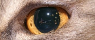
- Choroid - can be nodular, planar, diffuse and located in any zone of the organ. A characteristic feature of the formation is its dark brown color. It has a spongy structure and a rough surface.
As the disease progresses, it affects the main part of the retina and causes a sharp decrease in vision. Sometimes a person loses it completely. Most often, education is of a multiple nature. The pathology is not characterized by a very rapid development and is considered non-aggressive. It is not prone to early metastases.
Depending on the stage of development, melanoma is classified according to the following TNM parameters:
- Category B – in this case, the formation is located in the deep structures of the skin. It is insignificant, does not show activity and does not metastasize.
- Category N - shows the localization of the formation in the structure of the organ in combination with its partial germination into adjacent lymph nodes. These are where the first metastases occur.
- Category M - in this case, melanoma leaves the organ and grows into nearby organs and systems. In such a situation, metastases affect the internal organs, circulatory and lymphatic systems.
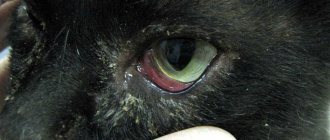
Melanoma of the eye in a cat
Ocular melanoma is more common in older cats. As a rule, it is a malignant tumor that is formed from pigment cells that produce melanin. It is a dark formation that has jagged edges. Sometimes light melanomas occur.
This pathology is characterized by early and rapid metastasis. Most often, the lymph nodes that are localized near the tumor are affected. The only method to combat formation in cats is to remove the eye. Before the operation, you need to perform an x-ray, which will help identify metastases in the lungs. If they are not, surgery will save the animal's life.
Complications of eye melanoma
If measures are not taken promptly, melanoma can cause dangerous complications.
These include the following:
- Increased pressure in the structure of the eye. As melanoma progresses, there is a risk of developing glaucoma. Symptoms include pain and redness of the eyes. There is also a risk of decreased vision clarity.
- Blindness. Large tumors often cause vision loss in the affected eye and can cause dangerous complications. These include retinal detachment, which can also cause blindness. Small tumors can lead to vision loss if localized in critical areas. In such a situation, there is a risk of impaired central or peripheral vision. Common formations can provoke vision loss.
- Spread of tumor outside the eyes. The tumor can metastasize to distant organs. These include the liver, bone tissue, and lungs.
Causes of eye melanoma
The exact causes that cause melanoma have not yet been established.
But doctors suggest that the risk of developing the disease increases under the influence of such factors:
- A large number of ultraviolet streams. This category includes both natural sun rays and the radiation that a person receives in a solarium. This reason is considered the main one.
- Light skin, brown hair, gray or blue eyes. People with this type of appearance react worse to external factors. Their tissues have difficulty resisting radiation and quickly transmit it through the skin. As a result, harmful radiation accumulates inside.
- Old sunburn. Even after many years, they can provoke the appearance of a tumor. A destroyed cell lattice can change at the molecular level, which leads to uncontrolled cell division. As a result, they become malignant. These processes provoke the formation of compaction. Over time, it turns into a tumor.
- Genetic predisposition. It has been proven that a tendency to cancer can be inherited even at the stage of fetal formation.
- Age. Natural aging of the body impairs tissue restoration; they become more susceptible to external factors. As a result, there is a risk of the formation of malignant tumors on the surface of the dermis.
Diagnosis of eye melanoma
Melanoma can be quite difficult to diagnose. At the initial stage of development, it is disguised as other processes. The optometrist should perform a general examination.
Based on its results, he prescribes additional procedures:
- Ophthalmoscopy is the main method for diagnosing melanoma and other eye pathologies.
- Fluorescein angiography is aimed at examining the ocular vessels. The procedure helps to identify changes in blood vessels, the level of development and structure of the network that feeds the tumor.
- X-ray – allows you to detect damage to the bone elements of the orbit. It can also be used to identify distant metastases in other organs.
- Radioisotope scanning is a diagnostic procedure that involves introducing radioactive isotopes into the bloodstream. The fact is that the metabolism of the tumor is significantly accelerated when compared with healthy tissues. Therefore, isotopes accumulate in it faster. Their high concentration is recorded using a radioisotope scanner.
- Biomicroscopy – eye tissues are studied using a microscope.
- Measuring intraocular pressure - deviation of this indicator in a certain direction can be an indirect symptom of a tumor.
- Computed tomography and magnetic resonance imaging are used to assess the extent of spread of the abnormal process to nearby tissues. They also help to find metastases of formation in other organs.
- Histological examination - it can be carried out only after removal of the tumor. Preliminary biopsy of fragments is dangerous. It can cause metastasis.