Dermatomycosis is a general category of skin diseases, commonly called “lichen.” The disease is caused by microscopic fungi that affect the skin, fur and even the claws of the animal. When the first signs of dermatomycosis appear in dogs, treatment should begin immediately, since it is easily transmitted to humans. Representatives of small breeds, as well as animals with reduced immunity, are most susceptible to infection.
Reasons for appearance
The sources of fungal infection are sick mammals, with direct or indirect contact with which pathogens enter the skin and penetrate through microscopic lesions. Not every dog can get sick, but only those at risk.
Provoking factors are considered:
- poor nutrition without a sufficient amount of natural meat and other necessary components in the diet;
- weakened immunity, typical for puppies, elderly or sick animals;
- metabolic, hormonal or vitamin imbalance;
- antibiotic treatment;
- helminthic infestations, especially of a chronic nature;
- lack of proper conditions of detention according to sanitary, hygienic and temperature requirements.
In addition, the cause of dermatomycosis can be constant trauma to the skin in pets sitting on a leash or leading a sedentary lifestyle in small enclosures.
Important! The incubation period of the disease ranges from 1 week to 1 month, but can last up to 3 months. Throughout this period, the dog is a carrier of the infection and poses a danger to humans.
Routes of infection
Sources of pathogens are sick animals (dogs, cats, rodents). Fungi penetrate the skin through wounds, scratches and cracks.
A pet can become infected in two ways:
- In direct contact with a sick individual;
- Through common objects on which fungi persist (beds, combs, ammunition).
Important. The incubation period of dermatomycosis lasts up to three months (on average 1-4 weeks). All types of the disease are dangerous for humans and other pets.
Characteristic signs
The symptoms of dermatomycosis are largely determined by the form of its course, which can be:
- atypical (weakly expressed);
- follicular (in-depth).
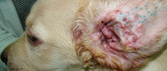
The first usually affects dogs with strong immunity, the second affects puppies and weak animals. At the same time, untreated atypical lichen quickly acquires a follicular form with all its symptoms. In any case, without adequate treatment, photos of dermatomycosis in dogs present a terrifying picture. The lesion can affect the entire body - not only the skin and coat, but also the internal organs.
In addition, dermatomycosis is divided according to the type of pathogen. Dogs are most often affected by two types of fungus:
- microsporia;
- trichophytosis.
Less common is the achorion, which causes the development of scab. Moreover, each type of disease is characterized by its own distinctive features.
Microsporia
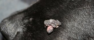
The symptoms of microsporia are manifested by the formation of small lesions with clearly defined edges and fallen or broken hair. Depending on the shape, the condition of the skin varies significantly:
- with atypical – redness, dryness and peeling appear;
- with follicular - it festers with the release of exudate, which after drying forms a crust.
Most often, lichen appears on the top of the head, near the ears, on the paws, around the tail and between the toes.
Trichophytosis
The signs of trichophytosis are similar to microsporia, but for this variety the follicular course is more typical. Trichophytosis is often perceived as a severe stage of microsporia.
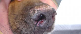
Under the influence of the fungus, abundant purulent discharge appears on the affected areas, forming dense crusts. Therefore, the main symptom of this type of dermatomycosis is hairless lesions covered with dried purulent crusts. In the final stage, the disease affects the paw pads and claws.
Scab
Scab is the most severe type of this fungal infection, which is characterized by the following symptoms:
- Not only the skin is affected, but also the bones, and in advanced cases, even the internal organs;
- the first lichens appear on the head, ears and around the claws;
- lesions become covered with scabs;
- the fur falls out completely.
A common symptom of any form and variety of dermatomycosis is severe persistent itching, burning and a small rash. At the severe stage, the dog becomes weak, exhausted and lethargic due to damage to the internal organs.
Important! Even in its advanced state, dermatomycosis is treatable. But it will not be possible to completely rid the dog of the fungus, so if any of the above provoking factors appear, the symptoms will resume again.
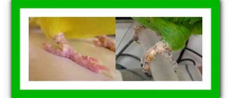
It is enough to look at a photo of dermatomycosis in dogs before treatment to present the general clinical picture of the disease and independently determine the symptoms of the disease at the first stage. Damage to the skin and coat in the form of lichen has pronounced characteristic signs that are difficult to confuse with other diseases. However, a final diagnosis can only be made using special diagnostic methods.
Rare systemic diseases
- Blastomycosis - the pathogen is found near water in sandy soils. The organs of movement, breathing, and visual system are affected. Multiple plaques and nodules throughout the body. Ketoconazole and amphotericin B are prescribed.
- Cryptococcosis - infection occurs through pigeon droppings. The central nervous and visual systems are affected.
- Histoplasmosis - the pathogen lives in damp places. Preserved in the droppings of bats and birds. Affects the gastrointestinal tract, respiratory and visual systems. Papules and nodules appear on the skin on all parts of the body. Treatment with ketoconazole.
- Aspergillosis - pathogens are part of the normal microflora of the skin, wool, and mucous membranes of the eyes. Ulcers and nodules appear on the mucous membranes of the eyes and on the skin. There is bloody discharge from the nose.
- Protothecosis - the source of infection is in sewage or standing water. Penetrates through wounds. Affects the central nervous and visual systems. The normal functioning of the gastrointestinal tract is disrupted. Treatment is amphotericin B with ketoconazole.
Systemic mycoses cause deep infections of internal organs.
Treatment Options
For any symptoms of dermatomycosis in dogs, treatment is carried out comprehensively. Treatment measures should include:
- vaccination or antibiotic administration;
- external skin treatment;
- strengthening the immune system with vitamin supplements.
Simultaneously with the treatment, the provoking factor is eliminated and the pet is provided with proper living conditions.
Vaccination
Traditionally, vaccines are used for prevention purposes, but in the case of ringworm, they can be an excellent treatment.
The main antimycotic drugs for dogs are:
- Polivak-TM – administered 3 times with an interval of 10–14 days;
- Vakderm - applied twice with the same interval;
- Microderm - usually 1 injection is enough, but if necessary, can be repeated after 2 weeks.
A lump often appears at the injection site, which resolves within 2–3 days. Vaccination is not carried out at elevated temperatures.
Drug treatment
In case of hyperthermia, when vaccines are prohibited, treatment of dermatomycosis in dogs is carried out using antibiotics in tablet form. The most effective drugs are considered to be:
- Itraconazole
- Ketoconazole
- Griseofulvin.
Simultaneously with any of the above internal treatment options, external treatment of the skin is performed. Before this, the hair around the lesions is shaved or clipped. Good results are obtained by using the following ointments 2 times a day:
- Clotrimazole.
- Nystatin.
- Ketoconazole.
In the follicular form of the disease, the dog is bathed every 3–4 days using special shampoos.
To speed up treatment, it is recommended to irradiate your pet with a quartz lamp. Such procedures quickly relieve redness, improve tissue regeneration, and strengthen the immune system. Quartzization procedures in most cases can fully replace the use of antibiotics, which negatively affect the animal’s body. After proper treatment, the symptoms of ringworm in dogs (pictured) gradually disappear, and the affected areas become overgrown with hair over time.
Preventive actions to prevent dermatomycosis in dogs
- Reduce the risk of ringwormyou can follow these simple recommendations:
- Regularly vaccinate your dog against microsporia and other fungal diseases.
- Avoid contact with stray animals.
- Use only individual pet care items and clothing. Periodically treat your dog's equipment with disinfectants.
- Strengthen your pet's immunity with a healthy diet, adequate exercise and good care.
- Regularly inspect your dog's skin and coat to begin treatment on time and protect family members from infection.
Timely treatment guarantees quick relief from fungal diseases in your pet. Our “YA-VET” veterinarians - dermatologists are sure that dermatomycosis in dogs is not a death sentence for communicating with four-legged friends, therefore, if necessary, they will come to your home to diagnose and prescribe treatment for a sick pet. The doctor will bring all the necessary equipment, medications, and required documents with him.
All you need is to dial the center’s phone number and consult with a doctor, set a day and time for your visit. You can also purchase veterinary drugs at competitive prices, vaccinate your dog against lichen, and much more. You can find out the prices for services and medications by calling us. We guarantee transparent pricing and no unnecessary, hidden costs.
Prevention measures
Since curing dermatomycosis is much more difficult than preventing it, it is recommended to take simple preventive measures. They include:
- vaccination once a year;
- ensuring proper nutrition and proper living conditions;
- avoiding contact with potential carriers of the fungus.
Dermatomycosis is not only unpleasant, but also a very serious disease that is easily transmitted to humans. Therefore, you need to carefully monitor your pet’s condition and if unusual bald patches appear on its skin, immediately contact a veterinarian.
Dermatophytoses of cats and dogs
N.I. Levyatova, Veterinary Clinic "Center", Moscow
Dermatophytoses are infectious diseases of keratinized tissues (skin, hair, nails) caused by fungi of the Microsporum, Trichophyton or Epidermophyton species. In cats and dogs, fungal infections are less common than other skin diseases, but due to the threat of zoonosis, they deserve special attention. In many people with normal immune systems (as in many healthy pets), when infected with dermatophytes, the clinical manifestations are limited to a limited scaly lesion that easily responds to topical therapy and tends to spontaneously remit within a few months. Some patients develop exudative, crusty dermatitis, which is usually very difficult to treat. This development of the disease is more common in newborns and young children, the very elderly and those in a state of immunosuppression (HIV-positive or undergoing chemotherapy) people. In animals, a predisposition to dermatophytosis with clear clinical manifestations is observed at the age of up to 12 months (imperfect cellular immunity, deficiency of nutrients, especially proteins, fatty acids and vitamin A, viral diseases) and in adult animals in a state of severe stress or immunosuppression (spontaneous and iatrogenic immunodeficiencies). Also factors predisposing to infection are parasitic infestations, high humidity and ambient temperature, and damage to the skin. The most common cause of disease in cats is Microsporum canis. In dogs, the disease is most often caused by Microsporum canis and Microsporum gypseum. Other, less common causes of dermatophytoses include Trichophyton mentagrophytes, Microsporum persicolor, Microsporum erinacei, Microsporum verrucosum. Dermatophytes can be isolated from the skin and hair of cats (especially those kept in nurseries and shelters that attend exhibitions) without visible lesions (more often in Persian kittens). In places where animals gather in large numbers, a serious problem is the spread of fungal spores that remain viable for 18 months (according to some sources up to 52 months) in the environment. There is a breed predisposition to the occurrence of dermatophytosis. For example, Jack Russell terriers are more often affected by Trichophyton mentagrophytes and Trichophyton erinacei, German shorthaired pointers M.gypseum, Yorkshire terriers and Pekingese M.canis. Longhaired Persian and Himalayan cats are more likely than other cats to be diagnosed as asymptomatic carriers of M. canis. After infection with dermatophytes, the animal's immunocompetent cells engage the cellular and humoral immune systems, which ultimately free the body from the infection. The inflammatory response leads to an increase in epidermal proliferation, which, in turn, leads to the cleansing of the epidermis from dermatophytes (in the process of desquamation of horn cells). Immune status does not guarantee absolute resistance, although with subsequent infection there is a more rapid onset of clinical manifestations and a tendency towards a shorter duration of the disease. During experimental infection, the lesion covers a maximum area after 5 weeks. CLINICAL PICTURE IN SMALL ANIMALS Clinical symptoms of dermatophytosis caused by Microsporum canis in cats can vary from asymptomatic carriage to skin lesions with eschar formation. The typical manifestation of the disease is single or multiple, rapidly spreading, ring-shaped lesions of round or irregular shape, accompanied by erythema, scaling and alopecia with a diameter of about 3 cm, most often found on the head and extremities. Itching and inflammation with localized lesions may occur but are usually minimal. Other manifestations of dermatophytoses caused by Microsporum canis include focal or generalized alopecia, papulocrustic dermatitis, localized subcutaneous granuloma, onychomycosis, and paronychia. In dogs, Microsporum canis in a generalized form causes a stronger inflammatory response than similar lesions in cats. Dermatophytosis caused by Microsporum persicolor is rare. Fungal hyphae infect the stratum corneum of the skin without affecting the hair. Clinically characterized by superficial minimal alopecia and inflammation, most often occurring on the scalp. Dermatophytoses caused by Trichophyton mentagrophytes and Microsporum gypseum cause a pronounced inflammatory response. Generalized skin disease is not uncommon. Lesions on the face can be surprisingly symmetrical and may be accompanied by alopecia, erythema, crusting and furunculosis. Itching can be expressed to varying degrees. Generalized lesions can affect entire parts of the body (for example, in chronic disease caused by M.gypseum or M.mentagrophytes). Diffuse scaly alopecia, according to some authors, occurs more often in Persian and Himalayan cats. Pseudomycetoma, more often recorded in cats of Persian breeds and Yorkshire terriers, is characterized by the appearance of a nodule in the dermis or subcutaneous tissue, resulting from the growth of dermatophytes in the tissues. The spores probably enter the tissue from the infected hair through destroyed hair follicles. In most cases, high body temperature inhibits the growth of fungal hyphae, and the corresponding immunocompetent cells remove them from the tissues as a foreign body (usually a boil is formed, which opens onto the surface of the skin). In some cats, granulomatous nodular reaction is accompanied by the growth of dermatophytes in the center of the lesion. Clinically, pseudomycetoma can be represented by single or multiple nodes, which rarely form fistulas and often recur after surgical excision. DIAGNOSTIC TESTS A careful clinical examination and possibly the presence of a zoonotic or anthropozoonotic lesion may hint at the presence of a dermatophyte infection, but treatment should never be initiated without a definitive diagnosis. Microscopic examination of a KOH preparation can help detect spores around the hair shaft, but this method produces many false negative results. Examining a cat in a darkened room using a Wood's lamp (which must be warmed up before testing) may reveal a green glow (fluorescence) in some cases of dermatophytosis caused by Microsporum canis. Culture of suspicious material on DTM-arap or Sabouraud agar is the only way to obtain a definitive diagnosis. 1. Inspection using a Wood's lamp. The characteristic apple-green glow can be caused by M.canis (less than 50% of strains), M.distortum, M.ferrugineum and the anthropophilic M.audouinii, as well as bacteria (Pseudomonas aeruginosa, Corynebacterium minutissimum), horny scales, soap and locally used medicinal products drugs. When carrying out the procedure, you need to remember that the fungal hyphae are located along the hair shaft. Fluorescence can only be observed when the invasion intensity is sufficient. Cases of infection of cats with the same strain of fungi have been reported, when some showed pronounced clinical signs and a bright specific glow during fluorescent diagnostics, while others were asymptomatic carriers of the infection, and the study gave a negative result. 2. Mackenzie method. It is based on combing the wool with a sterile toothbrush or comb, followed by inoculation on a nutrient medium. This method is recommended for examining animals in large colonies in order to identify asymptomatic carriers of infection. 3. Examination using a microscope. Before collecting material for microscopic examination and culture, the skin area is treated with 70% alcohol to reduce bacterial contamination. Damaged broken hair is used for research. Scrapings are carried out within the alopecia area, from crusts and papules. Before performing microscopy, it is recommended to keep the material treated with 10-20% KOH at room temperature for several minutes or warm it up slightly to accelerate the dissolution of free keratin and debris. The hyphae of the fungi swell and, even with a cursory examination, you will be able to notice thickened areas with uneven contours on the hair shaft. The spores form a “sheath” around the hair and give it a vague outline. The probability of detecting infected hair is quite low, so this study does not make it possible to completely exclude the presence of infection. 4. Fungal culture. Sabouraud dextrose agar is used as a culture medium. To conduct this study, there is a very convenient and informative test “Dermatophyte Test Medium” or DTM-arap, which contains Sabouraud agar, cycloheximide (inhibiting the development of saprophytic and systemic fungi), gentamicin and chlortetracycline (to minimize contamination with bacterial flora) and an indicator pH media phenol red. Dermatophytes primarily prefer to absorb proteins and, as a result, form alkaline metabolic products that change the color of the medium to red. They produce these metabolites as the colony grows, and a change in color of the medium occurs 2-7 days after sowing (sometimes this process takes 14 days). Saprophytic fungi prefer to metabolize carbohydrates, producing neutral and acidic metabolites that do not change the color of the medium. When the supply of carbohydrates is depleted, they can utilize proteins and as a result cause the color of the medium to change to red. The result of the study can be correctly interpreted only if the growing colonies are examined daily (or every other day). 5. Roth's flag method. It is carried out to make a final diagnosis and identify the pathogen. Based on the detection of macro- and microconidia in prints from grown colonies. 6. Histopathological examination. Fungal hyphae can be found in the stratum corneum, hair follicles and around the hair shaft. The number of fungal elements detected is usually inversely proportional to the severity of the inflammatory reaction. About 80% of patients with dermatophytosis have a positive skin biopsy. THERAPY It must be remembered that with an effective immune response, spontaneous self-healing is possible! Despite this, treatment is necessary to avoid infection of humans and other animals. LOCAL TREATMENTS For animals, local treatment is less important than for people. Animals are covered with hair, which reduces the effectiveness of procedures. The area of application of the drug should be much wider than the location of the visible lesion, and affect healthy tissue, because mushrooms can be cultivated from areas of fur and skin located at a distance of 6 cm from the lesion. Local therapy should be considered as an auxiliary treatment method. The drugs used to treat animals are: 2% lime sulfur solution, povidine iodide, 0.2% enilconazole solution, 2% miconazole in cream and spray form, clotrimazole and terbinafine creams, ketoconazole shampoo, etc. P. According to some practicing dermatologists, chlorhexidine is ineffective for cleansing the skin of dermatophytes and treating the external environment. Others recommend using shampoos and rinses containing chlorhexidine at a concentration of 2-4%. The use of creams, ointments and gels is not recommended (the exception is very localized lesions, such as kerion in dogs). In case of extensive lesions, it is considered more effective to wash the animals after preliminary cutting and destruction of the hair (this procedure is mandatory in long-haired cats and in all cases of generalized dermatophytosis). This procedure can significantly reduce contamination of the external environment with dermatophyte spores. Animals with minimal, limited demarcation barrier lesions do not need clipping. SYSTEMIC ANTIFUNGAL DRUGS GRISEOFULVIN (Gricin. Biogrisin. Fulcin) Griseofulvin was first isolated in 1939 and was used for fungal diseases of plants. It was introduced into medical practice in 1958 and was historically the first specific antimycotic for the treatment of dermatomycosis in humans. Griseofulvin is a fungistatic antibiotic. When exposed to it, actively metabolizing young fungal cells can be killed without compromising the integrity of the cell wall, and in more mature cellular elements the drug only causes inhibition of reproduction. The drug is very poorly soluble in water and its absorption in the gastrointestinal tract is variable and incomplete. Absorption can be improved by administering the drug along with fatty foods. The drug accumulates in the stratum corneum of the skin, its highest concentration is found in the superficial layers. In dogs, side effects when using the drug are vomiting, diarrhea, and a reversible increase in liver enzymes. Cats may experience anemia, leukopenia, vomiting, diarrhea, depression, itching, and sometimes ataxia. Cases of bone marrow dysfunction have been described. In cats with immunodeficiency virus, griseofulvin can lead to quite significant secondary neutropenia. Changes in bone marrow function occur due to individual intolerance to the drug and are not associated with the dosage regimen. Griseofulvin is a potential teratogen and should not be used to treat pregnant animals. In dogs and cats, the recommended dose can vary within a fairly wide range from 20 to 150 mg/kg per day, divided into 2 doses. Given the possibility of potential idiosyncrasy, it is recommended to conduct blood tests (with mandatory platelet count) before starting treatment and after 7-10 days of therapy. If signs of bone marrow suppression are observed, treatment should be discontinued and appropriate supportive therapy should be instituted, which may include blood transfusions (for very low platelet counts) and antibiotic therapy (for high white blood cell counts). When prescribing this drug to an animal, you should be very careful and be sure to inform the owner about external signs of anemia. Azoles for systemic use (ketoconazole, itraconazole, fluconazole) are well absorbed when taken orally. The bioavailability of ketoconazole and itraconazole may vary significantly depending on the level of gastric acidity and food intake. The antifungal effect of azoles is caused by disruption of the integrity of the fungal cell membrane and disruption of the synthesis of ergosterol, the main structural component of the fungal cell membrane. KETOCONAZOLE Ketoconazole is a synthetic broad-spectrum antifungal drug belonging to the imidazole group. It is a potent inhibitor of ergosterol synthesis. Ketoconazole is considered a fungistatic, but under anaerobic conditions and high enough concentrations it can have fungicidal properties. An acidic environment is required for optimal absorption. Used at a dosage of 5-10 mg/kg every 12 hours or 10-20 mg/kg once a day with food. When using the drug in dogs, the most common side effects are: lack of appetite, itching, alopecia and reversible lightening of the coat. Cats are more sensitive to the drug and may exhibit anorexia, fever, depression and diarrhea. Asymptomatic hepatitis with a reversible increase in liver enzyme levels may occur. More serious liver dysfunction is caused by an individual hypersensitivity reaction and occurs in 1 in 10,000 cats. In dogs, these side effects are even less common. Ketoconazole has a teratogenic and embryotoxic effect. ITRACONAZOLE (Irunin, Orungal) Itraconazole is successfully used to treat dermatophytoses in cats and dogs. Dosage 10 mg/kg 1 time per day. After 7 days of daily use, you can switch to the so-called pulse therapy (taken every other day or every other week) while maintaining the high effectiveness of treatment. Itraconazole is much better tolerated than ketoconazole and unwanted side effects with its use occur much less frequently. FLUCONAZOLE (Diflucan, Flucostat) All systemic azoles, except fluconazole, are metabolized in the liver and eliminated primarily through the gastrointestinal tract. Fluconazole differs from other antifungal agents in that it is excreted through the kidneys (mainly unchanged? - 80-90%) and can be used in animals with liver disease at a dose of 10-20 mg/kg every 12 hours. TERBINAFINE (Exifin, Lamisil) A human antifungal drug that can be used in small pets. Belongs to the group of aplylamines. It has a predominantly fungicidal effect. Unlike azoles, it blocks earlier stages of ergosterol synthesis. It has a wide spectrum of activity, but only its effect on pathogens of dermatomycosis is of clinical significance. Terbinafine is used in a dosage of 20-30 mg/kg daily once a day, and then pulse therapy (every other day). The experiment did not reveal any fetotoxicity or effect of the drug on the reproductive function of animals. Very effective in the treatment of onychomycosis. Inactivated by M.Canis, vaccines do not have sufficient efficiency and should be used as a component of therapy simultaneously with a systemic antifungal drug. Treatment should continue within 4-6 weeks and should not stop until the negative result of sowing is obtained. This is very important, since the result of sowing can be positive after a long time after the visible clinical recovery. Typically, treatment continues until clinical remission, which occurs often after 4-6 weeks of therapy. In the case of onychomycosis, its duration can reach 6-12 months, and with a severe lesion, the surgical removal of claws can be the only effective way. Then the study is carried out by the Mackenzie method (sowing the material after combing the sterile toothbrush). If the result of sowing is negative, 4 weeks after the cancellation of systemic antifungal therapy, another cultural study is carried out. Its necessity is dictated by the possibility of a false negative result in the first study (due to the high resistance of the drug in the hair and scales taken for analysis). Only with a repeated negative result is an animal considered clinically healthy. Unfortunately, in practice, many owners refuse to re -diagnosis and decide to finish the treatment based on a clinical examination and examination using Wood lamp in cases of infection with fluorescent strains. Environmental processing of the area (premises) in the house where the animal is located, it is necessary to vacuum daily to remove infected hair and dispute. Cells and other surfaces that are resistant to chlorine should be processed daily by household chloride 1: 10s water. Where possible, a formalin solution (10%), sodium, iodine or steam hypochlorite is used. Abroad to process the external environment, an epilconzole spray is used. Prevention of dermatophytosis in nurseries and animal shelters The occurrence of infection in places of accumulation of animals is an extremely serious problem. Despite the implementation of all possible preventive measures, we cannot guarantee their success. The reasons are high stability of the dispute in the external environment, a large number of false -negative results in diagnosis, low efficiency of premises, error when choosing drugs for the treatment of dermatophytosis, insufficient duration of therapy. The liquidation program for dermatophytic infection in nurseries and animal shelters. 1. The detection of infection (clinical inspection, research using Wood lamp, Mackenzie method, sowing) 2. Stop all reproduction programs. 3. To treat the entire livestock with a systemic antimycot. Animals with a negative result of sowing are prescribed therapeutic doses of the drug in order to prevent the spread of infection. 4. divide into three separate groups of animals a) infected, b) contacting with patients, c) nursing cats with their offspring. 5. Carry out a thorough multiple disinfection of premises 6. After 14 days, conduct a second examination of animals with a negative reaction in the first sowing. 7. Culling or treatment of animals with a positive sowing result 8. Strict quarantine of newly received animals before their placement in the nursery. Another option for getting rid of infection is to completely disband the livestock, carry out a thorough multiple disinfection and start work again with a “clean cat”. It is necessary to take into account the existence in some animals of a genetic predisposition to the transition of the disease to a chronic asymptomatic course. Key points 1. The clinical picture of dermatophytoses is very variable and it is impossible to exclude or confirm the presence of infection only on the basis of a physics examination 2. Do not forget about the high probability of erroneous diagnosis 3. Aware of the possibility of zoonosis 4. The duration of treatment should be based on the results of the fungal culture 5. Good is very important. Contact with Summary Levyatova Ni Veterinarian Dermatologist. Veterinary Clinic "Center", Moscow. Dermatophitoses of Cats and Dogs. In this Report Described Etiology, Diagnostic, Clinical Finding and Methods of Treatment Dermatophitoses of Cats and Dogs. ALSO, Described Products for The Systemic Treatment of Superficial Mycoses and Its Side Effects.
Materials of the XVI Veterinary Congress
Diagnostics
At the appointment, the veterinarian will examine the dog, illuminate the fur with a specially designed lamp, in which the existing fungus will glow in emerald color. However, not all varieties of fungus will be illuminated by this lamp - about 40% simply will not show a characteristic glow. When establishing a diagnosis, high-quality treatment will be required, and for this, an in-depth analysis is carried out.
A complete analysis is required to establish the type of fungus, its quantity, growth rate, and sensitivity to drugs. Basically what is required is:
- Blood test to determine allergic status . If your dog's skin becomes inflamed due to an allergy, the fungus will make itself known some time after treatment. In this case, only getting rid of the allergen will give a positive result.
- Blood test for bacteriology . The dog may be sensitive to bacteria, cocci, but the fungus is a secondary infection. It will be possible to cure your pet only after getting rid of the original source.
- Detailed biochemical analysis of urine and blood . The general condition of the dog is determined. Tests will show the presence of any malfunction in the dog’s body.
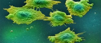
View of fungus under a microscope
Establishing diagnosis
A veterinarian can make a preliminary diagnosis after examining the dog using a Wood's lamp. When you point the lamp at the affected area of the skin, the fungi glow green.
To make a final diagnosis, a deep skin scraping is taken from the animal and laboratory tests are performed. To determine the pathogen, biological material is inoculated on a nutrient medium.
For a complete clinical picture, blood and urine tests are taken.