The central nervous system is the core of any organism. The life activity of humans and animals depends on the state in which it is located. It is for this reason that any pathology of the central nervous system is deadly.
A brain tumor, regardless of origin, can lead to disability in a dog and, in severe cases, to death. Fortunately, compared to neoplasms of other organs, brain tumors are diagnosed less frequently.
Causes of the disease
Brain tumors (BTT) are classified into benign and malignant. The first type includes slowly growing neoplasms with a clear localization and a clearly visible boundary between the affected and healthy tissue. Tumors do not metastasize, but if left untreated they can transform into cancer.
The second type is malignant tumors. Such neoplasms are characterized by rapid growth, the absence of a boundary between healthy and diseased tissues, and the presence of metastases. Almost always lead to the death of the dog.
Primary tumors develop directly in the brain when DNA mutations occur in normal cellular structures. Mutated cells begin to actively divide, destroying and displacing healthy ones. This leads to the formation of OGM.
Secondary brain tumors are commonly understood as neoplasms that develop in different organs and reach the brain through metastases.
The main reasons contributing to the development of OGM include:
- brain injuries;
- radiation, radioactive radiation;
- congenital pathologies;
- parasitic, viral diseases;
- bad environment;
- autoimmune diseases;
- exposure to chemical and toxic substances.
Tumors can occur in absolutely any dog, but the risk group usually includes animals over 5-7 years of age whose tumors arose under the influence of certain negative factors.
Based on the results of the studies, it was found that the most common brain tumors in dogs are astrocytoma, meningioma, oligodendroglioma, glioma, and ependymoma.
Malignant edema in dogs:
Malignant edema, in combination with other viral diseases in an animal, can be much more complicated and cause even more discomfort to the pet. In such cases, inflammatory lesions of bone tissue, muscles and internal organs are inevitable.
The bacteria that cause this type of disease can be found in the soil, rotting tissues of living organisms and the remains of natural secretions from the intestines. Thanks to natural secretions, these harmful microbes spread throughout the entire area.
The main distinguishing feature of malignant edema is the breakdown of protein and lactose in the body of an infected animal.
Despite these studies, this type of virus can manifest itself in any animal and person, including. The area where it spreads may be a territory with a significant overpopulation of people and animals, without observing standard sanitation rules. In an area contaminated with a significant amount of natural excrement of living organisms, bacteria multiply quickly. Then, entering through wounds, they beneficially multiply and develop in the body of the infected person.
The disease can also enter a pet’s body in any medical clinic that is negligent in disinfection and personal hygiene measures. This may occur due to improper castration, childbirth with heavy blood loss, or surgery. Entering the body through the mucous membranes and internal organs, without oxygen, the microbe begins intensive life activity in the animal’s body. In the victim’s body, they begin to release toxic substances that destroy organs. Without oxygen, together with foreign bodies or body particles, bacteria instantly destroy all healthy tissue.
Main symptoms
Determining that a dog has a brain tumor is quite difficult. Pathologies have a blurred clinical picture, symptoms often resemble other diseases of the central nervous system, in particular epilepsy.
OGM can be suspected based on the following signs:
- seizures;
- convulsions;
- glassy look;
- swinging gait (pendulum);
- uncontrollable vomiting;
- rotation in place;
- head tilt down;
- sagging lips and eyelids;
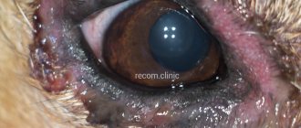
- blurred vision, nystagmus;
- hearing and smell impairment;
- increased thirst;
- frequent urination;
- cardiopalmus;
- labored breathing.
The dog's behavior also changes. He becomes aggressive or overly affectionate, he may refuse food or, conversely, absorb everything, even bags.
Unfortunately, most dogs go to the vet when the tumor reaches a significant size.
Predisposition to disease
A brain tumor can appear in individuals of any age or gender. The disease is most often diagnosed in dogs over five years of age. There is some breed predisposition to the disease. Doberman Pinschers, Pinschers, Golden Retrievers, Scotch Terriers and Bobtails are more likely to get sick.
The most common types of neoplasms are:
- Oligodendroglioma. It is localized on the frontal lobes of the brain. The disease affects dogs with a flattened and shortened muzzle - brachycephalics (bulldog, Pekingese).
- Ependymoma. Develops from epithelial cells of the dog's cerebral ventricles. This tumor often develops in older brachycephalic dogs.
- Gangliocytoma or ganglioma is a tumor of the cerebellum or medulla oblongata of a dog. Benign, slow-growing neoplasm. Young dogs are susceptible to the disease.
- Medulloblastoma or neuroblastoma is a malignant tumor. It affects the brain stem, the midbrain. Can grow into the cerebral ventricles.
- Meningioma is a benign tumor that is localized on the cerebral hemispheres. It is characterized by slow growth and has a favorable prognosis. This disease affects dogs with an elongated narrow skull - dolichocephals (collie, Russian greyhound). The most common type of disease.
Diagnostics in a veterinary clinic
To be able to make an accurate diagnosis, the veterinarian needs, in addition to a visual examination of the animal, to carry out a number of diagnostic measures.
An important point is the differentiation of the tumor from other diseases with similar symptoms (Aujeszky's disease, canine plague, etc.), for which appropriate studies are carried out.
Physical, neurological, and laboratory diagnostic methods make it possible to determine the etiology, size and localization of the tumor. The most complete information is provided by ultrasound examination, stereotactic puncture biopsy, computed tomography, magnetic resonance imaging.
If the doctor suspects a secondary tumor, procedures are performed to determine the location of the cancer.
Clinical picture, symptoms of cerebral edema
Cerebral edema can be local and widespread (generalized). Clinical manifestations of cerebral edema are varied and depend on the duration, localization, prevalence, and severity of the pathological process.
With cerebral edema, there are 3 main groups of symptoms:
- symptoms associated with intracranial hypertension;
- focal symptoms;
- stem symptoms.
Increased intracranial pressure is indicated by a bursting headache, nausea, vomiting, and decreased level of consciousness.
Intracranial hypertension can lead to the development of seizures.
Focal symptoms are loss of certain functions. When cerebral edema is localized in certain areas, the function of these areas will be disrupted, so those functions for which the affected area is responsible will be lost.
Focal symptoms include paresis, paralysis, sensitivity, vision, and speech disorders.
When the cerebellum is damaged, balance and gait disturbances occur.
The brain stem contains the vital centers for breathing and heartbeat. If cerebral edema affects this area, cardiovascular disorders develop, breathing and thermoregulation are impaired, reflexes fade and the level of consciousness decreases, and seizures may develop.
Diagnosis of cerebral edema is based on the patient’s complaints (if the patient is conscious), examination data, assessment of the neurological status, and the results of additional examination methods.
During a neurological examination of the patient, attention is paid to unconditioned reflexes, loss of functions, and disorders in various areas.
When examining the fundus of the eye, which is carried out by an ophthalmologist, it is possible to determine swelling of the optic nerve nipples - one of the symptoms of cerebral edema.
To assess concomitant pathology affecting the course of cerebral edema, it is necessary to conduct laboratory research methods. It is necessary to conduct a general blood test to determine the level of protein and electrolytes (potassium, sodium, magnesium, chlorine) in the blood plasma.
If cerebral edema is suspected, a spinal puncture, angiography of cerebral vessels, and computed tomography or magnetic resonance imaging of the brain are performed.
Computed tomography or magnetic resonance imaging of the brain will help determine the size and location of cerebral edema.
The list of additional research methods performed can be expanded depending on the leading cause of cerebral edema.
To eliminate hypoxia, it is necessary to carry out oxygen therapy - the artificial introduction of oxygen into the body through the respiratory tract.
For cerebral edema, local hypothermia is a simple and effective treatment. To do this, cover your head with ice packs or other sources of cold.
The outflow of excess fluid from brain tissue is facilitated by intravenous administration of hyperbaric solutions (10% sodium chloride solution, 40% glucose solution).
The use of medications that have a diuretic effect helps eliminate cerebral edema. Furosemide, Lasix, magnesium sulfate, urea, mannitol are prescribed.
Treatment method and prognosis
Treatment should be started as early as possible. It is important to understand that the count is not even in months, but in days. There is no effective drug therapy; no drug can promote tumor resorption. To date, the only way to slow down the development of a tumor is surgery followed by chemical and radiation therapy.
When it comes to cancer, chemotherapy methods give good results only at an early stage of the disease.
Radical surgery is carried out under the control of an ultrasound scanner and gives the most positive result. The procedure involves complete removal of the affected tissue (tumor), reducing intracranial pressure and eliminating perifocal edema.
Sometimes the location of the tumor is such that it is not possible to completely remove it. In such cases, palliative operations are performed - partial removal of the tumor. The main goal of the operation is to alleviate the dog’s condition and reduce the number of cancer cells.
After surgical partial or complete removal of the tumor, the dog undergoes chemotherapy or radiotherapy. These treatment methods are very effective, but cause serious side effects in the form of severe intoxication of the body.
In order to improve health and improve the quality of life, the dog is prescribed symptomatic treatment, which includes taking:
- diuretics (Mannitol), which relieve intracranial pressure;
- steroids that slow the growth of certain types of tumors;
- anticonvulsants (Phenobarbital, Bromide), eliminating seizures.
Infusion therapy is mandatory and a special diet is prescribed.
As for the prognosis, doctors give them extremely carefully. A favorable outcome depends on the age and individual characteristics of the dog, the size and location of the tumor, the quality and timeliness of treatment.
Dogs with benign tumors have a greater chance. However, it happens that in case of remission, an operated and treated dog can live to a ripe old age, and sometimes the dog dies without even living a year.
That is why it is so important to immediately show the animal to a specialist at the first alarming symptoms.
Symptoms of a brain tumor in a dog and methods of its treatment
A brain tumor is one of the most serious diseases of the nervous system in dogs with complex consequences. The mammalian nervous system is responsible for the coordinated functioning of all organs and systems. Therefore, any illness affects the functioning of the whole organism, and often ends tragically.
Classification of tumors
Brain tumors are classified according to several criteria:
- by localization;
- by the type of tissue from which they develop;
- by origin;
- by the nature of development.
Classification by localization
Relative to the cranial vault, brain tumors in dogs can be localized as follows:
- Intracerebral - localized in the substance of the brain.
- Extracerebral - localized in the meninges and bones of the skull.
Classification by origin
Based on their origin, tumor diseases can be classified as primary or secondary:
- primary ones are formed from cells located in the structure of the brain itself and its membranes;
- secondary ones are formed during the process of metastasis of tumors that arise outside the brain, behind the meninges.
Classification by tissue type (according to histological characteristics)
There are 7 main classes of tumors. Their main distinguishing feature is the type of source fabric:
- Glial tumors (grow from the so-called glia - auxiliary cells of the nervous tissue).
- Neuronal tumors (grow from neuronal tissue).
- Embryonic tumors (original tissue - embryonic stem cells).
- Tumors of the meninges (tissue of the meningeal meninges is involved).
- Lymphoma (localized in lymphatic tissue).
- Epidermoid tumors (localized on the epithelial membrane of the cerebral ventricles).
- Rare primary tumors of the central nervous system.
Classification by nature of development
No matter how many types of neoplasms there are, according to the nature of development they are all divided into two large groups:
- Benign. They are characterized by slow growth and do not form metastases. Their boundaries are clear, the pathological process does not extend beyond the affected tissue. They respond quite well to treatment and in most cases have a favorable prognosis.
- Malignant. They have exactly the opposite properties. They are characterized by rapid growth and spread the pathological process throughout the body through metastases. Their forecast is not so optimistic.
Predisposition to disease
A brain tumor can appear in individuals of any age or gender. The disease is most often diagnosed in dogs over five years of age. There is some breed predisposition to the disease. Doberman Pinschers, Pinschers, Golden Retrievers, Scotch Terriers and Bobtails are more likely to get sick.
The most common types of neoplasms are:
- Oligodendroglioma. It is localized on the frontal lobes of the brain. The disease affects dogs with a flattened and shortened muzzle - brachycephalics (bulldog, Pekingese).
- Ependymoma. Develops from epithelial cells of the dog's cerebral ventricles. This tumor often develops in older brachycephalic dogs.
- Gangliocytoma or ganglioma is a tumor of the cerebellum or medulla oblongata of a dog. Benign, slow-growing neoplasm. Young dogs are susceptible to the disease.
- Medulloblastoma or neuroblastoma is a malignant tumor. It affects the brain stem, the midbrain. Can grow into the cerebral ventricles.
- Meningioma is a benign tumor that is localized on the cerebral hemispheres. It is characterized by slow growth and has a favorable prognosis. This disease affects dogs with an elongated narrow skull - dolichocephals (collie, Russian greyhound). The most common type of disease.
Symptoms of the disease
Typically, a brain tumor in a dog has pronounced symptoms that develop over quite a long time (as the tumor grows).
It can be assumed that the primary symptom in dogs (as in humans) is headache. But since they cannot inform the owner about this, the appearance of this sign can be recognized by some changes in the animal’s daily behavior:
- decreased physical activity;
- the desire to hide in a dark place;
- the appearance of some uncleanliness.
As the disease progresses further, the owner may notice the appearance of neurological symptoms:
- convulsions involving the whole body or part of it:
- manege movements (monotonous running in a circle);
- some gait disturbances;
- ataxia (lack of coordination of movements);
- unnatural head tilt.
A pet with a brain tumor may suffer from uncontrollable vomiting and a complete lack of appetite. The animal will look depressed or, on the contrary, restless and aggressive. A sick dog may refuse to follow the owner's commands and may not recognize him.
If the owner notices something unusual in the behavior of his pet, he should contact a veterinary clinic as soon as possible. The earlier the disease is detected, the more confident we can say that the dog will live for quite a long time.
Symptoms that allow you to recognize the disease at an early stage
In some cases, a specialist will be able to determine the location of the tumor at an early stage, without additional examinations:
- when the olfactory and frontal lobes are affected, convulsions may occur;
- if the cerebral hemispheres are affected, the lip may sag, the head will tilt, the eyelid may sag;
- when the frontal lobe of the cerebral hemispheres is damaged, sensitivity disturbances in various parts of the body occur;
- visual disturbances may indicate damage to the visual center and pituitary tumor;
- hearing impairment indicates damage to the temporal lobe or nucleus of the brain;
- impairment of the sense of smell most often occurs due to damage to the temporal lobes of the brain;
- gait disturbances, falling to one side indicate damage to the vestibular apparatus;
- nystagmus (involuntary rhythmic vibrations of the eyeballs) is associated with damage to the cerebellum;
- endocrine disorders, problems with the adrenal glands, unquenchable thirst, increased urine production can be the cause of a pituitary tumor;
- disturbances in heart rhythm and respiratory function, sensory disturbances may indicate that the tumor is located next to the brain stem, compressing it.
But sometimes clinical signs may not correspond to the true location of the tumor. This is possible because changes in the brain caused by the tumor (hemorrhage, swelling, increased intracranial pressure) will have their own, more pronounced symptoms.
Practice shows that in most cases, the owner begins to worry about the health of his dog only when he already sees neurological manifestations of the disease. Therefore, the dog comes to the veterinarian quite late, at a time when the tumor has already reached a significant size.
Diagnostics
To make a diagnosis, the doctor collects an anamnesis, interviewing the owner in detail. Carefully recording all the features of the patient’s behavior, he compiles a medical history.
Then he examines the dog and prescribes a detailed blood and urine test. To clarify the diagnosis, a sample of cerebrospinal fluid is taken for examination and sent for an MRI or CT scan.
By comparing data from an external examination, laboratory tests and instrumental studies, the doctor is able to make an accurate diagnosis.
When diagnosing, it is necessary to differentiate AGM from other diseases with similar symptoms (rabies, nerve distemper).
Causes
A brain tumor is a mutation of brain tissue cells. It is not possible to determine exactly what caused the tumor in this particular case. But there is a list of factors that can trigger the onset of the disease:
- hereditary factors;
- factors determining the breed's predisposition to this disease;
- consequences of traumatic brain injuries;
- complications of viral infections, inflammatory diseases, parasitic infestations;
- age predisposition;
- exposure to radioactive substances;
- chemical burns;
- environmentally unfavorable living conditions;
- autoimmune diseases.
Under the influence of these conditions that are detrimental to health, brain cells mutate and begin to rapidly divide, giving impetus to the development of pathological neoplasms. A brain tumor in a predisposed dog progresses rapidly.
Treatment
The sooner treatment is started, the greater the dog’s chance of recovery.
Therapeutic measures are designed to solve the following main problems:
- eliminate the tumor or reduce its size;
- reduce the clinical manifestations of secondary signs of the disease.
Fighting neoplasm
The main way to solve this problem is surgery. Through which the tumor is completely or partially removed. But sometimes its location is such that complete removal is not possible.
Chemotherapy or radiotherapy is used to stop growth. Treatment with these methods has proven itself well, despite significant side effects (severe intoxication of the body).
Symptomatic therapy
Symptomatic therapy is intended to improve the animal’s well-being and improve its quality of life:
- high intracranial pressure is relieved by diuretics (“mannitol”);
- some forms of tumors slow down when given steroids;
- in the presence of seizures, anticonvulsants (“phenobarbital”) are prescribed;
- Infusion therapy and a special diet help improve the general condition of the body.
After completing the course of therapy and stabilizing the process, the dog’s health condition should be under close medical supervision.
Disease prognosis
The prognosis of the disease can be favorable only if the surgical operation to remove the tumor is successful. In the absence of metastases, such dogs usually have every chance of recovery and live quite a long time.
It is important for owners to follow all doctor’s recommendations. At home, you need to provide your pet with complete peace, protecting it from other dogs and children.
I really want to do everything so that after the operation the dog lives a full life. But we must remember that any brain surgery carries a certain risk.
It is possible that important nerve trunks may be damaged. This is fraught with partial loss of organ functionality. Relapses of the underlying disease are also possible.
Therefore, every 3-5 months the dog is brought to the veterinary clinic for a control examination.
Source: https://VeterinarGid.ru/dogs/bolezni/opuhol-mozga-u-sobaki-simptomy.html
Prevention measures
It is impossible to prevent the appearance of brain tumors, since the mechanism of development of this pathological process is not fully understood. It is not always possible to cope even with known factors. However, some pathological conditions can be prevented.
Before buying a puppy, it is advisable for the future owner to familiarize himself with its pedigree. If neoplasms have been recorded in your ancestors and the reasons for their appearance have not been established, then perhaps you should refrain from purchasing. However, this recommendation does not work in most cases - you cannot use cold calculation when choosing a future family member.
Diagnostics
The veterinarian will ask you to provide your pet's complete medical history.
, describe the symptoms and tell when exactly they began to appear. Then the doctor will examine the animal and prescribe a urine test, general and biochemical blood tests. Test results are usually within normal limits. For further testing, your veterinarian will take a sample of your pet's cerebrospinal fluid, which circulates around the brain and spinal cord to protect and nourish them.
Magnetic resonance imaging and computed tomography
- the two most effective methods for determining pathological changes and their location. Your veterinarian may also do a biopsy to make a full diagnosis.
Symptoms of the disease
Typically, a brain tumor in a dog has pronounced symptoms that develop over quite a long time (as the tumor grows).
It can be assumed that the primary symptom in dogs (as in humans) is headache. But since they cannot inform the owner about this, the appearance of this sign can be recognized by some changes in the animal’s daily behavior:
- decreased physical activity;
- the desire to hide in a dark place;
- the appearance of some uncleanliness.
Causes and types of traumatic brain injury
Pets can suffer injuries to the head area as a result of the following reasons:
- Road traffic accidents. Mobile and active pets often become victims of collisions with vehicles. Heavy parts of moving mechanisms cause serious damage to the bones of the skull.
- Falling from height. The reason is relevant mainly for miniature and dwarf breeds.
- Blows to the head due to animal cruelty.
- Hunting dogs often suffer severe skull injuries when colliding with foreign objects (such as wood) while pursuing prey.
- Gunshot wounds.
Depending on the force of impact on the bones of the skull, the type of damage, and the characteristics of the object, the injury can be open or closed. Experts distinguish between depressed, linear and stellate fractures of the cranial bones. One of the severe injuries is considered to be trauma to the base of the skull, which is associated with the risk of infection entering the brain.
If the head injury is not accompanied by a violation of the integrity of the bones, then the animal may develop a bruise, concussion, concussion, and swelling. The greatest danger to a dog’s life is edema and hematoma of the brain. As a rule, most traumatic brain injuries are accompanied by increased intracranial pressure.
We recommend reading about stroke in dogs. You will learn about the causes of the disease, the first signs of a stroke, emergency care for a pet, diagnosis and treatment. And here is more information about a spinal fracture in a dog.
Classification of tumors
Brain tumors are classified according to several criteria:
- by localization;
- by the type of tissue from which they develop;
- by origin;
- by the nature of development.
Relative to the cranial vault, brain tumors in dogs can be localized as follows:
- Intracerebral - localized in the substance of the brain.
- Extracerebral - localized in the meninges and bones of the skull.
Based on their origin, tumor diseases can be classified as primary or secondary:
- primary ones are formed from cells located in the structure of the brain itself and its membranes;
- secondary ones are formed during the process of metastasis of tumors that arise outside the brain, behind the meninges.
There are 7 main classes of tumors. Their main distinguishing feature is the type of source fabric:
- Glial tumors (grow from the so-called glia - auxiliary cells of the nervous tissue).
- Neuronal tumors (grow from neuronal tissue).
- Embryonic tumors (original tissue - embryonic stem cells).
- Tumors of the meninges (tissue of the meningeal meninges is involved).
- Lymphoma (localized in lymphatic tissue).
- Epidermoid tumors (localized on the epithelial membrane of the cerebral ventricles).
- Rare primary tumors of the central nervous system.
Classification by nature of development
No matter how many types of neoplasms there are, according to the nature of development they are all divided into two large groups:
- Benign. They are characterized by slow growth and do not form metastases. Their boundaries are clear, the pathological process does not extend beyond the affected tissue. They respond quite well to treatment and in most cases have a favorable prognosis.
- Malignant. They have exactly the opposite properties. They are characterized by rapid growth and spread the pathological process throughout the body through metastases. Their forecast is not so optimistic.
Main varieties
There are three main types of cerebral edema in dogs:
- Vasogenic.
- Cytotoxic.
- Interstitial (osmotic, hydrostatic).
Interstitial edema is very common with hydrocephalus, when intraventricular pressure is sharply increased. As a result, sodium and water penetrate through the ventricular wall into the paraventricular space.
Treatment
Complete cure requires surgery to remove the tumor.
, but this is not always possible.
Sometimes the tumor is in an inaccessible place or cannot be completely removed due to its invasiveness. In such cases, radiation therapy
. In addition, infusion therapy, a special diet and medications will help keep the pathology under control and stabilize the dog's condition.
The overall prognosis depends on the outcome of surgery. Most dogs whose tumors are successfully removed have a good chance of recovery.
. However, some animals continue to get sick due to tumor penetration into other tissues or other complications.
You will need to take your pet to the vet regularly
to monitor the course of the disease and the effect of medications.
During the postoperative period, dogs feel unwell. To relieve pain, veterinarians prescribe painkillers, which must be used with extreme caution
to avoid overdose. During the recovery period, try to provide the dog with peace and protect it from children and other animals. The dog may need to be crated to limit physical activity.
Tumors of the brain and spinal cord
Today it is a fairly common pathology in dogs and cats.
Tumors of the brain and spinal cord can occur in any breed, at any age, regardless of gender. Brain neoplasia
is more common in dogs than in cats.
It has also been established that dogs of brachiocephalic breeds are more predisposed to gliomas
(brain tissue itself), and doligocephalic dogs and cats are more prone to
meningiomas
(tissue of the membranes of the brain). Also, a brain tumor can be a consequence of a tumor of neighboring tissues (bone) or be metastases of tumors from other organs (breast and prostate glands, lungs, sinus mucosa). The clinical picture at the initial stage is asymptomatic. Then, as the tumor grows, a pattern of compression of individual areas of the brain develops, which depends on the location of the tumor growth site.
It could be:
- aimless wandering
- walking in circles
- convulsions
- paralysis
- vestibular disorders
- sudden blindness
- violation of the act of swallowing, etc.
Spinal cord tumors are characterized by slowly progressive para-, hemi- or tetraplegia. In the case of a tumor affecting the roots of the spinal cord, the main symptom is pain in the limbs or back.
German Shepherd with a cystic neoplasm of the spinal canal at the L1-L2 level (slowly progressive paraplegia of the pelvic limbs)
Tumors of the brain and spinal cord are characterized by a slow progression of the disease. More often, brain neoplasia is recorded in animals older than 5 years. The main diagnostic method is magnetic resonance imaging (MRI) and computed tomography (CT).
Glioma of the frontal lobe of the brain involving the olfactory bulb in a brachycephalic dog
Meningioma in the thalamic region of the brain in a cat
Cystic tumor in the spinal canal in a dog
Cystic tumor in the spinal canal in a dog
Treatment of tumors of the brain and spinal cord consists of: surgical treatment, chemotherapy and radiation therapy, symptomatic treatment. Surgery for benign tumors of the brain or spinal cord, such as meningiomas, has a favorable to fair prognosis. Complete healing of the animal is possible through surgery alone. In cases of glial tumors, chemotherapy is required after surgery to remove the tumor. Our clinic has accumulated sufficient experience in surgery for spinal cord and brain tumors.
Question answer
Question: What tests does a cat need to undergo before sterilization?
Hello! Tests are advisable, but are done at the discretion of the owner. The cost of biochemical and general analysis is about 2100 rubles. Ultrasound of the heart – 1700 rubles. The operation is performed by two methods - abdominal (5500 rubles) and endoscopic (7500 rubles). In both cases, both the uterus and ovaries are removed, but endoscopic surgery is less traumatic.
Question: my cat has bloody stool, what could be the reason?
Malignant brain tumors (MBTs) have a number of features that distinguish them from tumors of other locations.
Firstly, most AMCs are characterized by the absence of lymphogenous and hematogenous metastasis outside the central nervous system. The spread of the tumor occurs mainly along the meninges, liquor pathways of the brain and perivascular spaces.
Secondly, AMS is characterized by a particular severity of clinical symptoms. The relatively small volume of the animal’s skull and the spread of the tumor into the vital structures of the brain lead to the rapid development of a rather complex clinical picture, aggravated by hypertension syndrome due to cerebral edema and occlusion of the ventricular system.
Thirdly, the development of certain symptoms of the disease is largely determined by the localization of the primary tumor, the direction of its growth, and the peculiarity of the involvement of adjacent brain structures in the pathological process.
Treatment of malignant GMOs is complicated by a number of factors. These include the resistance of most tumors to traditional therapy, infiltrative spread into the brain parenchyma, and limited penetration of drugs into tumor tissue through the blood-brain barrier. Tumors of a high degree of malignancy, as a rule, have high proliferative activity (Lindenbraten L.D., Korolyuk I.P., 1993; Galperin E.K. et al., 1999; Kislitsyn Yu.V., 1999).


