| Increased intraocular pressure and optic nerve atrophy in glaucoma |
| This is how a healthy animal sees |
| Narrowing of the peripheral visual field in early glaucoma |
| This is how a dog with advanced glaucoma sees a butterfly. Sharp narrowing of visual fields. |
| Advanced glaucoma. Vision is practically absent. |
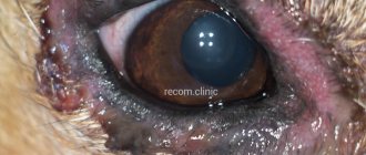
| Dog 8 years old, mixed breed with glaucoma. The eye is enlarged in size, the cornea is swollen and blue, the vessels of the sclera are tortuous and dilated. Vision is lost forever. |
Glaucoma is a group of diseases associated with a constant or periodic increase in intraocular pressure and impaired blood supply to the eye.
These disorders can gradually lead to damage (atrophy) of the optic nerve (the nerve fibers that carry information from the eye to the brain), which in turn leads first to loss of peripheral and then central vision and irreversible blindness. What makes this disease one of the most dangerous and insidious in veterinary and humanitarian ophthalmology. The main cause of damage to the retina and optic nerve in glaucoma is an increase in intraocular pressure, which causes compression of the blood vessels that supply oxygen to the nervous tissue. Which in turn leads to hypoxia and atrophy of the nerve fibers responsible for transmitting nerve impulses to the visual areas of the brain. The narrowing of the visual fields occurs imperceptibly, but quickly enough and the animal can completely lose vision in literally 2-3 months.
What is a cataract?
Cataract is an eye pathology characterized by clouding of the lens. This structure is normally a clear lens that sits in front of the pupil. Violation of transparency leads to a deterioration in optical properties and, as a result, a significant decrease in the pet’s vision, up to complete blindness.
The pathological process begins with a disruption of a number of metabolic reactions at the molecular level in the structures of the eye. The result is the accumulation of water and proteins in the lens, which are toxic to the retina. Excess moisture leads to swelling of the lens, destruction of fibers and clouding.
Advanced cataracts are accompanied by serious complications: uveitis or glaucoma. Taken together, these pathologies can lead not only to loss of vision, but also of the eyeball.
According to the nature of the development of the cataract process, there are:
- The initial stage can last for years. At this stage, with timely diagnosis and initiation of treatment, it is possible to preserve your pet’s vision without surgery.
- Immature – at this stage, the clouding gradually becomes larger, and the pet’s visual acuity decreases.
- Mature - the lens is cloudy white in color and occupies almost the entire volume of the pupil. The most convenient stage for surgical treatment.
- Overripe - the density of the lens is increased, the capsule is wrinkled. Such cataracts require prompt removal, as the risk of developing severe complications is high.
Causes of pathology in animals
- Diabetes causes diabetic cataracts. When blood sugar levels are elevated, a number of reactions in the body change. The pressure in the eyeball increases, and water and galactose begin to accumulate in the lens. This type of pathology is characterized by rapid development. Cloudiness develops from a day to two weeks.
- Congenital or hereditary cataract. It can appear in the first months of a puppy’s life and up to 6 years of age. Caused by a genetic predisposition to the disease. Breeds in which the disease is more common are: poodles, cocker spaniels, retrievers and terriers. It develops rapidly and is not associated with any associated causes.
- Age. Animals over 8 years of age develop senile cataracts. Progresses slowly.
- Eye trauma (bruises, fights with other dogs or cats) – penetrating injuries cause damage to the anterior chamber of the lens, resulting in the formation of traumatic cataracts. Depending on the nature of the damage, the pathology progresses from several hours to several days.
- Poisoning with heavy metals and poisons.
- Eye burns: thermal, chemical, light radiation.
- Eye diseases. Local inflammatory processes are accompanied by the release of toxins that affect the lens.
Kinds
The most common types of cataracts found in pets are hereditary, diabetic and traumatic cataracts.
Hereditary
This disease in dogs occurs in representatives of certain breeds that are prone to this disease.
Diabetic
Develops in an animal simultaneously with diabetes. This disease causes clouding of the lens.
Most often, dogs over ten years of age suffer from this disease. Although sometimes there are cases where young animals develop diabetic cataracts.
Traumatic
This type of cataract may, for example, be the result of a cat's claw hitting a dog's eye.
Symptoms of Cataracts in Dogs
Signs indicating the development of lens pathology:
- The appearance of a white-gray spot on the eye - first at the edges, and then completely in place of the pupil.
- Deterioration in coordination of movements in the pet, which is associated with deterioration in the quality and acuity of vision.
- Disorders of orientation in space.
- Photosensitivity - the animal squints and turns away from bright light sources.
- Cloudiness can occur in one eye or in both eyes at once.
- With complete loss of vision, the pet stops playing, mobility is reduced to a minimum, and the general condition is depressed.
If cataracts in a dog are a secondary disease, you should pay attention to the accompanying symptoms:
- Excess weight, increased thirst and urination may indicate diabetes.
- If the pet's organ of vision is damaged, visible damage around the eye (on the eyelids, mucous membranes), discharge, pain, swelling, and itching are detected.
- Eye pathologies are accompanied by anxiety of the animal, a depressed state, redness of the conjunctiva and eyeball.
Signs and symptoms
Dog owners can detect cataracts by looking into the dog's eyes. The affected eye appears bluish-gray and cloudy. As a dog ages, the lens of the eye can also become cloudy due to age-related changes known as nuclear sclerosis.
It is important to note that the signs and symptoms of cataracts and nuclear sclerosis are very similar, so an examination by a veterinarian is required to determine which of these conditions is actually present.
Differences from nuclear sclerosis
Most veterinarians can quickly tell the difference between nuclear sclerosis and cataracts with a test. It involves checking the cornea. If cloudiness is present on or behind the cornea, the problem is due to cataracts.
If the veterinarian looks deeper into the animal's eye and can clearly see the bottom of the retina, then it is nuclear sclerosis. If a dog is diagnosed with nuclear sclerosis, it will not significantly impair vision and no treatment is required.
Despite popular belief, nuclear sclerosis does not develop cataracts.
Diagnostic methods
Diagnosis of cataracts in animals includes general and highly specialized methods. Without ophthalmic devices, it is difficult to accurately diagnose. In addition, a thorough examination of the eyeball is necessary to assess the involvement of other ocular structures in the pathological process.
General diagnostics include:
- Collecting an anamnesis (information about housing and feeding conditions, information about deworming and vaccination, asking the owner about how long ago complaints about the pet’s health began), palpation and auscultation of the animal, a superficial examination of the organ of vision.
- Taking general clinical and biochemical blood tests.
- Conducting a clinical urine test.
- Determination of blood glucose levels.
Additional studies are carried out by a veterinarian-ophthalmologist using special equipment:
- Slit biomicroscopy - a method that allows you to check the tissues of the anterior and posterior chambers of the eye.
- Gonioscopy – assessment of the condition of the anterior chamber of the eyeball.
- Electroretinography analyzes the functional state of the retina. This research method is the most informative for cataracts.
- Tonometry – determination of intraocular pressure.
- An ultrasound examination of the organ of vision is carried out to assess the condition of the vitreous body and retina (its detachment).
Treatment of cataracts in dogs
The main method of treating the disease is cataract surgery. This procedure is not available in every clinic and has some nuances. The conservative method is practically ineffective.
Treatment of pathology at home is impossible even in the initial stages. The pet must be examined by a veterinarian-ophthalmologist to confirm the diagnosis and further recommendations.
Treatment of cataracts in dogs with folk remedies is also unacceptable and ineffective. Self-care for such diseases only leads to wasted time and deterioration of the animal’s condition.
Drug therapy
The use of medicinal drops is indicated only for the initial stage of cataracts. Drug therapy can stop the development of the process, but the drugs used do not cure the disease or restore vision.
Example of prescribed eye drops:
- Taufon
- Oftan katachrome.
- Vitafacol.
- Catalin.
The drugs are used over a long period of time. If there is a deterioration in the condition, the drugs are discontinued until the next visit to the doctor.
If cataract is a secondary disease, then concomitant therapy for the underlying cause is prescribed. The main method of treatment (surgical) will be available only after the animal’s condition has been stabilized (lowering glucose levels in diabetes, anti-shock therapy for injuries, etc.).
Surgery
Surgery is the only effective treatment for cataracts in animals. Before the operation, the surgeon assesses the patient, determines his readiness for intervention and the use of anesthesia. The age of the dog, blood sugar level (for diabetic type of pathology), and the degree of involvement of other structures of the organ of vision are taken into account. If both eyes are affected, the ophthalmologist gives recommendations in what order the manipulations should be performed. The interval between operations in such cases can be 2-3 months.
Before surgery, preparatory therapy is carried out, which includes the use of:
- Eye drops containing antibiotics - 4 days before the planned procedure (Tobrex, Tsiprovet, Floxal).
- Angioprotectors – 7 days before (Emoxipin).
- Local glucocorticosteroids – 7 days before (Tobradex, dexamethasone).
Cataract surgery in dogs involves removing the old lens and replacing it with a new plastic or acrylic one. With timely and correct manipulations, the animal’s vision is restored almost completely.
Postoperative care includes the use of local antibiotics and steroidal anti-inflammatory drugs. The animals are given microsutures to limit the mobility of the eyeball in the first days. The dog is put on a protective collar for the entire rehabilitation period, avoiding the possibility of self-injury with its paws.
Expert opinion
Kuzmenko Olga Olegovna
Information about the expert
Ask a Question
Local complications may occur 3-4 days after surgery. As undesirable consequences, pathologies such as uveitis, increased intraocular pressure, glaucoma, and dry eye syndrome may develop. In such cases, the doctor takes the necessary measures and adjusts further treatment.
If during the diagnosis it is revealed that the process extends to other important structures of the organ of vision, the doctor may decide to enucleate the eyeball.
The cost of cataract surgery for a dog is quite high. The procedure requires specialized skills from a specialist and the availability of ophthalmological equipment in a veterinary clinic. The cost of the manipulation depends on a number of factors - the weight of the dog, the general condition of the pet, the reputation of the institution, and the qualifications of the veterinarian. The price range starts from 40,000 rubles and above. In addition, preliminary diagnosis of pathology will also require certain costs.
Phacoemulsification
This method is widely used in modern medicine and is highly effective. Its use is possible even with advanced forms of cataract - in the stage of overmaturity. The procedure is carried out as follows:
- a special ophthalmological device – an ultrasonic flaw detector – is inserted into the eyeball;
- the lens is crushed;
- Through the resulting hole with a diameter of about 1.5-2 mm, the cataract is sucked out.
The process of suctioning cataracts during phacoemulsification surgery
To reduce the risk of complications, the veterinarian makes a puncture in the peripheral tissues of the eye cornea. Instead of the removed lens, an intraocular lens or a lens made of artificial materials is placed. The new lens, rolled up, is placed through a puncture into the lens bag, where it is placed in place of the damaged element.
Until recently, surgical intervention was allowed only for mature cataracts. Today, ophthalmologists have changed their views and agreed that phacoemulsification is quite effective at the initial stage of the disease. In this case, the “soft” lens is removed, which is less traumatic than in later stages of the disease.
To avoid possible mechanical injuries, viscous viscoelastics are injected into the animal's eye during preparation for surgery.
This is what a dog's eye looks like after phacoemulsification:
If veterinarians are faced with cataracts in both eyes, in which the lens of one eye has become cloudy and a sharp decrease in visual acuity has been recorded, and intensive progression of the disease is observed in the second eye, then it is recommended to initially perform surgery on the second eye, and on the first after 2.5-3 months.
This operation is indicated for animals from six months of age to the ripe old age of 18 years. Modern equipment allows such operations to be performed without the direct participation of a surgeon, and the risks of complications for the pet are minimal. After the procedure, the animal recovers quite quickly.
Prevention of disease in dogs
- A well-formulated diet. Industrial feeds must include a daily allowance of taurine, a component that is essential for maintaining eye health. When feeding animals naturally, they are given vitamin supplements containing the amino acid.
- Regular examinations by a veterinarian once a year for young, old and pets with eye problems.
- Maintain a healthy weight for the animal and prevent the development of obesity. If it becomes noticeable that the dog has gained weight, it is necessary to gradually increase physical activity and reduce the calorie content of the food.
- Selection – do not allow animals with congenital cataracts to be bred.
- Animals diagnosed with diabetes should be examined by an ophthalmologist every six months. As a preventative measure, such patients are prescribed eye drops containing antioxidants. They will not cure cataracts, but they can slow down the rate of development of the pathology.
- Avoid direct sunlight entering the eye.
- Securely hide household chemicals, fertilizers, insect and rodent repellents.
Cataracts in dogs are a pathology that leads to blindness of the animal. Externally, the disease looks like a cloudy white spot instead of a pupil. The problem cannot be treated therapeutically, and the sooner professional help is provided, the greater the chance of preserving the animal’s vision.