Glaucoma, an increase in intraocular pressure (IOP), is one of the leading causes of irreversible blindness in dogs. This is a disease accompanied by increased intraocular pressure and enlargement of the eyeball. There is also dropsy of the eye, which differs from glaucoma in that it is a consequence of inflammatory processes, primarily of the vascular tract.
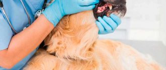
Since glaucoma can lead to blindness within 24-48 hours of onset, it requires urgent aggressive drug therapy and immediate attention to a veterinary ophthalmologist. It is easily undetected clinically. It should be a rule to examine all animals with hyperemia of the eyeball, in which ulcers and infections of the cornea and purulent processes have been excluded, for the presence of glaucoma.
Description of the disease
Keratitis is an inflammatory disease of the cornea. The cornea is a clear, biconvex lens that sits at the front of the eye. Its main functions are light refraction and protection. It is thanks to the cornea that animals distinguish the brightness, shape and size of an object.
Most often, keratitis affects dogs of brachycephalic breeds (boxers, pugs, bulldogs, Pekingese, etc.). Due to the unusual structure of the skull, the eyes are not sufficiently moistened, resulting in inflammation.
Its varieties
Based on the degree of damage to the cornea, there are:
- Superficial keratitis in dogs - the process affects the external part.
- Deep – inflammation spreads to the inner layers. This type of pathology is accompanied by the formation of ulcers and scars, which remain even after treatment, worsening the quality of the animal’s vision.
Depending on the nature of the development of the pathology, the following types of keratitis are distinguished:
- Infectious. It develops when pathogenic microorganisms (fungi, bacteria) come into contact with the cornea. This inflammation is accompanied by profuse yellow-green discharge from the eyes.
- Vascular. Occurs when there is excessive growth of blood vessels in the tissue of the cornea. The front part of the eye becomes reddish and lumpy.
- Pigmentous keratitis occurs in brachycephalic dogs. With this type of pathology, brown pigment accumulates in the cells of the altered epithelium. This causes the cornea to become cloudy and then darken. There are local, subtotal and total.
- Dry keratitis develops, as a consequence, with pathologies of the lacrimal glands, with dry eye syndrome.
- Interstitial. A blue-white cloudy film appears on the cornea. The nature of this type of pathology is usually infectious (hepatitis, parvovirus, etc.)
- Phlyctenulous keratitis accompanies poisoning or chronic allergic reactions. The presence of irregularities is noted in the eyes, the third eyelid becomes inflamed and swollen.
Trauma as a cause of deviation
Although the eyes are a paired organ, it happens that one eye of a dog becomes cloudy. This may indicate the formation of a cataract. Such a defect often appears as a result of injuries from a blow, burn, insect bite, etc. This type of thorn can be treated and treated successfully. The therapy is not complicated, but requires timeliness, so you can achieve the complete disappearance of the cloudy film on the surface of the visual organ.
Age-related eye degeneration cannot be avoided, but you can make life easier for your aging faithful friend with vitamin drops and other doctor’s recommendations. But pathological processes caused by diseases cannot be ignored categorically. If the owner loves his pet, he will never let any deviations in his health take place. But you love it, don’t you?
Causes of disease development in animals
There are several causes of inflammation of the cornea of the eye, including:
- Breed predisposition in brachycephalic dogs. In animals, the eyelids do not close completely when blinking, which is why the central surface of the cornea is not sufficiently moistened. Also, in pets of these breeds, pathologies of the lacrimal duct are more often noted; as a result, the eyes do not receive the necessary amount of fluid for normal functioning.
- Chronic corneal injury. When the cornea is irritated, a kind of protective reaction is triggered in the body. In response to long-term damage, tissues become denser, but at the same time lose their main function - light transmission. This occurs in animals with abnormal eyelash growth, long-haired pets (especially if they have bangs), or if the dog is kept in a dusty room.
- Dry eye syndrome. For various reasons, little tear fluid is produced, nutrition and breathing of the corneal surface are disrupted. Keratitis in this case will be a secondary problem.
- After surgical interventions on the organ of vision.
- After viral keratoconjunctivitis (previous viral infections: canine distemper, hepatitis, parvovirus enteritis).
- Allergic reactions.
- Diabetes.
Film
White cloudy
If a dog's eye is covered with a white film, then most likely this is a sign of problems with the third eyelid. If there is a thickening, which is a symptom of adenoma of the third eyelid, the involvement of a surgeon will be required .
What to do if a film appears - a trip to the veterinarian to determine the nature of this formation and identify visual disturbances at an early stage will be the best way out.
Transparent
A transparent film on the eyes without signs of inflammation is amyloid corneal dystrophy . May be caused by an unbalanced diet, a violation of fat metabolism. There is no need for treatment, since it does not create any inconvenience for the animal and has no consequences. There is a possibility of extinction.
After examination, the doctor may prescribe Emoxipin , 1 drop 3 times a day, for 1-2 months.
Blue
If your eye is covered with a blue film, then you should immediately go to the veterinarian! A bluish film on the membrane of the eye appears only when the eye is swollen, which occurs for several reasons and, depending on the causes, is treated using different methods. Having such a disease can be extremely painful for a dog. The treatment must be chosen by a doctor.
How long does keratitis last in dogs?
The duration of the disease depends on the degree of damage to the cornea and the timely and correct initiation of treatment. In the superficial form, the shell recovers quickly. Therapy will take 7-14 days.
With severe inflammation, the process can drag on for several months. However, it is not always possible to completely restore vision function.
Expert opinion
Kuzmenko Olga Olegovna
Information about the expert
Ask a Question
The speed of recovery is influenced by many accompanying factors: nutrition, the age of the pet, the state of immunity, the presence of chronic diseases.
Pigmentary keratitis in pugs • Zoo-Vision™ Zoovision™
Pigmentary keratitis is the most common eye disease in pugs, in which the clear part of the eye becomes covered with an opaque pigment film. Pigmentary keratitis is the main cause of blindness or poor vision in pugs, Pekingese, Shih Tzu and some other dog breeds.
A dog's cornea must be perfectly transparent for normal vision. And not only dogs - all living creatures who look at the world through their eyes. Animals with an opaque cornea experience vision deficits. Some people just don’t see well, others don’t see at all. According to one study, about 80% of pugs suffer from pigmentary keratitis.
What is pigmentary keratitis in pugs?
Pigmentous keratitis (also known as corneal melanosis) is a disease in which brown pigment is deposited on the cornea. At first, these spots are pale, translucent, completely superficial, growing from the inner corner of the eye (from the medial corner of the eye).
As the spots grow, they acquire a more pronounced brown color, occupy a larger area, and grow deeper into the cornea. As pigment spots grow, dogs begin to experience vision deficits and may lose their vision. At first, do not notice small objects, then large ones. Then the dog may see worse at dusk, lose its owner, or crash into obstacles.
Typically the disease begins to progress by age 3. However, the disease can also occur in puppies up to six months of age.
Where does my dog get pigmentary keratitis?
The exact cause of pigmentary keratitis is not completely clear. A genetic basis is assumed, but so far this is at the stage of speculation without direct evidence.
It was previously assumed that pigmentary keratitis is a consequence of medial entropion of the eyelids in pugs and Pekingese.
However, studies have shown that in the presence of entropion of the eyelids in almost 100% of pugs, pigmentous keratitis develops in 80%.
What causes pigment growth on the cornea?
Any factors that cause the cornea to send an “SOS” signal. For example, eye injuries, entropion of the eyelids, improperly growing eyelashes, neoplasms on the eyelids, dry eyes. These factors accelerate pigment growth more than others.
How is pigmentary keratitis treated?
Treatment of this disease always begins with diagnosing the underlying cause. Or the main factor influencing pigment growth. Dogs with dry eye syndrome should first be treated for tear deficiency. Those who have suffered an eye injury should first treat the cornea.
If pathological eyelashes (for example, ectopic) are to blame, surgical treatment of these eyelashes is required. It also happens that the main cause is inversion of the eyelids. Almost 100% of Pugs have an inversion of the eyelids in the inner corner of the eye. This is where the pigment begins to grow.
In this case, the dog is indicated for eyelid surgery.
The pigment itself can also be removed. But this is carried out when the main factors of the disease have been eliminated. That is, a dog with dry eye syndrome should not have pigment removed until tear production is restored. Otherwise, at best, the pigment will grow back in 2 weeks. And in the worst case, you can get a corneal abscess.
Eyelid surgery in a pug. Whether it is necessary?
If this plastic surgery is performed according to indications, then it is definitely necessary. Sometimes this is the only thing that can protect a dog from blindness. But in order for this plastic surgery to be performed exactly according to the indications and have the desired effect, you should seek help from a highly specialized veterinarian, an ophthalmologist.
Green arrows point to the area where the eyelids turn in. Red – on the pigment growth zone.
Green arrows point to the area where the eyelids turn in. Red – on the pigment growth zone.
Pug 1 month after eyelid surgery. The palpebral fissure is of normal size, there is no longer any fur in the corner of the eye.
1 month after surgery. The eyelids are rounded and fit tightly to the eye. There is no more fur in the corner of the eye.
What about anesthesia? Pugs cannot be given anesthesia.
Pugs are the most common dogs that can do anything. And anesthesia as well. They once earned themselves a bad reputation because of their anatomical structure.
The long velum palatine (this is what causes them to grunt and snore) could be blocking the breathing of a sleeping, relaxed dog. Why the dog could have died. However, for more than 10 years this problem has not been relevant, because today all pugs are operated on with an oxygen-gas mixture.
And during operations they are intubated. That is, a tube is inserted into the trachea, thanks to which the dog breathes even easier than in normal life.
In addition, the dog will always undergo a heart and general medical examination before surgery. You will never go to the operating table without tests, will you? So your dog is not allowed. This way we get a patient about whom we know enough to decide whether he can be operated on.
What to do if your dog has pigmentary keratitis?
Regardless of whether you have a pug or another breed, pigmentary keratitis is a reason to contact a veterinary ophthalmologist. The illness is not an emergency and therefore does not require immediate attendance to a doctor. However, the visit should definitely not be delayed. Superficial pigment is much easier to remove without relapse. While total pigmentation of the cornea is very difficult to deal with.
Join us on social networks
Similar
Source: https://www.zoo-vision.com/thw_companion/pigmentnii-keratit/
Symptoms of pathology
You can recognize the signs of keratitis in a dog at home and seek help from a veterinarian as soon as possible:
- Loss of vision. The pet moves uncertainly and bumps into surrounding objects.
- Fear of light. The dog hides in dark places and avoids daytime walks.
- Redness of the eyes and mucous membranes, blood vessels are dilated and filled with blood.
- Discharge from the eyes. Accumulations of pus, a large number of yellow-green crusts on the fur around the eyelids.
- Itching. The dog scratches its face with its paws or rubs against external objects.
- A cloudy white film appears before the eyes.
- General apathy. The animal stops playing and constantly sleeps.
It is more difficult to detect pigmentous keratitis, since the eyes of dogs are dark and against such a background it is difficult to see when the cornea becomes not transparent, but acquires a brownish color.
Symptoms
The clinical signs of the disease are quite specific. O and “blackening” of the cornea.
Pigmentous keratitis in a pug
In Pugs and Pekingese, pigmentous keratitis is usually observed in the medial sector of the cornea (closer to the nose). This is due to constant irritation of the eyelids and skin folds of the eyeball by hairs. As a result of this chronic process, damage (erosion) is formed, and in advanced cases, a corneal ulcer. During the healing process, pigment cells “appear” on the damaged areas and pigmentous keratitis develops.
In severe cases, pigment is deposited on a large part of the cornea, which negatively affects visual functions in animals.
Diagnostics
Diagnosis is made by visual examination by a veterinary ophthalmologist, ophthalmoscopy, and slit lamp examination of the anterior segment of the eye. To identify corneal defects, it is stained with a 1% fluorescein solution.
Treatment
Treatment of pigmentary keratitis in dogs consists of eliminating the cause that causes this pathology. Thus, it is recommended to perform surgical correction of entropion of the medial canthus in dogs of brachiocephalic breeds.
Complex therapy is carried out aimed at protecting the cornea from damaging factors. Medicines are also used to reduce the amount of pigment on the cornea, which can ultimately lead to its disappearance.
In advanced cases, surgical treatment of pigmentous keratitis is performed: superficial keratectomy, barrier keratoplasty, cryodestruction, etc.
Categories: Black Eye, Corneal Diseases
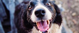
It just so happens that eye diseases occur quite often in our pets. One of the most common ailments affecting the eyes is keratitis in dogs.
– inflammation of the cornea, which often leads to decreased vision and sometimes blindness, which is why if such a disease develops, it is important to visit a veterinary ophthalmologist as soon as possible.
How to treat keratitis in a dog?
Treatment always begins with the use of local medications. Surgery is indicated for animals if there is no positive response to conservative therapy.
Medication
Therapy is aimed at:
- elimination of pathogenic microflora;
- reduction of toxic-allergic state;
- restoration of hydration of the ocular surface.
Groups of medications used to treat keratitis in dogs:
- Antibacterial drugs (Tsiprovet, Floxal, Normax, Tobrex).
- Lubricants (the action of this group of drugs is aimed at moisturizing the ocular surface). These can be drops, gels or ointments. The doctor must determine which form of release to choose during the examination. In each case the approach is individual. Examples of prescribed drugs: Hyphenlez, Hilabak, Systeine.
- Glucocorticosteroids (drops or ointments: Dexamethasone, Nevanak, Maxitrol) are prescribed with caution, as these drugs reduce the local immunity of the eye and worsen the process of regeneration of the cornea. Long-term use of the products can lead to corneal ulcers.
- Cytostatics (Optimmun ointment) are milder in their action, but a long course of administration also carries a number of side effects.
Before instilling the drops, remove all dirt from the eyes. A clean cotton pad soaked in freshly brewed tea or chamomile decoction is suitable for this. Rub the eye from the outer corner to the inner (towards the nose).
If it was not possible to remove all the discharge at one time, use a new cotton pad for each subsequent treatment.
To administer eye drops, the dog's head is fixed. The drug is applied by slightly pulling the lower eyelid, then make 3-4 blinking movements using your fingers. If several drugs are prescribed, the interval between treatments should be at least 15 minutes.
Application of eye gel is similar. Place a strip of gel under the lower eyelid and blink several times so that the medicine is distributed evenly.
If a dog intensively scratches its eyes, a special protective collar is worn throughout the course of treatment to avoid the development of secondary infection and self-injury.
By performing an operation
Surgical intervention is indicated in cases of:
- If keratitis progresses during therapeutic treatment.
- When allergic reactions to the drugs used occur.
- When a corneal ulcer occurs during treatment with drops.
- In case of total pigmentous form that does not respond to treatment.
The operation is performed in specialized clinics and consists of removing the pathological area of the cornea. The problem is solved surgically when keratitis occurs against the background of improper eyelash growth and other pathologies that are the root cause.
Treatment of glaucoma and other eye opacities
Choosing the right treatment depends on an accurate diagnosis. Drugs that lower intraocular pressure are prescribed (pilocarpine drops 0.5%, eserip 0.5%, physostigmine salicylate 1%, etc.). To prevent drying of the cornea, ointments are used - xeroform, antibiotic, yellow-mercury, etc. In case of excruciating pain, anterior chamber puncture or enucleation of the eyeball is performed. In acute glaucoma of the seeing or partially seeing eye, the goal of treatment is to maintain vision and comfort for as long as possible, using all medical and surgical treatment methods. In acute cases, IOP must be reduced quickly and effectively.
Glaucoma is treated as an emergency and then the patient is referred to an ophthalmologist. The goal of treatment is to reduce intraocular pressure (IOP). This can be achieved in two ways:
- By reducing aqueous humor production:
- Carbonic anhydrase inhibitors: dichlorphenamide, methazolamide;
- Sympathomimetics: epinephrine, dipivephrine (epinephrine derivative);
- Cyclocryotherapy: (ophthalmologist).
- By increasing the drainage of aqueous humor:
- Cholinergics: pilocarpine;
- Sympathomimetics: epinephrine;
- Surgical treatment: (ophthalmologist).
Attention: paracentesis is contraindicated.
Drug treatment is temporary until surgical correction of the process. Thoracic glaucoma is controlled with medication. In the case of primary glaucoma, a 0.5% timolol solution, 1 drop 2 times a day, can be injected into the second eye to prevent progression of the disease.
Urgent Care:
- Mannitol (1-1.5 g/kg IV): 5.0-7.5 ml/kg 20% solution over 15-20 minutes helps significantly reduce IOP within an hour.
- Carbonic anhydrase inhibitors: dichlorphenamide (Daranide) 2-5 mg/kg PO 3 times daily in dogs and 1 mg/kg PO and methazolamide (Neptazane) 1-2 mg/mg PO 3 times daily in dogs and cats. Side effects include metabolic acidosis, vomiting and shortness of breath. Dorzolamide 2% (Trusopt) is a topical carbonic anhydrase inhibitor. Administer 1 drop 3 times a day.
- Pilocarpine 2%: administer 1 drop every 30 minutes until the effect appears (miosis), then every 6 hours. Contraindicated in anterior uveitis or severe iritis.
- Prednisone: This is used for moderate anterior uveitis and to prevent the breakdown of the blood-aqueous humor barrier caused by the miotic drug.
- Sympathomimetics: dipivefrin HCL (Propine), an epinephrine derivative, administered 1 drop 2 times a day.
- Timolol 0.25%, 0.5% reduces the production of aqueous humor. Administer 1 drop 2 times a day. May cause bradycardia.
Combination treatment with pilocarpine and epinephrine has been shown to be very effective in dogs. Most often, with glaucoma, topical application of atropine is contraindicated.
Table. Drugs used to treat glaucoma in dogs
| Group of drugs | Drugs | Dosage, features |
| Hyperosmotic solutions | Manitol, 25% | 1 -2 g/kg. IV, for 20 minutes, after administration, deprive of water for 4 hours, repeat after 4 6 hours |
| Glycerin (50%) | 1 -2 g/kg s.c. May cause vomiting, do not administer to dogs with diarrhea | |
| Systemic carbonic anhydrase inhibitors | Dichlorphenamide | 4 mg/kg, every 8-12 hours, strongest IR, with few side effects in dogs |
| Methazolamide | 2-4 mg/k, every 8-12 hours, available in 25 mg tablets, which is convenient for dosing for small dogs | |
| Acetazolamide | 4-8 mg/kg every 8-12 hours, cheapest IR, but has many side effects | |
| Cholinomimetics | Pilocarpine solution (2%) | every 6-12 hours, contraindicated in uveitis, local irritation |
| Pilocarpine gel, 4% | every 24 hours | |
| Echothiophate iodide 0.06%, 0.125% | every 12-24 hours, do not use with organophosphorus preparations against parasites | |
| Devmekarium bromide, 0.125%. 0.25% | every 12-24 hours | |
| Adrenergic blockers | Timolol maleate (timoptic) 0.5% | every 12 hours, also available as a 0.25% solution, which is ineffective for dogs and may cause bronchospasm |
| Betaxolol | every 12 hours | |
| Sympathomemetics | Dipevefrin 0.1% | every 12 hours |
| Potassium preparations | Potassium chloride | 600 mg tablet per 25 kg dog with food every 24 hours |
Eye restoration with keratitis in dogs
If you consult a veterinarian-ophthalmologist in a timely manner, keratitis can be completely cured. It is important to comply with the prescribed treatment and monitor the dynamics after the start of therapy. If there is no effect after 10-14 days, you need to come back for a second appointment. The doctor will conduct an additional examination and change the treatment. In some cases, a positive response occurs after a month or more.
In case of severe damage, complete restoration of the cornea is impossible. Scars and ulcers remain on the membrane, which significantly impair the pet’s vision.
If you do not consult a veterinarian, keratitis can be aggravated by the development of glaucoma, cataracts, and complete loss of vision.
Cataract
This eye disease is associated with aging of the lens, a violation of its transparency - subnuclear (nuclear) sclerosis.
The development of cataracts is most likely at the age of 8 years, when the elasticity of the lens decreases, it hardens and ceases to adjust vision to different distances. It is more common in Golden Retrievers, Poodles, Yorkshire Terriers and Boston Terriers. The disease progresses over the years.
Cataract photo:
Causes
Less commonly, the causes of cataracts are heredity. Acquired cataracts occur after trauma, after inflammatory diseases, diabetes, etc.
Symptoms, treatment and diagnosis
An external sign of cataract is clouding of the lens. The cloudiness of cataracts is uneven. It is possible to recognize cataracts on your own. When a beam of light hits the affected eye, the clouding will shrink in size. In low light, on the contrary, it increases.
In some cases, this dangerous disease can develop for a long time without visible symptoms. But a dog owner may notice decreased vision by analyzing the dog's behavior.
The most reliable way to diagnose an eye disease called cataracts remains to be examined by a specialist.
Treatment without surgery
How to treat cataracts without surgical intervention at home? But no way. The use of medicinal drugs does not allow us to hope for the recovery of our four-legged pet. At best, the progression of the disease will slow down. It is possible to use drugs only at the very beginning of the development of cataracts, when surgery is not yet required. Vitamins and enzymes are usually prescribed.
Cataracts cannot currently be prevented. Do not fall for the tricks of deceivers who recommend Smirnova eye drops, vitaiodurol, vicein, catachrome, vitafacol for the treatment of cataracts . These drugs cannot prevent, slow down, or even cure clouding due to the fact that the nature of the disease is not clear.
When is cataract surgery necessary?
The only effective way to treat cataracts is surgery to replace the clouded lens with an artificial one. From a technical point of view, this operation is very complex. Its result largely depends on the stage at which the application was made to an ophthalmologist. Obviously, earlier treatment guarantees a better result.
The modern technology of lens removal using ultrasound - phacoemulsification - is finding more and more fans. But it is also effective in the early stages.
Prevention
Disease prevention
To protect your pet from keratitis, you need to follow a number of recommendations:
- Strict adherence to vaccination schedules according to the dog’s age.
- Brachycephalic dogs should be seen by an ophthalmologist annually.
- If pathology of the tear ducts is detected, the pet’s eyes need to be moisturized artificially with special means. For some animals, this procedure is lifelong.
- High-quality feeding, exclusion of harmful and allergenic foods or treats from the diet.
- Timely treatment of any eye pathology.
- In case of injuries to the organ of vision, it is necessary to undergo examination by an ophthalmologist, even if all visible causes (foreign bodies, dirt) have been eliminated.
- Control and treatment of chronic diseases.
Keratitis in dogs is a fairly common pathology of the visual organ. The process affects the cornea, but in severe cases it can also affect other structures of the eye. Inflammation leads to deterioration or complete loss of the animal’s vision, so it is important to recognize the problem in time and seek quality treatment. The sooner you start therapy, the easier your pet’s recovery will be.
Reasons that contribute to clouding of the corneal layer
I would like to draw your attention to the fact that, according to statistics, very often the cornea becomes cloudy due to some ophthalmological diseases. The disease may also be associated with the accumulation of pus, excess fluid, vascular ingrowth, and the appearance of excess calcium or cholesterol.
The list of pathologies that are characterized by corneal opacities is very large and includes diseases such as:
- Keratitis is a disease that is accompanied by inflammation of the corneal layer of the eye. The animal's vision begins to decline sharply. Keratitis occurs most often due to infectious diseases;
- conjunctivitis - leads to an inflammatory process and the appearance of pus on the surface of the sensory organ;
- Glaucoma is a disease characterized by increased pressure inside the sensory organ. This dynamics leads to clouding of the corneal layer. Urgent medical attention is required;
- erosion, ulcer - can have different depths and sizes. Such formations can arise due to the appearance of bacteria or viruses and due to injury to the organ of vision. The dog constantly squints his eyelids. The animal's eyes turn red;
- degeneration process - leads to the formation of calcium, amyloid and cholesterol crystals in the corneal layer;
- Scars - appear after an injury, ulcer or burn.
Important! Cloudiness can also occur after cataract extraction. The old age of the dog also leads to the fact that the corneal layer begins to gradually become cloudy. Gradually, new layers form behind the walls of the cornea, which after some time begin to roll around the lens. Therefore, with age, the pet’s eyeball begins to become cloudy.
You may be interested in: How to recognize and treat keratitis in rabbits in time?
Prevention measures
Prevention of keratosis in dogs involves following simple recommendations:
- Regular vaccination of the animal against adenoviruses and herpes.
- It is necessary to try to prevent possible traumatic injuries to the pet's eyes.
- Contact a specialist promptly in case of conjunctivitis or other eye diseases in your dog.
- It is imperative to monitor the animal’s balanced, rational diet to prevent the development of vitamin deficiency and weakening of the immune system.
- Provide your pet with proper care, regularly comb its fur and trim the fur around the eyes.
- Regularly wash your dog's eyes with cotton pads soaked in chamomile decoction, tea leaves or boric acid solution.
Keratosis in dogs is quite treatable. By following the recommendations of an animal care specialist, you can prevent the development of undesirable consequences, achieving a complete cure for your pet - without complications or relapses!