- home
- Diseases
»
09.20.2018Diseases 1 comment
There are diseases that initially do not pose a serious danger, but with prolonged inactivity, the harmless nature of the disease can lead to dire consequences. This is exactly what a cyst in a dog refers to.
general characteristics
A cyst is a cavity that contains a dense secretion or fluid. Sometimes the contents are dead tissue or a collection of parasites. Bubbles form in internal organs or as small growths on the body.
For some time, the hollow formation does not cause discomfort to the dog. But you can’t ignore the problem - it can get bigger and eventually burst. The released fluid will spread to other internal organs and cause further infection of the body.
It is important! A cyst is not a malignant tumor. But if nothing is done about it, it can cause cancer.
Types of tumors
Among veterinary specialists, it is customary to distinguish the following types of pathological blisters:
A pathological vesicle appears immediately after the ovulation process. As a rule, progesterone cysts are characterized by small sizes up to 3 cm. Corpus luteum cysts account for no more than 5% of all pathologies of this kind in dogs.
According to veterinary experts, this type of cavities is not a disease.
In some cases, so-called paraovarian cavities are detected in four-legged patients. These cysts develop between the gonads and the uterine horn.
Types of cysts
Most often, the epidermal form of the anomaly develops. It does not cause pain and rarely exceeds 5 cm. It is located in the upper layers of the epidermis.
A congenital disease includes a dermoid cyst in a dog. The growth grows on the body in the form of a soft bubble. Poses a threat to the dog's entire nervous system.
A small growth on the body (up to 3 cm) is called the follicular form. Occurs due to the accumulation of dead skin cells.
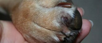
Furunculosis or interdigital cyst in a dog manifests itself in the form of small growths on the paws. The causes are allergic reactions, ingrown fur, and polluted environments.
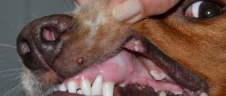
The most common pathology of the oral cavity is a salivary gland cyst in a dog. The exact causes are unknown, but it can appear due to injury. With prolonged inactivity it becomes a tumor.
An ovarian cyst in a dog is a dangerous disease in bitches, as it causes other pathologies. Almost always leads to the appearance of fibroids, false pregnancy, infertility or a tumor. Deaths are common.
Serious pathologies that precede mastopathy include a mammary gland cyst in a dog. It appears as a growth near the nipple and causes white or greenish discharge. Signals about hormonal imbalance.
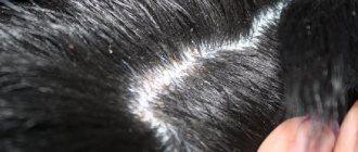
Skin cysts
Svetlana Belova, Estonian University of Life Sciences, www.vetderm.eu Photos of the author are used in the article
A cyst is a pathological cavity (in our case in the skin) with a wall lined with epithelium. The contents of the cyst depend on the secretory activity of this epithelium. Cysts can be acquired (for example, blockage of the excretory duct of the skin gland) or congenital.
In the skin, there are cysts of sweat and sebaceous glands, follicular cysts and dermoid cysts/sinuses.
A sweat gland cyst is a small (several millimeters) sized cavity with a bluish tint (photo 1). It is filled with clear liquid (sweat) and protrudes above the skin level. It is relatively rare in both dogs and cats. Single sweat cysts, as a rule, do not cause discomfort and do not require treatment.
Multiple cerumen/ceruminal (modified sweat) gland cysts in the external auditory canal and pinna of cats are a relatively common occurrence and are called ceruminal cystomatosis . While the cysts are small in size (they look like dark gray, almost black smooth elevations a few millimeters in diameter), they do not cause any discomfort (photo 2). However, when they reach a certain size and quantity and block the lumen of the ear canal, they cause external otitis media with secondary infections, soreness and itching (photo 3). Cysts can degenerate into adenomas and adenocarcinomas of the sulfur glands. Treatment consists of surgical excision (preferably laser excision).
Sebaceous gland cysts are small (1–3 mm) elevations of a whitish or yellowish hue (photo 4). They are relatively common, especially in dogs. They do not cause discomfort and do not require treatment.
Follicular cysts (FC) are most often formed by the epithelium of the mouth of the hair follicle and are a cavity filled with dead keratinized epithelium - keratin. FC occurs quite often in dogs (predisposed breeds are German Shepherds, Pekingese, Shih Tzu and Boxers) and rarely in cats. Clinically, FC appears as a rounded raised area of grayish skin ranging from a few mm to 6 cm, often with a hairless and comedonal surface (Figure 5). FC is often mistakenly called a sebaceous gland blockage. They can be single or multiple and have a progressive course. Follicular cysts that have reached significant sizes often have a “pore” on the surface of the skin through which a curdled content (keratin) of a grayish tint is released. When drying, keratin forms a kind of cutaneous horn on the surface of the skin (photo 6). Secondary bacterial infection of the FC is also not uncommon (especially after an attempt to squeeze it out!), in which erythema is noticeable and pain and itching appear (photo 7). Main differential diagnosis: abscesses, tumors. Diagnosis is made based on history, clinical presentation, cytological examination of cyst aspirate (keratin, cholesterol crystals +/- signs of infection) or histology. Treatment consists of surgical excision. Retinoids (isotretinoin 1.5–3 mg/kg/day) may be effective in inhibiting the growth of old cysts in dogs and preventing the formation of new ones.
A dermoid cyst (DC) is a congenital anatomical anomaly in which a fully functioning piece of skin with all appendages (hair follicles, sweat and sebaceous glands) is enclosed in a closed cavity. DC becomes noticeable already in puppyhood, most often on the midline of the head (photo 8). It looks like one or more soft, round or oval-shaped bumps that can be infected and have a “pore” on the surface, from which a tuft of hair often sticks out. Treatment is surgical excision. A dermoid cyst may be connected by a tubular tubule to the spine (dermoid sinus), which naturally creates a risk of bacterial infection of the nervous system. Rhodesian Ridgebacks are especially predisposed to this anomaly, in which the presence of a crest is predetermined by a mutation of certain genes inherited as an autosomal dominant trait and is associated with the formation of dermoid sinuses (photo 9). The diagnosis is made on the basis of anamnesis, examination, contrast radiography, magnetic resonance imaging. Treatment is surgical excision (photo 10).
| Photo 1. Sweat gland cyst | Photo 2. Cysts of sulfur/ceruminal glands |
| Photo 3. Ceruminal cystomatosis | Photo 4. Sebaceous cyst |
| Photo 5. Follicular cyst | Photo 6. Dried keratin on the surface of a follicular cyst |
| Photo 7. Infected follicular cyst | Photo 8. Dermoid cyst (photo by D. Graham published with permission of the author) |
| Photo 9. Rhodesian Ridgeback dermoid sinus fistula at the base of the tail (photo by S. Rüfenacht published with permission of the author) | Photo 10. Surgical excision of the dermoid sinus (photo by S. Rüfenacht published with permission of the author) |
Bibliography
1. Salmon Hillbertz N and Andersson G (2006) Autosomal dominant mutation causing the dorsal ridge predisposes for dermoid sinus in Rhodesian ridgeback dogs. Journal of Small Animal Practice 47: 184–188.
2. Salmon Hillbertz N et al (2007) Duplication of FGF3, FGF4, FGF19 and ORAOV1 causes hair ridge and predisposition to dermoid sinus in Ridgeback dogs. Nature Genetics 39: 1318 – 1320.
3. Kiviranta AM, Lappalainen AK, Hagner K, Jokinen T.. Dermoid sinus and spina bifida in three dogs and a cat. J Small Anim Pract. 2011 Jun;52(6):319-24.
4. Fleming JM, Platt SR, Kent M, Freeman AC, Schatzberg SJ. Cervical dermoid sinus in a cat: case presentation and review of the literature. J Feline Med Surg. 2011 Dec;13(12):992-6.
5. Muller and Kirk's Small Animal Dermatology, 7th Ed. Miller W, Griffin C, Campbell C. WB Saunders, 2012.
6. White A, Stern A, Campbell K, Santoro D.. Multiple (disseminated) follicular cysts in five dogs and one cat. Vet Rec. 2013 Sep 21;173(11):269.
SVM No. 6/2013
Rate material
Like Like Congratulations Sympathy Outrageous Funny Thoughtful No words
Signs of a cyst in a dog
In most cases, a hollow formation on internal organs has no symptoms. However, sometimes the following symptoms may appear:
- loss of appetite;
- lethargy and weakness;
- elevated temperature;
- loss of consciousness;
- bloody discharge (a sign of internal bleeding);
- pain (the dog whines and behaves unnaturally).
Skin growths have common symptoms:
- growths increase in size;
- do not cause pain or discomfort;
- loss of appetite;
- general lethargy, apathy;
- temperature increase.
If several signs are present, the dog must be shown to a veterinarian to rule out oncology. You can also look for a photo of a dog’s cyst on the Internet and compare it with what your pet has.
Prevention of occurrence
The following recommendations and advice from veterinary specialists will help the owner avoid the development of a cyst-like cavity in the ovaries of the pet:
- If the dog is not of breeding value, it should be sterilized before the onset of its first heat. This approach negates the risk of developing many genital diseases, including the formation of follicular and other types of cysts.
- Hormonal veterinary drugs should not be used to suppress sexual heat and protect the animal from unplanned fertilization. Uncontrolled use of such products dramatically undermines the reproductive health of the pet.
- Ovariohysterectomy should be performed by a highly qualified specialist in order to avoid the development of remnant syndrome.
- A balanced diet and prevention of obesity are a guarantee of good animal health.
And here is more information about whether a dog can have an abortion.
Ovarian cyst in dogs is a chronic disease without pronounced symptoms, often detected during routine ultrasound diagnostics or during sterilization. The disease leads to infertility, which makes it difficult for the breeder to work. In rare cases, a ruptured bladder can cause peritonitis. The radical treatment method is ovariohysterectomy.
Most people know that a cyst is a cavity that is filled with fluid and forms in a certain organ not only in humans, but also in animals. As a rule, the main causes of the occurrence of such neoplasms are pathological processes in the body and internal organs.
There are several varieties of these formations. Their main classification depends on the location, formation, and structure of the neoplasm.
Cyst treatment
For small tumors, drug therapy is prescribed. Sometimes small growths are left undisturbed and left under observation. It is noted that treatment of cysts in dogs with drugs practically does not bring a positive result. And only in rare cases does it go away on its own.
If the abnormal cavity continues to increase in size and causes discomfort to the dog, then surgical intervention is required. Surgery is the most effective method of getting rid of pathology.
It is important! A cyst can develop into a malignant tumor, so self-medication should be completely avoided.
Why are cysts dangerous?
Follicular varieties and dermoids are filled with a soft, casein substance (a derivative of the sebaceous and sweat glands). The danger is that this content can be contaminated with secondary microflora, as a result of which a full-fledged putrefactive process will develop in the cyst cavity. This is a direct threat of developing sepsis, peritonitis or something similar. The same salivary gland cyst (if large) simply prevents the dog from eating normally, and the risk of inflammation should not be discounted. Again, this risks periodontal disease.
Disease prevention
The exact causes of cysts have not been identified. There are only general recommendations to reduce the risk of developing pathology. These include regular veterinary checkups, good dog hygiene and a healthy diet. It is also important to independently examine your pet’s skin.
Regardless of the location of its occurrence - be it a neoplasm of an internal organ or a paw cyst in a dog, this pathology is a fairly serious disease and can cause more dangerous complications. In some cases, a putrefactive process may develop in the blisters, which will become a direct threat of peritonitis or sepsis.
You should not attempt treatment on your own - this may cause additional harm to your pet. Therefore, it is extremely undesirable to postpone a visit to the doctor, even if the dog’s cyst is very small.
Treatment and prevention
The doctor examines the pet and in certain cases does not even prescribe treatment. But this is only the case when the growth does not cause inconvenience to the dog and does not threaten the health. But in any case, the owner is recommended to monitor the development of education. If the tumor continues to grow, then treatment is prescribed. Medicines rarely help the animal, but there are drugs that are injected under the skin and dissolve the growth. But only small growths are treated with these methods. If it progresses, then such treatment is useless. Therefore, the most reliable method of treatment is removal through surgery.
This operation can be performed under general local or general anesthesia. It all depends on where the growth needs to be removed. And after the operation, you need to listen carefully, or better yet, write down all the recommendations that the veterinarian will give for a speedy recovery. Since the disease is not fully understood, veterinarians give general recommendations to prevent such cases. Balanced nutrition, dog hygiene, and brushing. This will help reduce the risk of the accumulation of microorganisms on the wool.
But, in any case, you should immediately contact a veterinarian when the disease manifests itself. Keep an eye on your pet, as you are now responsible for it. And if something happens to him, then first of all the responsibility falls on the shoulders of the owner.
Currently reading:
- What do tumors on a dog's body and tail mean?
- 8 ways to treat papillomas on a dog’s body
- We fight warts on the face and body of dogs
- Causes of kidney stones in dogs and methods of treatment
⚠️ Classification of ovarian cysts
1️⃣ Follicular cysts appear from the so-called graaf follicles. The size of one tumor is usually several centimeters. In polycystic disease, they form groups whose diameter can be about ten centimeters. These cysts are very often observed in females, especially bitches over six years old, as well as in nulliparous bitches. The reasons for the development of follicular cysts can be partial removal of the ovaries, taking drugs to suppress estrus, long-term (longer than a month) sexual heat, lack of pregnancy after several matings, termination of pregnancy, too rare or frequent estrus, as well as abnormal interest in male dogs in the wrong period. The contents filling the cyst cavity are usually rich in estrogens, the amount of which gradually decreases. A symptom of the presence of a follicular cyst is the continuous estrus of the female dog.
2️⃣ Luteal cysts are characterized by the fact that their cavity is filled with luteal tissue. These formations usually contain large amounts of progesterone. In terms of their method of action, they are similar to the corpus luteum of the dog’s reproductive cycle. Both luteal and follicular formations appear due to inadequate secretion of the hormone during the absence of ovulation. At the same time, the follicles continue to actively develop, enlarge, but do not ovulate.
3️⃣ Corpus luteum cyst is formed immediately after the ovulation process. It is formed from the corpus luteum and is observed quite rarely. The corpus luteum cyst is not large - its size is usually several centimeters. Like luteal cysts, these formations contain progesterone.
4️⃣ Paraovarian cysts are formed from the remains of the Wolffian ducts. They can be discovered during sterilization.
The main danger of ovarian cysts is that they have an extremely negative effect on fertility, as well as the dog’s reproductive cycle. The formed cavities can compress organs, which, of course, leads to the loss of their normal functionality and severe pain in the area of compression. Gradually filling, the cyst may rupture, which will lead to the formation of new cysts, and sometimes to the development of peritonitis, the development of which can be fatal.