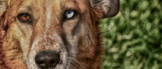
What is an eyesore on a dog?
An eyesore usually appears in older dogs, but can also be diagnosed in puppies, characterized by all the same signs as in young dogs. In general, this term refers to a scar formation on the cornea, which from a distance resembles a dense thick milky or gray spot with translucent edges.
In addition to scar areas, there are other pathologies that are mistakenly mistaken for a thorn:
- corneal ulcer (the surface is affected almost completely, and the deeper the impact, the less painful it is);
- various forms of keratitis (inflammatory process on the surface of the cornea);
- dystrophies of the organs of vision caused by disturbances in the metabolism of the organ;
- degenerative processes characterized by the deposition of calcium salts and cholesterol;
- endothelial edema (“blue eye”, sometimes appearing after a dog’s vaccination);
- accumulation of protein and pus in the anterior chamber of the eyeball, provoked by the inflammatory process in them;
- congenital luxation of the lens (typical of certain breeds of dogs, for example, minibull and many varieties of terriers);
- cataract and glaucoma.
Did you know? Dogs are much better than people at distinguishing all shades of gray, while blue-green, bluish or light green colors are perceived by them as white.
Animal vision cannot be called color in the usual sense of the word, but it is not black and white either.
Description and reasons
The second name for cataract is leukoma. This term refers to a scar or scar that forms on the cornea of the eye after an injury or illness. A dog's eyesore appears for various reasons:
- age-related changes (most often the cataract appears in older dogs);
- injuries leading to the formation of ulcers on the cornea;
- congenital pathologies (with some of them the eyelids do not close completely, which means the cornea constantly dries out and is easily injured);
- inversion of the eyelids (eyelashes constantly irritate, scratch and injure the cornea of the eye);
- exposure to the eyes of smoke, chemicals , etc., which can also lead to poisoning of the animal;
- unsuccessful operation;
- diseases leading to inflammation and damage to eye tissue (glaucoma, keratitis, conjunctivitis, etc.);
- autoimmune diseases;
- neoplasms in the eyeball.
There are several types of leukoma:
- peripheral (only the rim of the eyeball becomes cloudy);
- central (the center of the eye turns white);
- total (the entire surface of the cornea suffers).
In all cases, there is a high risk that the animal will never be able to see in the affected eye.
Why does it appear
There may be several reasons for the appearance of an eyesore in a domestic dog, however, regardless of the nature of the origin of this disorder, the animal still needs treatment.
The most common reasons for the development of cloudiness in the eye may be:
- injury to the cornea of the organ;
- inflammatory processes;
- complications of other ophthalmological diseases (a cloudy cloud can be a consequence of aseptic processes, keratitis, ulcers);
- the result of improper treatment of purulent inflammation;
- the use of potent medications or contact with the eye of chemically active substances (salts of heavy metals, such as lead, zinc or silver, have a negative effect in this regard);
- congenital pathologies, in particular those associated with breed predisposition.
The nature of the specific cause of the appearance of a cataract largely influences the strength of the manifestation of the disease, and therefore the choice of a method to solve the problem.
For what reasons can this formation appear?
Dogs' eyes become cloudy for a number of reasons:
- congenital pathology (extremely rare);
- due to injury;
- inflammatory process;
- consequence after an illness;
- the result of improper treatment of purulent inflammation.
Spots and clouds appear most often due to improper home treatment. A cataract is a consequence of purulent inflammation. There are also medications that contain lead, silver and zinc - after their use, small stains appear and they cannot be treated.
Attention! It cannot be ruled out that some breeds are predisposed to cloudy eyes. These include pugs, Pekingese, Labradors, and bulldogs.
What are the symptoms of leukoma?
The appearance of a spot on a dog’s eye largely depends on the root cause of its appearance. Thus, a thorn after keratitis is characterized by clouding and whitening of the cornea, which, in the superficial form of the disease, returns to normal within 1–3 days. If the disease has affected the deep layers of the eyeball, the cloudy pupil and dots on it persist much longer, and the dog experiences discomfort due to a sharp decrease in vision: the animal becomes restless, looks confused, and its gait seems awkward and uncertain.
Important! Do not try to remove plaque from the surface of your pet's eyeball yourself. Additional impact on the cornea will only worsen the situation, causing irritation and the development of an inflammatory process.
The pet may avoid bright light, and the veil on the surface of the cornea often becomes blue or yellowish, although it may well simply turn white over time. A white spot in the eye often occupies both the central part of the eyeball (next to the pupil) and is localized closer to the edge of the iris, but this area will become cloudy in any case.
It is the appearance of such a characteristic spot of cloudy or white color that is considered the main sign of a cataract, which can be supplemented by other manifestations:
- cloudy discharge from the eyes, sometimes mixed with pus;
- photophobia;
- corneal roughness;
- protrusion of the white spot outward;
- the appearance of redness as a result of burst capillaries (the dog constantly rubs its head with its paw, trying to remove the film lining the eye, thereby only increasing irritation).
Causes
The treatment and subsequent prognosis for leukoma depend on the causes of the appearance of the cataract.
Most often, the formation of a cataract is associated with:
- with a genetic predisposition to eye diseases;
- with severe pathologies (for example, diabetes mellitus);
- with mechanical damage;
- with poor nutrition and poor living conditions;
- with the presence of infections and inflammations.
Among the most popular causes are the following pathological conditions.
Keratitis
This is an inflammatory process in the cornea, as a result of which vision is reduced or completely lost. Keratitis can be caused by hepatitis, poisoning, or severe intoxication of the animal. Home treatment and therapy with folk remedies is impossible.
Erosion (corneal ulcer)
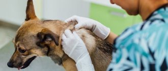
The appearance of a cataract depends on the width and depth of the defect. Ulcers occur with burns, injuries, viral and bacterial infections. As a result of an allergic reaction, chlamydia. You can determine the symptoms of erosions visually, independently, by sufficiently retracting the lower eyelid. If there are defects, the dog gets worried, rubs its eye with its paw (which provokes additional injury), the cornea and white have a reddish tint.
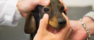
How is the diagnosis carried out?
Ophthalmological diagnosis for vision problems in dogs is based on several step-by-step steps. First of all, the veterinarian conducts a thorough examination of the eyes and determines the strength of the “growth” of the cataract, for which an ophthalmoscope (a device for determining the passage of light) is usually used. To verify the presence of a problem and determine its nature, a cytological examination of a smear from the cornea of the affected organ is performed.
You will be interested to know what patella is in dogs, signs and treatment of the disease.
Considering that the use of this technique is quite traumatic for the organs of vision, it is used only in extreme cases, for example, if the doctor suspects a bacterial infection is joining the inflammatory process. To establish the exact cause of the appearance of an eyesore, other types of instrumental and laboratory tests would be appropriate.
The list of common actions includes:
- collecting fluid from the internal cavity of the eye for a broader study for the presence of pathogenic microflora;
- serological blood test for the presence of fungi;
- diagnosis and assessment of the condition of the fundus;
- electroretinography;
- Ultrasound of the eyeball (available only in large veterinary clinics due to the complexity of the procedure).
An integrated approach to the issue of diagnosing cataracts ultimately allows you to select the most effective methods for solving the problem, thanks to which the dog will preserve its vision and will not experience discomfort.
Diagnostics
Veterinarians most often resort to the following diagnostic methods:
- cytology (scraping from the cornea of the damaged eye);
- analysis for the presence of herpes, since this pathology contributes to the appearance of a cataract;
- swab from the cornea of the eye;
- intraocular fluid sample for analysis;
- serology (if a fungal infection is suspected);
- X-ray of the retina;
- Ultrasound (necessary in advanced cases, when a cloudy film has completely covered the surface of the eye).
Based on the results of tests and other studies, it will become clear what caused the appearance of leukoma (infectious disease, injury, surgery, etc.). And depending on the cause, the specialist will prescribe treatment.
At the early stage of the disease, veterinarians, as a rule, recommend drug treatment: antibacterial ointments (Tetracycline, Korneregel, Oftagel, etc.) and eye drops (Dexamethasone, Oftan, etc.). But in advanced cases, surgical removal of the leukoma, and sometimes the eyeball itself, is indicated.
Treatment of a cataract in a dog
Depending on the complexity of the case and the presence of concomitant ophthalmological ailments (the surface can become cloudy for a variety of reasons), the doctor will prescribe the most appropriate method of treating leukoma: using only medications or through surgery to remove the affected area of the eye. Each of these cases has its own characteristics that are worth knowing about in order to quickly get rid of cloudiness.
Did you know? According to statistics, dogs of small and medium breeds live longer than large animals. For example, pets weighing about 9 kg live about 11 years, while their relatives weighing more than 40 kg live on average to 8–10 years.
With the help of drugs
Drug therapy is always effective only in the first stages of the formation of a cataract, while the dog does not yet experience serious discomfort from its appearance.
The conservative treatment regimen in this case will include the following important actions:
- Removing secretions accumulated in the eye using cotton wool soaked in peroxide (move the sponge in the direction from the outer corner to the inner).
- Place the ointment in the conjunctival sac (drugs containing an antibiotic will be effective).
- Rinsing the damaged eye with furatsilin solution to prevent inflammatory processes.
- Injection of vitreous humor for effective resorption of the cataract.
Among antibiotic-based ointments, the ophthalmic tetracycline and chloramphenicol varieties are best suited for use at home. Oftan and Lidaza drops have antioxidant properties, and to relieve inflammation from the affected area, medications based on corticosteroid substances, for example, hydrocortisone ointment or suspension, would be appropriate. Of course, no one can cure animals in one day, but with patience, the owner can achieve what he wants.
Important! You can instill eye drops or put ointment into the conjunctival sac only after cleaning the eye with antiseptic agents. If purulent discharge is not removed, it will not be possible to achieve a positive treatment effect.
When is surgery necessary?
Leukoma in dogs can also be treated surgically, but it is only used in very serious and advanced cases, when the stain has spread throughout the entire eye and damaged most of the cornea. During the operation, the doctor removes the affected area of the eyeball, sometimes replacing it with an artificial membrane (relevant for those cases where the destructive process has affected the deep layers of the cornea). Before performing surgery, it is important to thoroughly examine your pet, paying special attention to its cardiac function. Older dogs with a large number of chronic ailments are much more difficult to tolerate surgery, so before doing it, it is advisable to undergo at least an ECG and a general blood test.
Is it possible to use folk remedies?
Folk remedies often prove effective in a variety of cases, so it is not surprising that certain methods of alternative medicine have found their application in the case of a cloudy spot in the eye. However, in order to quickly eliminate the existing disorder, it is better to use such recipes in combination with classical medications, especially if we are talking about the rapid progression of the disorder.
The most successful folk medicines will be the following:
- A mixture of half a teaspoon of honey and 100 ml of warm boiled water. After combining the ingredients, the finished solution is applied to the cornea 1-2 drops 2-3 times a day.
- A decoction of chamomile or calendula is used to wash the affected eye before performing basic therapeutic measures.
- A mixture of celandine and propolis (the juice of the plant is combined with a solution of propolis in a ratio of 1:3, after which 2 drops are dropped into the eyes every 2 days for a week).
- Drops from fir resin (drip 1 drop once a day).
- Rinsing the affected organs with boiled honey (1 teaspoon is stirred in a glass of warm water and heated in a water bath). After cooling, the finished substance is dripped into the conjunctival sac 2-3 times a day.
- Powdered sugar is another “medicine” of traditional medicine. It is blown into the animal’s eyes, achieving the natural process of lacrimation.
Sometimes onion pulp is used to remove a cataract, but given its aggressive effect on the mucous membrane, you should think twice before treating it, risking your dog’s vision.
Find out the causes of hearing loss in dogs, treatment of deafness.
Progress of treatment: medications and methods
Therapy for cataracts is aimed at eliminating the root cause of the development of leukoma and relieving the inflammatory process. A timely visit to the clinic gives you a chance to restore your vision in full.
Glucose (amp.)+Levomycetin (drops)+vitamin drops
Wash your eyes with Miramistin and warm water. Apply 1-2 drops of Levomycetin after 15-20 minutes. 1-2 drops of glucose, after 10 minutes. – vitamin drops. The course of treatment is until the thorn completely disappears, procedures are carried out 5-6 times a day.
Actovegin + Tetracycline ointment + Taufon
Rinse the eye, apply Tetracycline ointment in the morning, half an hour later - 1-2 drops of Actovegin. Taufon (absorbable sore) is used 2-3 times a day. At night, Actovegin is first dripped, then Tetracycline ointment. The course is until the eye condition improves.
Prevention measures
Preventive measures to prevent the appearance of cataracts are largely based on general preventive recommendations:
- preventing mechanical injuries to the dog’s visual organs (try not to walk your pet in places with a lot of thorny plants and control its interaction with other animals);
- regularly examine your pet, focusing on squinting of the eyes, photophobia and spatial disorientation, which may indicate deterioration of the animal’s vision;
- undergo a veterinary examination at least two or three times a year, promptly eliminating any problems with the dog’s health;
- comply with the requirements of sanitary and hygienic standards for keeping your pet, regularly cleaning its resting place and washing all bowls before the next dispensing of food;
- promptly remove accumulated discharge from the eyes, especially in dogs of brachycephalic breeds with a flatter muzzle and bulging eyes (such animals often have copious discharge from the conjunctival sac).
To care for your pet's face, you can use gauze pads soaked in peroxide, but special antiseptic preparations sold in pet stores are no less relevant. It is better not to use alcohol-based compounds so that their vapors do not further damage the iris of the eye, causing severe irritation.
Disease prevention
You need to know how to cure an animal's eyesore. But also be able to figure out how to prevent the occurrence of diseases. To do this, you will need to perform certain preventative measures:
- Constantly monitor the condition of your pet. Examine your eyes frequently. If you notice a white spot, you should contact your veterinarian.
- Carry out vaccinations on schedule, which will prevent possible disease.
- Strictly follow the recommendations for caring for the dog, create appropriate conditions, provide a balanced diet, and frequent walks.
- For preventative purposes, visit a veterinarian. Thanks to this approach, the doctor will be able to promptly recognize the signs of any disease and take all measures to eliminate it immediately.
Any pathologies are treated without delay. If you take a responsible approach to caring for your pet, you can avoid many problems and not endanger the animal’s health.
What to do at home?
Home treatment is best combined with professional veterinary treatment. It is prohibited to select medications on your own, since the dog owner cannot know the causes of such a pathology.
It is important to constantly monitor your dog's eyes to spot signs of deterioration. The following deteriorations may be observed:
- severe redness;
- change in the nature of discharge from the eyes;
- strabismus;
- partial loss of vision.
If your dog has completely lost his vision, you need to try to minimize stress. It is important that the animal does not scratch or injure its eyes. In some cases, it is better to wear a special collar or cone.
It is prohibited to use any medications or folk remedies without consulting a veterinarian.
Treatment of keratitis in pugs
It is believed that there are no ways to radically eliminate this disease. Overgrown pigmentation is difficult to respond to both conservative and surgical techniques.
In the treatment of pugs, both approaches are combined - a surgical scalpel and exposure to drugs . Local therapy is designed to stop the inflammatory process, often associated with pain. The four-legged patient is prescribed:
- cyclosporine;
- tacrolimus;
- corticosteroid drugs.
The first two drugs are equally effective, and the choice of one of them depends on the indications/contraindications of a particular dog. For pigmentary keratitis, the doctor usually prescribes corticosteroids, most often dexamethasone or prednisolone (in the form of direct injections into the conjunctiva of the eye).
But these medications have a significant disadvantage - they suppress the immune system, which begins to “miss” eye infections. Still, steroids are indispensable in the treatment of brachycephalic breeds prone to corneal ulcers.
Important! Occasionally, beta irradiation is used, which is justified in case of a large area of melanotic cataract and significant loss of vision. Removing pigmentation with a scalpel is considered ineffective, since after some time the spot appears again.
Eyelid surgery also helps to slow down the developing pigmentary keratitis, after which the inner corner of the lower eyelid is not able to cover the cornea. The doctor himself chooses the type of surgical intervention:
- Hotz-Celsus method;
- dissection of the medial canthal ligament;
- medial canthoplasty.
Pug owners pass on a recipe to each other, thanks to which the growth of pigment stops, and the thorn noticeably decreases in size.
First, the contents of the lidase ampoule are diluted with water for injection (5 ml). Next, dexamethasone (1 ml) and sea buckthorn oil (1 ml) are added to 1 ml of the resulting solution. Before instillation, done three times a day, the solution is shaken vigorously. Pugs tolerate the procedure calmly, as the liquid does not burn or sting.
After a course of instillation (about a month), take a week's break, since dexamethasone is a strong hormonal agent.
Return to content
Treatment
The most common cause of the disease is a bacterial infection. Having taken a smear and identified the pathogen, the doctor will select an antibiotic and draw up a treatment regimen. Medicines are used in the initial stages of the disease and after surgery. There are standard treatment regimens:
- Removal of exudate. To put it simply, you need to wash your dog’s eyes regularly. You can take regular boiled water or chamomile decoction, but it is better to use miramistin or furatsilin for these needs.
- Placing tetracycline ointment under the eyelid. This is the drug most often prescribed by veterinarians.
- Catarrhal lesions are treated with ointment or solution of chloramphenicol, as well as a suspension of hydrocartisone.
- To prevent the spread of infection, Tobrex drops are instilled into the eye.
- If degenerative changes occur in the eye, you cannot cope with it on your own. You will need the help of a veterinarian who will prescribe injections.
Attention! Surgical intervention is carried out if medication methods do not allow the process of pathological changes to be preserved.
Puppies
An eyesore is quite rare in puppies; it is a pathology of adult dogs. Treatment of a fragile body has its own characteristics and contraindications. Puppies can often injure their eyes with their claws, so a special cone or collar may be required.
Levomecithin solution can be used to treat the disease in a puppy. In this case, the dosage is reduced.
Tetracycline ointment should be used with caution. This safe antibiotic can cause a number of side effects in case of overdose:
- vomit;
- diarrhea;
- loss of appetite;
- lethargy.
In rare cases, puppies may experience an allergic reaction.