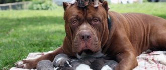
Keratitis is an eye disease in dogs that damages the cornea. The pathology is very common among domestic animals; both purebred dogs and mongrels are susceptible to it.
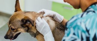
At the first symptoms of the disease, you must contact a veterinarian; you cannot start treatment on your own, since keratitis can cause serious complications.
What is this?
Keratitis is an inflammatory disease of the cornea caused by infection or injury. The disease can lead to significant vision impairment and even complete blindness. Possible complications of the disease:
- glaucoma;
- thorn;
- corneal perforation;
- cataract.
All pets, including dogs, can get keratitis. With a timely diagnosis, treatment of the disease takes from 1 to 2 months.
Treatment and prognosis
The inflammatory process in the cornea of the eye in the initial stages responds quite well to therapy. In the absence of timely treatment, keratitis can move from the acute stage to the chronic stage. It is extremely important to promptly notice characteristic changes in the cornea and begin treatment.
The treatment tactics for keratitis directly depend on the factors that provoked the inflammation and the type of pathology. The main task of a veterinary ophthalmologist is to eliminate the factor that provoked the development of the inflammatory process. Only after this, symptomatic treatment is developed. This tactic allows you to quickly achieve positive results, saving the animal from suffering.
Mandatory therapeutic measures for diagnosed keratitis are daily rinsing of the cornea with antiseptic solutions.
Depending on the type of inflammation and the individual characteristics of the body, the doctor develops a separate treatment regimen. For superficial types of inflammation, the doctor prescribes chloramphenicol drops and hydrocortisone injections. If the form of keratitis is purulent in nature, it is necessary to prescribe a course of broad-spectrum antibiotics. In addition, when treating purulent keratitis, special ointments with antimicrobial components are prescribed.
Treatment of keratitis caused by allergies includes eliminating the allergen and prescribing a special diet. It is important to prescribe histamine blockers. Therapy for pigmentary keratitis in dogs also requires eliminating the underlying factor that caused the inflammatory process. Due to the fact that the pigmentary type of keratitis is more often observed in dog breeds with brachycephalic syndrome, the doctor may recommend surgery to correct the palpebral fissure.
In the case of advanced forms of pathology, the animal is prescribed specific procedures and techniques, including tissue therapy. To quickly resolve the resulting areas of scarring, various ointments are used.
The general course of therapy for keratitis diagnosed in a dog ranges from several weeks to 2 months. This depends on the degree of neglect of the pathological process.
The prognosis in the initial stages of the disease is favorable. If keratitis is in an advanced form, the prognosis is cautious. Since the animal may completely lose vision.
Kinds
There are several types of the disease in dogs. They differ in the mechanism of keratitis initiation, its development and manifestations.
Ulcerative
It manifests itself as clouding of the surface of the eye and occurs due to ulcerative damage to the visual organ.
In advanced cases, open ulcers form on the cornea.
Pigmentary
Keratosis pigmentosa (melanosis of the cornea) can appear for the following reasons - dry eye syndrome, trauma, irritation, inflammation, excessive folds on the face (in some breeds), brachycephalic ocular syndrome, inversion of the medial corner of the eyelids, developmental defects during intrauterine formation.
With this type of keratitis, the eye membrane darkens, becomes cloudy or even blackens.
Superficial purulent
Appears as a result of injury. Characterized by photophobia, purulent discharge, lacrimation.
Vascular
Appears due to the proliferation of blood vessels and their ingrowth into the cornea. The color of the eyeball becomes red, the apple itself becomes lumpy.
Parenchymatous
The disease occurs either against the background of microbial infections - toxoplamosis and canine distemper, or due to injury.
It is characterized by swelling of the cornea, tearing, and the appearance of dots and spots. Often accompanied by other diseases - conjunctivitis and iritis (inflammation of the iris).
Spot
A rather rare and little-studied type of keratitis. It is characterized by the appearance of pearlescent cloudy spots on the eye, which do not affect vision in any way and do not cause any discomfort to the animal.
Phlyctenulous
This type of illness manifests itself against the background of poisoning with toxic substances or allergies. Accompanied by the appearance of grayish-white bubbles on the cornea.
In advanced cases, the bubbles may burst, turning the membrane of the eye reddish-gray. At the same time, the eyelid thickens and characteristic bumps form on it.
Phlyctenulous type
This form of the disease develops as a result of toxic poisoning or against the background of an allergic reaction. Experts say that shepherd dogs and collies are more predisposed to such keratitis.
A characteristic sign of phlyctenulous keratitis is the appearance of large gray-white bubbles on the eye cornea. If left untreated, these blisters will coalesce and then burst, causing the cornea to become red. This species is also characterized by tuberosity and thickening of the third eyelid.
[custom_ads_shortcode1]
Causes
The main factors that provoke keratitis are:
- foreign body getting into the eye;
- injury;
- inflammation;
- burn;
- diabetes;
- avitaminosis;
- allergy;
- hepatitis;
- weakened immune system;
- heredity;
- failure of the endocrine system;
- canine distemper;
- enteritis.
Symptoms
Symptoms indicating keratitis, despite the large number of varieties of this disease, are almost always the same. The main signs of this disease are:
- frequent blinking;
- rapid development of photophobia;
- excessive tearfulness;
- discoloration and clouding of the eye membrane;
- hypermia of the white membrane of the eye;
- the appearance of edema;
- yellow, gray and white spots on the cornea;
- redness;
- roughness of the eyeball;
- dark smudges, mainly in the inner corners of the eyes.
How long do they last?
Keratitis is characterized by a rather rapid development of symptoms. After just 2 hours, the eyeball becomes cloudy and the animal’s behavior changes. The dog hides from the light, becomes nervous, irritable, lethargic.
If you experience any symptoms of the disease, you should immediately consult a veterinarian.
How is the treatment carried out?
Therapy for keratitis in a dog must be carried out under veterinary supervision: the disease requires a professional, competent approach. The first step is to get rid of the dangerous inflammatory process occurring in the cornea. To do this, rinsing the eyes with disinfecting solutions is usually used:
- rivanol;
- furatsilin composition;
- boric acid.
It is important to establish whether viruses or bacteria caused keratitis in this case. The prescribed therapy will depend on this: for bacterial pathology, antibiotics are used, for viral pathology, immunoglobulins and antivirals are used.
If superficial catarrhal keratitis is diagnosed, instillation of a special sodium sulfacyl solution (20%) is prescribed. In addition, injections with the hormones hydrocortisone, ointments with antibiotic components, and the introduction of novocaine are necessary. For superficial types of the disease, dogs are injected with vitreous humor - a course of treatment of three to four weeks. In addition, injections of placental suspension are also indicated.
Treatment of keratitis in dogs of a phlyctenulous nature is carried out by instillation of vitamin solutions and pyridoxine injections. With pathology of this type, the dog is recommended to eat a diet without salt and with a minimum content of carbohydrates.
Allergic inflammation is treated with appropriate antihistamines. The main difficulty here is identifying the allergen - it is important to exclude this substance from the dog’s diet or treatment as early as possible. Diseases of a purulent nature are necessarily treated with antibiotics, and steroid ointments are also used.
If the pathology is caused by congenital entropion of the eyelid, then this defect cannot be corrected by anything other than surgical intervention. Surgery to correct an entropion of the eyelid takes a matter of minutes and entails a short recovery period. If the operation is not performed, the dog may suffer from keratitis all its life.
If the dog’s disease is already in an advanced stage, tissue therapy is used. In severe cases of the disease, the cornea becomes scarred, and yellow mercury ointment or lidase is used to resolve the scars. When scars cannot be reabsorbed, surgical intervention - keratectomy - may be prescribed.
The average course of treatment for keratitis in a dog lasts about one to two months. With timely initiation of treatment and competent, professional therapy, the outcome is usually favorable.
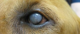
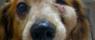
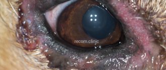
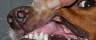
Photo
In the photo below you can see various manifestations of keratitis in dogs.
How dangerous is the disease?
If you do not begin timely treatment of keratitis in dogs, the disease will lead to unpleasant consequences, the safest of which is the ingrowth of inflamed vessels into the cornea, after which it will become lumpy.
More dangerous complications from untreated keratitis are glaucoma, perforation of the ocular cornea, cataracts, partial loss of vision, and blindness. If your pet has noticed the first signs of the disease, it is necessary to take it to a veterinary clinic for examination as soon as possible to make a diagnosis and prescribe treatment.
Experts have identified several types of keratitis, which differ in some symptoms and nature of occurrence. When examining the dog, the veterinarian will determine the type of disease and prescribe appropriate treatment.
Diagnostics
You can identify eye disease yourself using the above signs, but only a doctor can make an accurate diagnosis. For this purpose, the clinic conducts the following studies:
- visual inspection;
- Schirmer test (examination of tear fluid);
- analysis of the eye condition after fluorescein instillation;
- establishing the sensitivity of the eye shell.
During diagnosis, anesthesia is required, which helps the animal undergo all procedures absolutely painlessly. Based on the examination results, the veterinarian makes a diagnosis and prescribes appropriate treatment.
Breed predisposition
Some dog breeds have a genetic predisposition to keratitis, due to the structure of the head and the presence of excess skin folds. The following dog breeds are at risk:
- Pekingese;
- pugs;
- French bulldogs;
- boxers.
Also, collies and shepherd dogs most often suffer from phlyctenulous keratitis.
In pregnant women, lactating women and puppies
In pregnant, lactating dogs and puppies, the disease manifests itself in the same way as in all other animals. Diagnosis of the disease is also no different.
The only difference is the specially selected treatment for this category. It should be borne in mind that this category has weaker immunity, so you need to be more careful about your pet’s health. Veterinarians recommend during this period:
- monitor the safety of the animal;
- provide your pet with a balanced diet;
- walk regularly;
- avoid vitamin deficiency.
Complications after surgery
If treatment of keratitis in dogs at home is not started on time or was carried out incorrectly, the disease can be complicated by conjunctivitis or more severe pathologies: cataracts, glaucoma, inflammation of the iris. Inflammation of the old form leads to the germination of tissue fibers into the cornea, as a result of which an incurable thorn appears on the dog’s eye. Because of this seemingly initially quite harmless disease, a four-legged pet may eventually lose its sight for life.
Purulent keratitis sometimes leads to severe inflammatory processes in the eyeball, and as a result, to the complete removal of this organ. No owner would want such a fate for a pet.
To minimize the likelihood of your dog getting sick, you should carefully monitor the cleanliness and hygiene of your pet. The animal's eyes require regular washing - with a solution of boric acid or a bactericidal decoction of chamomile. In addition, it is important to vaccinate your dog against adenovirus and herpes infection on time.
Pay attention to the animal's injuries. In case of conjunctivitis or other processes occurring in the organs of vision, immediately contact a veterinarian. Monitor your pet's diet. The dog must receive all the minerals and vitamins its body needs: it is important that beneficial substances are included in the dog’s daily menu. This measure will help prevent dangerous vitamin deficiency and strengthen the immune system.
It is important to keep the dog away from dusty places: roads, attics, basements. In such places, a dog can easily pick up dirt and get an infection in the eyes. Brush your pet's fur with caution: small fibers may well get into the eyes and then cause inflammation, including the salivary glands. If a dog has a congenital entropion, the condition of its eyes should be monitored especially carefully.
If there are alarming symptoms, even mild ones: red eyes, irritation, not to mention severe cases, such as injuries, immediately take your dog to see a veterinarian. The sooner professional help is provided, the better for the pet and the peace of mind for its owner.
So, we have learned what this disease is and how this pathology can be dealt with. As you can see, despite all the seeming harmlessness, advanced keratitis can lead to sad consequences in the form of complete blindness, so measures to treat this disease must be taken urgently and adequately. Be sure to take your dog to the veterinarian: professional diagnosis and treatment will help you cope with the disease quickly and eliminate dangerous consequences.
Keratitis in dogs occurs due to injuries, infectious and parasitic diseases, as a result of allergies, and vitamin deficiencies. Inflammation of the cornea has a wide variety of forms - from superficial to phlyctenulous. Most often, purulent and ulcerative keratitis is observed in dogs. For diagnosis, special ophthalmological methods and tests are used.
Treatment includes local use of anti-inflammatory drops (for example, antibacterial drugs Levomycetin, Tsiprovet, Tobrex, Maxidin) and ointments (Levomycolic), allergy medications (Tavegil, Suprastin) may be prescribed. According to indications, the animal can undergo surgery - keratectomy.
Prevention
It is easier to prevent any disease than to treat it. To avoid keratitis in your dog, it is necessary to follow preventive measures. Rules to follow:
- timely vaccination against herpes and adenoviruses;
- avoid eye injuries;
- for any symptoms of the disease, you must immediately contact a veterinarian;
- provide your pet with a balanced diet;
- properly care for your dog (regularly comb and trim the hair around the eyes);
- Periodically wash your eyes with chamomile decoction or boric acid.
By adhering to all of the above rules, you can avoid the occurrence of keratitis. At the first symptoms of the disease, you need to consult a specialist, since the earlier the problem is identified, the faster the treatment will help.
Treatment of corneal ulcers in dogs
The disease involves several stages of treatment: therapeutic, medicinal and surgical. The use of conservative therapy is justified when the disease has just begun to develop. The veterinarian recommends taking topical antibacterial agents that are dripped into the damaged and healthy eye. For treatment, the active substances of Tobrex, Iris (drops) and Tetracycline, Optimmune (ointment) are prescribed.
Owners should remember that treatment is carried out under the strict supervision of a doctor and according to the prescribed regimen. It is prohibited to change or adjust the dose of medications on your own. If you are unable to apply eye drops or ointment yourself, you should contact your veterinarian.
Why do the drops need to be used so often (every 240 minutes)? The outer membrane must be kept as hydrated as possible, and this occurs by bringing it into contact with tears. This leads to premature washing off of the active substances and further spread of ulcers. The use of drops according to the prescribed scheme accelerates the achievement of clinical results.
However, this rule is incorrect to apply to pharmacological agents that contain a plant alkaloid. Drops or ointments are often prescribed for the treatment of eye diseases; they quickly reduce itching and spasms. When the plant alkaloid gets into the eyes, the pupils of the eyes sharply expand, and the animal practically goes blind, so it can be used 2 times in 24 hours.
Ulcers should not be treated with steroid hormones, which are successfully used in the treatment of other inflammations. Active substances, when they enter the affected membrane, begin to have the opposite effect, and ulcers begin to develop at double speed.
In advanced forms of the disease, veterinarians insist on surgical intervention. Depending on the type and technique of execution, there are several types of operations:
- Cutting the membrane of the eye. The operation is simple; during it, the outer shell of the eye is cut and smoothed out. The dog receives a dose of local anesthetic, and the incision site is further processed. In 99% of cases, after the interventions, visual activity is completely restored;
- Removal of part of the cornea of the eye. Surgery for a corneal ulcer in a dog is complex and traumatic; a laser beam is used to perform it. It eliminates all injured areas of the outer shell. The dog falls asleep for the duration of the treatment, but after the operation, vision returns in 99 percent of cases. The operation is performed by a veterinarian, which guarantees results in 100% of cases.
After the intervention, the operated eye is covered with optical glass, preventing possible relapses.
Surgical treatment of corneal ulcers is often performed using a trimming technique. To do this, the zone is removed from the outer shell of the affected eye a few millimeters from its base. Elimination of the zone is carried out without deformation of the nutrient vessels and should be proportional to the healthy area.
The place for attaching the zone on the outer shell is prepared in accordance with the norms of ophthalmic veterinary medicine. The area is attached to the cornea with material for making sutures used for ophthalmological effects. After the procedure is completed, the lesion site is temporarily isolated for further regeneration of tissues and blood vessels. Drug treatment is prescribed for two weeks, and the sutures are removed under local anesthesia.
Before surgery, a selection of tests (blood, feces and urine) and cardiac therapy (heartbeat reflection and electrocardiography) are carried out, which should confirm the absence of contraindications for the administration of anesthesia or general anesthetic. After surgery, therapy should be continued in the form of effective and restorative treatment.
Price
Treatment of wounds on the outer eye shell must be started immediately, since it can significantly increase in size, filling the entire cavity, this will significantly affect the cost of surgery. The cost of the operation can range from 1,500 to 20 thousand rubles. The price does not include anesthesia and all necessary medications for rehabilitation. Also, the price will increase if the eyes are affected by concomitant diseases or the infection affects other areas of the face.
During conservative treatment and after surgery, the owner must provide his dog with complete care and rest, as these are the basic rules for a speedy recovery.
Corneal ulcer is a serious disease. It can lead to various complications, loss of vision and death, so the pathology must be treated in the early stages and follow the doctor’s recommendations.
Diagnostic procedures
To make a correct diagnosis, the veterinarian must conduct the following examinations: An external examination of the eyeball is carried out with a flashlight, a microscope with maximum magnification and a slit lamp. At the same time, the frequency of blinking, the reaction to changes in lighting, and the hypersensitivity of the apple are calculated.
Staining with fluorescent dye. The affected area will appear clearly and glow with a light green hue. Schirmer test, sampling of the ocular coating and staining with other dyes.
Preventive measures
Look at the dog's behavior. Avoid walking in dangerous areas and dry grass, and avoid contact with aggressive dogs, games with which can lead to injury to the eyeball.
Monitor the condition of your dog's eyes. If you find red spots or hemorrhages, tears begin to flow from one or both eyes of the dog, pus is released, if in the morning the dog cannot open its eyes on its own due to the formation of crusts, it is better to immediately contact the clinic. Even if it is a classic inflammation, it is better to eliminate its manifestations at the first stage than to treat later the resulting wounds on the outer shell of the eye.
Follow the vaccination schedule, because most infectious diseases are the root cause of an inflammatory reaction on the eye membrane, which can develop into a wound. If you follow all the rules, your dog will not get sick and will delight you with his cheerful and kind games. If any of the symptoms are detected, you should contact a veterinary clinic.
Currently reading:
- How to cure stomach ulcers in dogs
- Treatment of proptosis for eye loss in dogs
- A warning sign for a dog with cloudy eyes
- Critical vision loss due to cataracts in dogs