Inflammation of the paraanal glands in dogs
Inflammation of the paraanal glands in dogs.
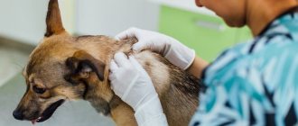
Treatment. Inflammation of the anal (paraanal) glands in a domestic dog is a fairly common problem, which appears in most cases due to the fault of the owner. Let's figure out what these glands are, what they are needed for and how to prevent them from getting sick.
A little biology Anal glands, also called paraanal sinuses (if translated literally from Latin, it will sound like near the anus) are a paired secretory organ in the form of small muscular sacs located on the sides of the anus. The filled glands are shaped like beans (beans) of medium size, their ducts exit at the entrance to the anus, and not into the rectum, as you can read on some sites.
Why does a dog need sinuses? The main function of the glands is to secrete a secretion that has a specific and unique smell, unique like the smell of human sweat, that is, unique only to a given individual. During the act of defecation, feces passing through the rectum squeeze out part of the secretion, and it ends up in the feces - thus, the dog marks its territory. Also, you probably noticed how your pet meets familiar people, while actively wagging its tail, at this time the smell spreads from the anal glands, the dog says - I’m glad to see one of my pack. When two male dogs meet, they sniff each other’s anus, exchanging information, and the paraanal glands take a direct part in such communication. As we can see, the functions of the organ we are interested in are diverse and important for the social life of the dog. The modern dog leads a sedentary, sofa-like lifestyle, does not eat properly, the muscles involved in the contraction of the sinuses have lost their tone, as a result the secretion stagnates, thickens and our pet gets a problem, and we along with it.
Symptoms of inflammation of the anal glands Oddly enough, but most often the symptoms of inflammation of the anal glands are located far from the source of the pathology and manifest themselves as follows: 1. The animal experiences constant itching , bald patches appear in the area of the croup, hind limbs and sides. Note that this is one of the frequent signs of the disease, which is not associated with the glands, but is treated for allergies, unfound mites, lice, lichen, eczema, that is, anything. 2. Protrusion of the anus , which is associated with blockage of the excretory ducts and overflow of the sinuses. 3. Discomfort in the anus , the animal sits on its fifth point and rides on the ground, trying to get rid of the accumulated secretion.
Find the answer
Do you have any problem or question? Enter “Breed” or “Name of the problem” into the form, press Enter and you will find out everything about the issue that interests you.
Fistulas are of origin:
- Congenital;
- Purchased.
Congenital fistulas are rare. They are formed due to improper intrauterine formation of the fetus. The wound can connect organs to the body surface. The case requires surgical experience. Without surgery, the puppy will die.
Acquired fistulas are common. They arise due to injuries, cuts, bites, rectal problems, and infectious complications.
Loading …
By type:
- External;
- Internal.
The pus is drained out through the external openings. Timely treatment can eliminate the problem.
Internal purulent openings are more dangerous. They allow bacteria access to internal organs. Wounds take longer and are more difficult to heal.
Fistulas occur:
- Near the anus;
- On the paw;
- On the stomach;
- On the head.
Near the anus, a fistula occurs if the paraanal gland becomes inflamed. The pain may radiate into the anus. In this place the pet feels severe pain. Pathology worsens the animal's health and behavior. Perianal fistula occurs due to poor nutrition, impaired metabolism, genetic predisposition, and lack of hygiene. Characteristic of some breeds - setters and shepherds. The resulting pathology should not be confused with hemorrhoids or other rectal disease.
Purulent channels can form on the paws. The cause may be a cut, bite, or bone damage. An abscess usually occurs due to a cut. The infection does not allow the abscess to heal - a fistula forms.
A fistula in the abdomen may extend to the abdominal organs. A purulent canal on the abdomen is dangerous. The cause of inflammation is a formation on the mammary gland or skin.
Often purulent canals appear on the animal’s cheeks. This happens due to oral diseases. Inflammation appears on the cheek.
Paraanal glands in dogs: problems and solutions
This behavior is mistakenly taken for helminthiasis. 4. In advanced cases, hair falls out completely at the base of the tail root , and subsequently purulent fistulas open. When an animal tries to empty the glands to no avail, a pyogenic microflora develops in their cavity - pus is formed, which melts nearby tissues and comes out.
Prevention measures As we said at the beginning of the article, the blame for this disease lies with the owner because the glands need to be cleaned regularly. This procedure can be performed independently in several ways.
First method: Let's call it internal. Before the procedure, we put on medical gloves, lubricate the index finger with lubricant (vaseline, baby cream) and insert it into the rectum. We feel for one sinus and pinch it between the index, thumb and middle fingers. That is, the index finger is on the inside, and the other two help it by pressing on the outside through the skin. With the other hand we fix the dog, holding it by the root of the tail. We proceed in the same way with the second sine.
Second method: External, this method is more suitable for small breed dogs. When using it, we also put on a glove, but do not insert a finger into the rectum, but grasp two glands at once, between the index and thumb, and gently squeeze out the secretion. In order to quickly find the sinuses, imagine that the exit from the rectum is the center of the dial, then the glands will be located: one at 16:00, and the other at 20:00. All manipulations must be carried out carefully and gently, trying not to cause additional pain to the animal. If the inflammatory process has gone far and you see that the dog has fistulas, then it is better to seek help from a veterinarian. Conclusion The frequency of cleaning the anal glands depends on several factors: breed, age, lifestyle (including diet). It is recommended to carry out sanitation at least once every 6 months, which will avoid inflammation of the glands. Remember - this manipulation must be carried out regularly if you want to see your pet cheerful and attractive.
CONSULT A VETERINARIAN (812) 607-68-62
Home » Health and care » Diseases » Dog diseases
Treatment of the disease
Experts strongly do not recommend treating dogs at home. The owners can only remove the most painful symptoms, but not the cause of the disease. As a result, a fistula can occur in any other place on the body, causing a lot of inconvenience to the dog.
That is why at the first sign of a sore, the dog should be taken to a veterinary clinic. There he will be examined and prescribed therapy that will eliminate the very root of the disease. For example, if the fistula occurs on the cheek or under the eye, then a diagnosis of the dog’s teeth and possible removal of tartar will be required. After this, the pathology can be eliminated.
If the disease is caused by blockage of the paraanal glands, then the animal can only be cured by removing them. Conservative treatment does not provide any long-term results, and the barking fidget is at risk of relapse at the most inopportune moment. The same applies to fistulas formed in other parts of the body. In this case, surgical intervention is performed according to one scheme: the fistula is completely eliminated, the cavity itself is thoroughly cleaned and only then sutured. The dog is prescribed a course of antibiotic drugs to eliminate the risk of infection.
The choice of medications for the period after surgery depends on the gender, age and current condition of the pet. Your doctor may prescribe painkillers or antihistamines, courses of vitamins, or antipyretic medications to relieve a high fever. Owners are not allowed to treat animals with medications intended for humans. To speed up the healing process, the dog should not be allowed to lick the stitches. A special collar around the neck can prevent this. Sutures should be regularly treated with an antiseptic.
Finally, I would like to say that the fistula protects the dog’s body from the accumulation of excessive amounts of pus in the tissues. However, it is quite problematic to treat, and the pet runs the risk of contracting a dangerous infection through it. Fistulas are most effectively dealt with through surgical intervention. If the pathology is ignored, it will lead to sepsis and possible death of the dog.
Due to pathologies of the rectum, feeding disorders and other reasons, a perianal fistula often occurs in a dog; how to treat such a disease is surgically. Conservative medicine does not have the desired effect, but only allows you to level out the process and reduce symptoms.Fistula in dogs: what to do?
Such a phenomenon as a fistula in medicine is otherwise called a fistula (from the Latin word fistula - tube, channel). This is the result of the formation of a pathological channel between hollow organs, or the focus of the disease and the surface of the dog’s body. Fistula is not a fatal disease if left untreated, but it is painful and causes a lot of anxiety to the animal.
07 October 2013
Author: Lando Anastasia
Usually it looks like a narrow canal lined with epithelium (healed smooth skin) or granulations (loose surface of the wound), constantly secreting pus, mucus, bile, urine, and feces.
What causes it to form
The word "fistula" comes from the verb "to whistle". It means emptiness, a defective hole. Another name for fistula.
A fistula is an opening through which pus comes out. Outwardly it resembles an abscess, but is more dangerous. The fistula acts as a channel from the inflamed area to the surface of the skin. Through it, the component of the abscess comes out. The fistula wound is tube-shaped, with smooth walls, and formed.
Unlike an abscess, a fistula heals more slowly. The open wound constantly leaks pus. Clearing the cavity of pus does not help healing. A fistula is dangerous because pathogenic microbes can enter the body through it. This will further worsen the dog's condition.
There are two types of fistulas in dogs:
- Congenital fistulas. They are formed as a result of incomplete healing of embryonic fissures and ducts during fetal development (cervical, umbilical fistulas). Fistulas of this type are quite rare.
- Acquired fistulas. They most often occur due to inflammatory processes, accompanied by suppuration and breakthrough of pus. They are accompanied by a number of diseases that are often found in dogs.
For example, with osteomyelitis , an inflammatory disease that usually develops in animals as a complication of purulent periodontitis and dental pulpitis . In dogs, the lower jaw is usually affected. Over time, the source of inflammation develops into a fistula, which opens into the oral cavity and is accompanied by purulent discharge. All this causes the dog severe pain. The animal refuses to eat and loses weight. Treatment is carried out by a veterinarian and includes a set of measures (cleaning the fistula canal from pus, prescribing antibiotics). At home, help consists of rinsing the dog's mouth with potassium permanganate.
A fistula can also form due to arthritis - inflammation of the joint. In its purulent form, a fistula is formed around the joint of the animal, releasing pus. At home, you can remove the hair around it and lubricate the skin with iodine. You should definitely contact the clinic.
Causes
Veterinarians distinguish two types of fistulas, each of which has its own characteristic features. This includes congenital and acquired types of pathology. The first includes cases that arise due to disruption of the normal development of the fetus in the womb. They appear in puppies, but, fortunately, they do not occur often. Such a fistula may well connect any internal organ to the surface of the body, creating an extremely life-threatening situation for the pet. Without surgical intervention, the animal will not live even a week.
Fistulas that a dog acquires during its life are much more common. They occur in such vulnerable areas of the body as:
- On the dog's face. The localization of the pathology is under the animal’s eye, and it occurs due to bad teeth. Often the inflammatory process manifests itself through the dog’s cheek. It is an extremely dangerous type of fistula, as it threatens not only the appearance of the pet, but also its vision.
- On the stomach. The fistula can communicate with the internal organs or abdominal cavity of the dog, which makes it easier for viruses and bacteria to enter the body.
- On the neck. The pathology is dangerous because the neck is in close proximity to the brain, and the main blood channels also run through it. Microorganisms that enter the animal’s body will spread throughout the entire body in a short time, leading to serious consequences.
- On the limbs. Most often, the reason for this is various mechanical damage to the paws, for example, while walking the dog got stuck on a rusty nail. An infectious agent enters the wound, causing an abscess that does not heal persistently, forming a through hole.
Postoperative fistula
Postoperative fistulas deserve special attention. Veterinarians have a special name for the pathology caused by this reason, namely, ligature fistula. It owes its appearance to the fact that the surgical intervention on the animal was not performed as needed.
Especially often, the sore appears after sterilization of the pet, if the seam with which the pet was sewn up was not sufficiently processed or became contaminated during the operation. As a result, the tissues around the infected thread begin to become inflamed, and purulent exudate is produced in the cavities of the body. Over time, this will lead to the formation of a fistula. Therefore, carefully monitor whether painful tubercles appear at the site of the sutures, in which, after 3-4 days, holes will appear that secrete pus.
It is important for owners to understand that the dog must be sterilized according to all the canons of veterinary science. It is unacceptable for a non-professional to do this. If the disease manifests itself, then treatment must be started immediately.
Treatment of inflammation of the anus in a dog
The doctor, as a rule, prescribes antibiotic therapy, novocoin blockade, glucocorticosteroids, etc.
Another disease in which, in severe cases, the formation of a fistula is possible is paraanalytitis . This is an inflammation of the pockets at the entrance to the dog's rectum. In this case, fistulas are formed at the entrance to the anus. At home, you can help the animal by massaging the pockets and squeezing out their contents. The procedure is extremely unpleasant and painful. Therefore, during this procedure, the animal should be muzzled to avoid bites. The massage is performed by inserting a finger into the anus. It is also necessary to follow a diet: exclusion of rough food (bones) and raw meat. Include rice porridge with water in your diet. Be sure to contact your veterinarian.
Fistulas can occur after injuries and wounds , especially when foreign bodies get stuck in the tissues and suppuration around them.
Most fistulas are not life-threatening . Their treatment is different and depends on the causes. In most cases, complete healing of the fistula occurs during the treatment process. Fistula tracts lined with epithelium are not prone to healing. They are treated through surgery. A method of excision of the fistula is used (many cuts are made on the surface of the tissue and after that the tissue is fused with the help of various medications).
For fistulas that extend outward, care is important to prevent irritation and infection of the skin around them.
Fistulas are dangerous mainly for homeless animals, which do not receive help when a fistula forms, which leads to blood poisoning. A pet, even if a fistula appears during an illness, will recover with proper care.
All publications -> Section: Other -> Topic: Plastic surgery
Methods of treatment and emergency assistance
The main point in the treatment of a fistula is to eliminate the cause that caused and maintains suppuration. It is necessary to ensure rapid release of the abscess and canal from pus and prevent its further formation. The easiest way would be to rinse with an antiseptic solution, but this does not always bring the desired effect.
The canal may be curved, contain pockets and additional cavities in which pus accumulates. It is difficult to remove exudate from such areas, so it is necessary to resort to surgical intervention - the canal is dissected along its entire length so that there are no obstacles to the outflow of pus. The abscess cavity is examined for the presence of pockets, which are dissected after detection. The wound is thoroughly washed with antiseptics, sulfonamide drugs and antibiotics are placed in it.
Antiseptic ointments and other means that clog the canal should not be used for treatment; this will prevent the drainage of exudate and lead to complications of the disease.
It is important to preserve the channel for the first time so that pus and other contents can freely drain from the wound. Sutures can only be placed on aseptic wound surfaces. If there remains a possibility of developing a purulent process, then a sutured wound will lead to a recurrent abscess. When the purulent focus is eliminated and all pathological tissues are removed, it is necessary to freshen the wound - the granulation and epithelial tissue is dissected and sutures are applied. Otherwise, the edges will not grow together.
Surgery is the only effective method of therapy. In this case, only after complete excision of the fistula tract and pathological cavity is recovery possible. Modern methods using electrocoagulation and freezing with liquid nitrogen do not show adequate effectiveness. But during the operation you need to be careful not to damage the surrounding tissue. Thus, when treating a fistula of the paraanal glands, injuries to the anal sphincter and rectum often occur.
Antiseptics:
- potassium permanganate solution 0.05%;
- furatsilin solution 0.02%;
- hydrogen peroxide 3%;
- migstream;
- ethacridine lactate.
Additional therapy includes a general course of antibiotics - bicilin, tetracycline. Local novocaine blockade of the affected area has a good effect; penicillin or streptomycin is diluted in novocaine. Intravenous infusions of physiological solutions and glucose should relieve intoxication in the body.
Clinical case of surgical treatment of chronic fistulas of the perianal region in a shepherd dog.
Chronic purulent paraproctitis (perianal fistula) is a chronic degenerative ulcerative disease of the perianal sinuses and the soft tissues surrounding this area.
A perianal fistula is also known as a perianal fistula and is a long-term non-healing pathological passage through which purulent discharge from the perianal tissue is released around the anus.
The most common manifestation and complex course of the disease is observed in German shepherds, which are prone to autoimmune skin diseases.
Pathology occurs in dogs regardless of their gender and age.
There is a hereditary predisposition to this disease.
Signs of primary tissue damage are characterized by deep organ necrotic-purulent damage to the skin and soft tissue around the anus with the formation of perianal fistulas and pathological scar-connective tissue.
Spontaneous healing, as a rule, does not occur.
Basically, the disease is asymptomatic for the body. Clinical signs are characterized by local manifestations of the pathological process in the anal area, causing irritation, itching and interest in this area in the animal (licking).
In addition to problems with licking the perianal area, some dogs may exhibit other clinical signs consistent with purulent inflammation. Such signs include increased body temperature, refusal to feed, forced position of the body and tail, which may be accompanied by constipation, pain, diarrhea, and cachexia.
In some animals, with extensive damage to the soft tissues of the perianal area, there may be damage to the sphincter, in the form of the development of rigidity or insufficiency, as a result of which they are not able to adequately close the anal ring, which leads to impaired defecation.
The cause of the development of perianal fistulas has not been fully elucidated. There are many theories that reflect the essence of the pathological process.
Research has been conducted to determine the possible causes of the disease: immunological, bacterial, endocrine and anatomical features of the development of the disease.
When conducting morphological studies, the histological picture of the studied pathological tissues of the perianal zone is characterized by elements of chronic nonspecific inflammation, with the exception of those cases where the cause of the pathological process is neoplastic processes (adenocarcinoma of the hepatoid glands).
Differential diagnosis should include mandatory studies to exclude the neoplastic nature of this disease.
Today, more and more experts agree that the cause of the disease is immunological, and not the anatomical features of this breed of dog, and therefore a specific approach to the treatment of this type of pathology is necessary.
Treatment methods for this type of disease are constantly being improved and updated. The opinions of various authors regarding treatment are divided into the use of conservative therapy and surgical methods.
Some authors propose the combined and staged use of treatment methods, including a combination of conservative and surgical tactics.
Conservative treatment methods.
Drug therapy involves the use of anti-inflammatory and immunosuppressive drugs.
Corticosteroids (prednisolone) are commonly used as an anti-inflammatory and immunosuppressive drug.
Treatment begins with high doses of prednisolone, gradually reducing them to the minimum dose that has a therapeutic effect.
Studies that have been conducted have shown that only one third of dogs respond positively to the use of prednisone, however, complete healing of the defect is practically not observed.
Corticosteroids have a number of side effects such as increased thirst and appetite, polyuria, and negative effects on the endocrine and muscular systems.
Due to the fact that an immune-mediated etiological factor in the development of the disease can be traced, the use of immunosuppressants in the form of cyclosporine A (Sandimmune) and azathioprine (Imuran) is justified and widely used in practice.
Other types of immunosuppressive drugs are also used.
Complications of this type of therapy are processes associated with decreased immunity, such as bacterial, viral and fungal infections.
With long-term use of cyclosporine A, complications such as nephropathy and hepatopathy are observed, which are not as pronounced as in humans.
The use of azathioprine can lead to chronic myelosuppression and anemia.
Surgery.
There are several methods of surgical treatment of perianal fistulas.
The chemical method involves removing the pathological surface or fistulous tract using chemicals, where the wound surface heals by secondary intention.
It has been established that this type of treatment is successful in more than 80% of cases, but 20% of patients experience complications of the technique used in the form of relapses and sphincter insufficiency.
Cryosurgery involves local injection of nitrogen into the fistula tracts. Success rates range from 50-85%. Sphincter insufficiency and stenosis resulting from this operation are minor compared to some other surgical methods.
Surgical excision is a fairly complex operation. It includes the removal of all the affected tissue with a significant part of the adjacent one, however, surgical manipulation is not always possible due to the extensive skin defect and anatomical features. Sphincter insufficiency occurs in 20-60% of cases as a result of damage to the nerves and tissues of the sphincter.
When using laser surgery to solve this problem, positive success reaches 90%.
Amputation of the tail involving the lateral folds of skin up to the second or third caudal vertebra does not objectively solve the problem of this pathology.
Clinical case.
A 6-year-old shepherd dog was admitted with clinical signs of chronic fistulas in the perianal region.
It was found that the duration of the disease is more than 8 months from the onset of the disease. The animal was repeatedly given courses of antibacterial and immunomodulating therapy, corticosteroid drugs and other medications were used. During the entire period of the disease, six surgical interventions were performed to surgically remove pathological tissues and suturing the defect. Unfortunately, the above measures were ineffective, and the size of the defect increased.
Double histological examination of the material indicated a chronic nonspecific inflammatory process.
| Photo 1. Perianal fistulas in a dog after treatment. |
It was decided to start conservative therapy using cyclosporine A (Sandimmune) at a therapeutic dose of 7 mg/kg per day. The response to treatment was insignificant, but the pathological cavity and signs of the inflammatory process clinically decreased ( photo 1
).
Taking into account the history of treatment of this type of disease in this animal, based on the local manifestation, it was decided to carry out surgical intervention in the amount of excision of pathological tissue within healthy tissue and covering the defect with a skin-muscular autograft on a vascular pedicle.
The surgery was performed under general anesthesia, using combined and combined anesthesia. After positioning the animal and preparing the surgical field, the pathological tissues of the perianal area were excised within the healthy tissues using generally accepted methods ( photo 2
).
The second stage of the surgical intervention was the maximum possible suturing of the own tissues of the area of the excised defect to reduce the surface of the surgical wound ( photo 3
).
The third stage was the formation of a musculocutaneous flap from the caudal surface of the right pelvic limb in such a way that the length and width of the musculocutaneous flap corresponded to the size of the soft tissue defect we created ( photo 4
).
| Photo 2. Perianal zone. Pathological tissue was removed. |
| Photo 3. Perianal zone. Suturing the edges of the wound defect. |
The final stage was covering the defect with inversion of the autograft by 180o, followed by suturing the edges of the surgical wound ( photo 5a, b
).
| Photo 4. Borders of the musculocutaneous autograft. |
| Photo 5a. Moving the musculocutaneous autograft 180°. |
In the early postoperative period, local treatment of surgical sutures and a course of antibacterial therapy were recommended.
In the early postoperative period, no signs of inflammation in the surgical area were found.
| Photo 5b. The edges of the wound are sutured. The surgery is completed. |
| Photo 6. Perianal area. 6 months after surgery. |
The act of defecation was painless and preserved. Signs of lameness on the right pelvic limb were noted for 18 days. The stitches were removed after 12 days.
After 6 months, the animal was re-examined, where no signs of an inflammatory process in the perianal area were found. There was a slight deformation of the “anal ring” with normal sphincter tone during rectal examination ( photo 6
).
The animal's condition is satisfactory, no signs of disease were detected.
Conclusion.
The purpose of the presented surgical intervention was to cover the pathological cavity with an autograft on a vascular pedicle; elements of muscle tissue are not only a volumetric natural formation for covering the bottom of the wound, but also a donor of blood circulation.
The obtained result requires further observation and allows the use of autograft techniques of one's own tissue for this type of pathology, as an alternative surgical method of treatment.
Literature
Clinical case of surgical treatment of chronic fistulas of the perianal region in a shepherd dog.
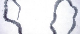
Day MJ, Weaver BMQ Pathology of surgically resected tissue of 305 cases of anal furunculosis in the dog. Journal of the Small Animal Practitioner. 1992 33, 583-589.
2. Dudsberg SC, Spurgeon TL, Liggitt HD Anatomic predisposition to perianal fistulae formation in the German Shepherd Dog. American Journal of Veterinary Research 1985 46, 1468-1472.
3. Harkin KR, Walshaw R., Mullaney TP Association of perianal fistulas in dogs. Journal of the American Animal Hospital Association 1987 23, 95-100.
4. Harkin KR, Walshaw R., Mullaney TP Association of perianal fistula and colitis in the German shepherd dog: response to high-dose prednisone and dietary therapy. J Am Anim Hosp Assoc. 1996 32(6), 515–520.
5. Killingsworth, CR, Walshlaw, R., Dunstan, RW et al. Bacterial population and histologic changes in dogs with perianal fistula. American Journal of Veterinary Research 1988 49, 1736-1741.
6. Misseghers BS, Binnington AG, Mathews KA Clinical observations of the treatment of canine perianal fistulas with topical tacrolimus in 10 dogs. Can Vet J 2000 41, 623-627.
7. Patricelli AJ, Hardie RJ, McAnulty JF Cyclosporine and ketoconazole for the treatment of perianal fistulas in dogs. Journal of the American Veterinary Medical Association 2002 220, 1009-1016.
Author: Vorontsov A.A., Mordas E.M., Guzeeva E.V. (12.2007)
In some cases, dogs may develop inflammation of the anal glands. They are located near the anus and have the characteristic appearance of “bags” located under the skin. Without regular cleaning, the gland ducts often become clogged, a large amount of secretion accumulates in the cavity and inflammatory reactions develop.
Causes of inflammation of the anal glands in dogs
It is believed that in dogs, the PJ is a rudiment that does not perform its original function. Due to non-functionality, secretion stagnates with subsequent inflammatory reactions.
Among the most common causes of inflammation of the pancreas are::
- Inactivity, poor diet. Dogs with diseases of the gastrointestinal tract, as well as those not exposed to regular physical activity, are more likely to have problems with PAD.
- Heredity. Small breed dogs are at increased risk.
- Obesity.
Overweight dogs need to have their glands cleaned regularly to prevent congestion. - Weak immunity. The development of pathogenic microflora can also cause inflammation of the paraanal glands in dogs.
- Traumatization. Cracks or wounds in the anal area are the gateway to infection.
- Eating bones.
- Irregular hygiene or lack thereof.
- Excess protein in the diet.
- Pregnancy, active mating.
Considering the number of possible factors leading to the development of the disease, it is necessary to carefully monitor the animal’s condition by regularly visiting a veterinarian.
Symptoms of pathology
The intensity of the symptoms of the inflammatory process in the paraanal glands directly depends on the degree of damage:
- I degree – the dog is bothered by itching and scratches the anal area. The secretion is secreted in small quantities, its color varies from light transparent to watery-whitish or yellowish.
- II degree – the secretion becomes even smaller, it is characterized by a thick consistency. The symptoms are complemented by other signs - alopecia (hair loss) develops, a rash in the form of pustules and vesicles appears on the skin of the inner thigh area. The pet begins to feel stiffness in the lower limbs.
- III degree - the secretion thickens to a creamy state, it becomes even smaller. In addition, the secretory fluid becomes darker in color. A neurological syndrome appears and the itching intensifies.
- IV degree - the consistency of the secretory fluid of the gland becomes similar to clay - thick, granular, from dark brown to black, paralyzes the pelvic girdle or half of the body.
In addition, a sick pet exhibits a number of other signs:
- The behavior of the animal changes - the dog may become aggressive, habits change.
- Appetite decreases or is completely absent.
- Soreness is felt in the anus, caudal base and between the anus and testicles.
- The skin and coat become damp.
- The pathology is accompanied by hyperkeratosis - hypertrophied compaction of the epidermis.
- Hypesthesia manifests itself - increased sensitivity.
- Pain when touching the skin.
- Excessive pigmentation.
- Erythema of the skin, which manifests itself as redness.
- Rectal-cutaneous fistulas (perianal sinuses) are formed.
- Ulcerations and eroded areas appear on the skin.
- The temperature rises and a febrile state occurs.
- Scratchiness, the pet intensively scratches the painful area.
- The anal area swells noticeably.
- Spasmodic pain in the anus.
- Depression, depressed mood, lethargy, fatigue.
At the very beginning of the disease, you can help your pet yourself by cleaning the paraanal glands. A similar procedure is carried out for preventive purposes and when blockage of the glands becomes a regular occurrence. But how to clean them correctly?
Development mechanism
The process takes place in several stages. First, there is a disruption of the outflow followed by overflow of the glands. Due to this, the secretion thickens, the outflow becomes even worse, and a local inflammatory reaction develops. Subsequently, pathogenic microflora joins, which leads to increased inflammation. At the peak of the disease, the skin festeres, an abscess forms, and the body temperature rises. Over time, the abscess opens, causing a fistula to form, connecting the environment with the cavity of the gland.
If the disease has reached the stage of fistula, then in the future, despite the regularity of cleanings, there is an increased risk of relapse.
Inflammation of the paraanal glands in dogs symptoms
Typical symptoms include:
- Constant itching in the anal area. A characteristic symptom that is often mistaken for allergic phenomena or helminthic infestations, which leads to improper treatment. The dog regularly “rides his butt” and rubs against different surfaces. This causes baldness or rubbing of the anal area and lateral thighs. Attempts at self-cleaning come down to constant licking of the problem area, creating the feeling that the animal is catching non-existent fleas.
- Protrusion of the anus.
Inflammation of the paraanal glands in dogs treatment
The choice of treatment method depends on the severity of the process.
Typically, therapeutic cleansing is used, which alleviates the condition and confirms the diagnosis. After it, medicinal suppositories with anti-inflammatory and antibacterial components are prescribed for several days. Severe cases at the fistula stage require the installation of drains or surgical removal of the glands to prevent relapses.
Therapy can be supplemented with medications:
- Broad spectrum antibiotics. At the stage of abscess formation, antimicrobial drugs will reduce the intensity of inflammation, improving the animal’s well-being; in the initial stages of the disease, they will prevent the development of inflammation and the accumulation of pus.
- Antiseptic solutions. Prescribed for washing the glands.
- Novocaine blockades. Can be prescribed for severe pain syndrome.
How to treat inflammation of the paraanal glands in dogs at home
At home, treatment comes down to treating problem areas and monitoring medication intake. Before you independently treat inflammation of the paraanal glands in dogs, you should consult a veterinarian. The focus should be on prevention with regular cleanings.
A fistula in a dog is a narrow channel, internally covered with epithelium or granulation (depending on the type of fistula), connecting a hollow organ or purulent focus in the tissues with the lumen of another organ or surface, forming an outlet in the form of an orifice on the mucous membrane of the organ or into the epidermis of the animal.
Paraanal sinusitis
This is the medical term for inflammation of the anal glands.
. Inflammation can be on only one side or on both. The glands become inflamed when they become infected. Usually these are different types of staphylococci and E. coli.
Paraanal sinusitis is often accompanied by phlegmon. This is the name for the diffuse presence of pus in the tissues of the subcutaneous tissue.
However, a dog of any breed and at any age can get sick. More often, the glands become inflamed in dogs with a sedentary lifestyle. This is due to the fact that inactive dogs have reduced muscle tone. The muscles that empty the sinuses of secretions are also weakened.
A genetic predisposition to sinusitis and gland abscesses has been identified. The development of the disease is also influenced by improper feeding and infestation.
When the anal glands become inflamed, the dog begins to roll its butt on the floor or frequently lick under its tail. There are problems with stool, refusal to sit on the butt, aggression when examining the painful area.
Symptoms of a fistula in a dog
Visually, only external purulent blind fistulas can be determined, since the fistulous tract comes out and forms a granular fistula ostium, which is a small hole, or an epithelized tract in the form of a funnel, which often reaches quite large sizes. From such holes there is a partial or complete release of exudate, which is released more intensely when you press on the area of the fistula mouth or when the animal moves, which, upon examination, indicates the localization of the fistula, and the color and consistency of the purulent discharge may indicate the age of the inflammatory process.
Inflammation of the paraanal glands in dogs
At the appointment, we observed a copious amount of pus and blood. dirt particles. All these processes, even with the use of systemic antibiotic therapy, led to osteomyelitis of the incisive and nasal bones. What is confirmed by x-ray examination
The etiology of oronasal fistula is periodontitis, trauma, neoplasia, electrical, chemical burns and iatrogenic diseases due to maxillofacial surgery. Animal owners often contact our clinic after unprofessional extraction of maxillary canines.
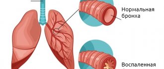
Inflammation associated with periodontitis leads to osteoclastic bone resorption. Periodontitis destroys the thin plate of bone and the normal attachment of the tooth. As a result, the oral and nasal cavities are connected. Oral bacteria, as well as food particles and other foreign bodies enter the nose through this fistula and this leads to inflammation in the nasal cavity, sinusitis.
Treatment of oronasal fistulas depends on the degree of tissue damage and the stage of the inflammatory process. The client was offered a two-stage surgical plan. During which the nasal and oral cavities are sanitized using the cavitation method, non-viable teeth and tissues of the upper jaw are extracted. Plastic surgery techniques, patch technology, osteosynthesis and modern materials are actively used in modern veterinary surgery.
Prevention of oronasal fistula.
Annual examination by a veterinary dentist.
Proper sanitation of the oral cavity of all subgingival calculus deposits with subsequent grinding and polishing of the enamel is possible only under general anesthesia.
Prevention of injuries to all organs of the oral cavity.
Extraction of affected teeth with closure (we put sutures on the gums) without tension.