Author:
Luzhetsky S. A., veterinary ophthalmologist.
Veterinary clinic of neurology, traumatology and intensive care, St. Petersburg.
Medial inversion of the eyelids
- a problem that, for unknown reasons, is not widely publicized among veterinarians and animal owners. At the same time, this problem is very relevant for some breeds of dogs and cats. What is medial entropion?
This is a condition when the eyelid is positioned incorrectly and the hair on the skin side has the opportunity to touch the cornea. The name “medial entropion” does not fully reflect the essence of the problem. It would be more accurate to call this pathology “dysplasia of the medial canthus,” i.e.
e. impaired development of the medial canthus.
Turn of the century
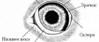
- a condition in which a normally developed eyelid, for one reason or another, turns in and as a result of this, contact of the hair with the conjunctival mucosa or the surface of the cornea occurs. With medial inversion of the eyelids, the situation is somewhat different: the eyelid in the inner corner of the palpebral fissure does not have a normal anatomical structure (Fig. 1).
Rice. 1. Inversion. If we simply describe the anatomy of the eyelid, it consists of the skin, muscle layer, mucous membrane and edge of the eyelid.
The rib of the eyelid
is the area where the skin of the eyelid meets the conjunctival mucosa. It is due to the presence of the rib that the eyelid can fully perform all its functions, and with medial inversion of the eyelids, the rib of the eyelid is absent in the inner corner of the palpebral fissure. There are two breeds of pets (pug dogs and Persian cats) that have 99.
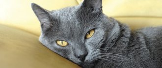
In 9% of cases, medial inversion of the eyelids occurs. Everyone knows that the eyes of these animals constantly require care and observation; they more often than others suffer from any diseases of the visual analyzer.
There is an opinion that “dirty” eyes
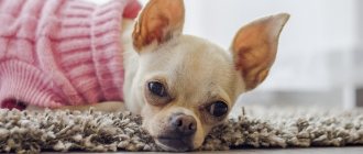
– a normal feature for these breeds. And it is these deep-rooted misconceptions that need to be destroyed. In this article we will talk specifically about pug dogs.
In my practice, I have never seen a single pug without medial inversion of the eyelids; 100% of animals of this breed that came to see us had this pathology. Every pug owner can independently detect this problem; you just need to know where to look. Finding the problem will require a pug, good light, and satisfactory vision on the part of the examiner.
Look carefully at the dog's lower eyelid; you will see that the sections of the lower eyelid from the side of the ear and in the center come into contact with the cornea precisely in the area of the eyelid rib. There is no fur in this place. Your gaze moves along the lower eyelid towards the inner corner of the palpebral fissure, and you should notice changes: the edge of the eyelid “disappears” and instead of it, fur begins to come into contact with the cornea.
This is the medial inversion of the eyelids. Do you have a pug and don't see this problem? Most likely, you really just don’t see it.
I, Sergey Aleksandrovich Luzhetsky, would be happy to look at a pug without medial inversion of the eyelids. Show me a dog like this and you will receive a free appointment with all the necessary diagnostics. Plus I will recommend your dog and your kennel (if you have one) as a good choice.
So, we have proven the fact of contact of hair with the surface of the cornea, which leads to disruption of the normal functioning of the auxiliary apparatus of the eyeball and in itself is an indication for plastic surgery of the medial canthus. Wool in contact with the cornea causes chronic irritation and creates conditions for the successful development of secondary microflora. Chronic irritation, in addition to direct damage to the cornea, contributes to the development of pigmentary keratitis (Fig.
Rice. 2a. Inversion.
Rice. 2b. Inversion.
Rice. 2c. Inversion.
Pigmentary keratitis is an attempt by the cornea to protect itself. Normally, the surface of the cornea is covered with multilayered epithelium, which has a number of properties, but most importantly, it is transparent. With chronic irritation, the corneal epithelium is not able to perform its functions normally, and as a result, it is replaced by the so-called “pigmentary keratitis”.
This tissue does not have the normal properties of corneal epithelium, but it is very resistant to external irritants. Essentially, it is a kind of “callus” on the surface of the cornea. Why is pigmentary keratitis bad?
Because it changes the normal properties of the cornea (Fig. 2b).
Pigmentary keratitis opaque
– the cornea loses transparency. The more area of the cornea is affected by pigmentary keratitis, the more the dog’s vision decreases (Fig. 2c).
The areas of the cornea covered with pigment are practically or completely insensitive. Normally, the cornea is extremely sensitive. This is necessary for her normal functioning.
- Wool that comes into contact with the cornea causes chronic irritation and creates conditions for the successful development of secondary microflora;
- Chronic irritation, in addition to direct damage to the cornea, contributes to the development of pigmentary keratitis;
- The presence of pigmentary keratitis reduces the sensitivity of the cornea. What happens when corneal sensitivity decreases? The production of “reflex tears” stops.
If a foreign body gets on the surface of the eye, the eye begins to water because as a result of irritation of the surface of the cornea by the foreign body, a large amount of tears begins to be produced. This is a normal and necessary reaction, since the foreign body must be washed out of the conjunctival sac as soon as possible. It is this tearing that we call “reflex tears.”
The cornea affected by pigment loses this ability. The level of reflex tears decreases or disappears completely. The result is keratoconjunctivitis sicca - the eye is simply not hydrated enough.
- Wool that comes into contact with the cornea causes chronic irritation and creates conditions for the successful development of secondary microflora;
- Chronic irritation, in addition to direct damage to the cornea, contributes to the development of pigmentary keratitis;
- The presence of pigmentary keratitis reduces the sensitivity of the cornea;
- Decreased corneal sensitivity leads to a decrease in the amount of tears produced.
The surface of the cornea dries. A dry cornea becomes an even more favorable environment for microflora; more damaged as a result of contact with wool; is in even worse conditions than at the initial stage of the process; experiences much more chronic irritation. It is much more difficult for a Pug dog to fully close his eyelids when the cornea is dry.
- Wool that comes into contact with the cornea causes chronic irritation and creates conditions for the successful development of secondary microflora;
- Chronic irritation, in addition to direct damage to the cornea, contributes to the development of pigmentary keratitis;
- The presence of pigmentary keratitis reduces the sensitivity of the cornea;
- Decreased corneal sensitivity leads to a decrease in the amount of tears produced;
- Decreased hydration creates conditions for chronic irritation;
- Pigmentary keratitis develops;
- Sensitivity decreases;
- Tear production decreases;
- Pigmentary keratitis develops.
The circle is closed. And this process develops to one degree or another in 100% of the pug dogs that we see. It is considered “normal” for a pug dog to have “dirty eyes”.
In our opinion this is not normal. This is the result of thoughtless breeding. The pathology “medial entropion of the eyelids” was fixed in this breed at the time of its breeding, and little importance is still attached to this pathology.
Breeders should pay attention to this pathology and try not to use dogs with pronounced medial entropion for breeding. There is no therapeutic solution to the problem of medial entropion.
You can support the cornea with symptomatic therapy, but the cause cannot be ruled out with therapeutic methods. No drops can remove hair that injures the cornea. It is a fact.
The only complete solution to the problem is plastic surgery of the medial canthus, the purpose of which is to remove the altered portion of the rib. Classic operations to eliminate entropion of the eyelids cannot give satisfactory results. The success of treatment directly depends on the stage of the pathological process.
If medial entropion is corrected early, then, with a high degree of probability, such a dog will not require any special care associated with medial entropion and its consequences throughout its life. If medial entropion has already caused serious changes in the cornea and decreased tear production, then the result of the operation may be a dog that does not experience irritation from contact of hair and cornea, but requires lifelong use of topical medications. The intensity depends on the degree of damage to the cornea.
- The presence of medial entropion in a particular dog;
- The ability of a particular dog to undergo surgery to correct medial entropion without problems.
The optimal age for performing such an operation in pug dogs is 7-10 months. As a rule, at this age the first signs of pigmentary keratitis begin to appear. It should also be noted that plastic surgery of the medial angle shortens the palpebral fissure.
This feature reduces the risk of eyeball prolapse. Eyeball prolapse is a serious condition that often occurs in dogs of brachycephalic breeds as a result of fights, injuries, and road accidents. We will not delve into the technique of the operation.
Anyone can find it on my youtube channel. com. This operation is low-traumatic for the dog and does not require special care from the owner.
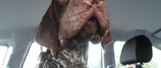
Scheme of surgery to correct medial entropion. After 14 days, the protective collar is removed from the animal, and the dog does not need any care for turning up the eyelids (Fig. 3a, b.).
Rice. 3a. Photo of the dog on the 14th day after surgery.
Rice. 3b. Photo of the dog on the 14th day after surgery.
Any owner who has chosen breeds that are predisposed to any diseases should familiarize themselves with them before purchasing a pet. For example, the owner of an English bulldog, as a rule, is aware of the pathology of “third eyelid gland prolapse” and knows how to properly solve this problem. Most likely, he received this information either from the breeder, or from a veterinarian, or from the Internet.
It is easy for English Bulldog owners to find adequate information about “third eyelid gland prolapse” because there is a lot of this information in the public domain. The owner of a pug finds himself in a much more difficult situation: there is practically no information about medial entropion. I hope this article will fix this problem.
Entropion is a condition in which the eyelid turns inward, injuring the cornea and mucous membranes. With a long course of the disease and lack of timely treatment, the condition worsens and leads to visual impairment. In particularly severe cases, the animal may not only go blind, but also lose an eye. This form of the disease is more common in dogs than in cats.
Causes
Entropion of the eyelid may have physiological causes caused by the specific structure of the muzzle in dogs with an abundance of skin folds on it. Wool in constant contact with the sclera and mucous membranes injures them, causing irritation and inflammation. As a result, erosion first forms, and then ulceration. It can be accompanied by an infection, which causes conjunctivitis and other dangerous diseases.
Among them is keratitis, leading to clouding of the cornea, its vascularization, and weakened vision. Over time, without treatment, the dog may become partially or completely blind.
The causes of keratitis are:
- injuries of the eyelid, eyes;
- ophthalmological diseases, inflammatory processes;
- damage to the cornea, causing the dog to constantly squint;
- systemic diseases;
- congenital or acquired change (reduction) in eye size.
The number of diseases does not include age-related entropion of the eyelid, which occurs in older dogs due to weakening of the ligamentous apparatus.
[custom_ads_shortcode1]
Useful video
Watch this video about the symptoms and treatment of entropion in dogs:
Similar articles
- Retinal atrophy in dogs: what provokes...
The cause of retinal atrophy in dogs is a gene, that is, the pathology is inherited, as well as cataracts, glaucoma, trauma and poisoning. If this is a progressive disease, then very soon the dog may become completely blind. Read more - Bloating in a dog: the main reasons why...
How to treat bloating in a dog. In the treatment of bloating in a dog, the root cause of the disease is of no small importance. In a situation where the cause of the pathology is gastric torsion or rupture of the organ wall, only emergency surgical intervention can help the animal. You can’t do without surgery either… Read more
- Eye injury in a dog: causes, treatment, dog after...
One of the most common reasons for visits to the veterinarian is an eye injury in a dog. There may be damage to the lens, cornea, or eyelid. Treatment is sometimes superficial, sometimes removal is necessary, after which the dog can live... Read more
- Stye in a dog: does it happen on the upper, lower eyelid...
It is quite common to find stye in a dog. It occurs on the upper and lower eyelids, internal and external. It is better to entrust treatment to the eye to a doctor, as it can lead to more serious diseases. Read more
- Tumor of the eyelid in a dog: is it really dangerous on the upper...
A tumor of the eyelid in a dog can occur due to a number of reasons: heredity, carcinogens and others. It can be on the lower, upper and third eyelid, benign and malignant. The sooner you contact a veterinarian... Read more
Main symptoms
An inversion of the eyelid begins with attacks of photophobia. The dog suffers from a sharp reaction to light, its eyes become watery and inflamed. The discharge from them first looks like mucus, then becomes thick and purulent. The animal tries not to go out into the light, hiding in the shadows.
Caring owners will pay attention to the strangely changed look of their pet. He begins to look a little askance, because the falling fur causes acute pain. At this point, you need to go to the veterinarian as quickly as possible, because the next stage will be severe inflammation, ulceration of the cornea and keratitis. Treating the disease is extremely difficult and troublesome.
[custom_ads_shortcode3]
Symptoms of entropy in dogs
Entropy usually manifests itself in dogs up to one year of age. Such animals are not allowed for breeding or participation in exhibitions, although after the restoration of the century, the animals live the usual life of a pet.
Dogs, experiencing discomfort from corneal irritation, constantly squint. The eyes become watery and light sensitivity increases. Sick animals attempt to rub their eyes. The conjunctiva becomes inflamed, resulting in redness of the eyes.
Scars and ulcers are the result of chronic irritation of the eye and, in addition to discomfort, cause pain to animals.
Diagnostics in a veterinary clinic
Short-faced dogs often experience habitual lacrimation, so it is very easy to miss the onset of the disease. A careful examination and regular visits to the clinic will help.
At the hospital, the dog is examined by dropping a special painkiller into the eyes. To detect erosions and ulcers, solutions with fluorescent properties are injected. By illuminating the surface of the eye with a source of ultraviolet radiation, any lesions and damage to the integrity of the membranes can be identified.
If the disease has progressed and an infection has appeared, the veterinarian will take a sample for bacterial culture. This will reveal the type of microorganisms that caused the inflammatory reaction and will help you choose the right drug for treatment.
[custom_ads_shortcode1]
Treatment method and prognosis
If the disease can be detected in the early stages, then it is enough to limit yourself to drug therapy. The veterinarian will prescribe drops with an analgesic and anti-inflammatory effect, as well as gels for the treatment of the eyelid, which have bacteriostatic and antiseptic properties.
A dog that is constantly trying to scratch its eyes should be put on an “Elizabethan collar” and monitor its behavior, not allowing the situation to worsen.
If there is a severe volvulus or an advanced disease, you will have to resort to surgery. The surgeon excises the fold that folds inward and places sutures to prevent it from folding back and keep the ligaments in the correct position.
Most often, specialized suture material is used that does not need to be removed. It simply dissolves after the surgical wound heals, which helps to injure the animal less. In older dogs or severely affected dogs, several surgical procedures may be required to completely resolve the problem or to improve the animal's condition.
[custom_ads_shortcode2]
Possible complications
The most dangerous complications are keratitis, conjunctivitis, prolapse of the third eyelid, ptosis (sagging of the eyelid due to impaired functioning of the ligaments). These diseases are difficult to treat and may require several complex multi-stage operations that do not guarantee 100% success.
[custom_ads_shortcode1]
Prevention measures
You can avoid serious illness if you create a comfortable and safe environment for your dog. It is necessary to remove all objects that are a potential cause of injury to the eye area, and limit access to cosmetics and household chemicals. Regular examination will help identify the problem at an early stage and begin treatment immediately. For dogs prone to watery eyes, eye care is an important procedure that should not be neglected. Periodic cleaning and rinsing with special preparations will help avoid dangerous consequences. This precaution is necessary for dogs with folds in the eye sockets.
To ensure that the animal does not suffer or lose its sight, owners need to carefully monitor its condition and promptly consult a doctor.
Authors of the articles: Belanta Clinic team
Veterinarians call entropion of the eyelids a common defect in dogs. It does not threaten life, but makes it incomplete, causing pain and discomfort. Delayed treatment leads to blindness.
A characteristic of the disease is a change in the position of both the lower and upper eyelids. This pathology leads to the fact that the edge of the eyelash directly touches the eyeball. Pet owners should take their dog to the clinic immediately at the first sign of symptoms. If you ignore a visit to the veterinarian, then blindness cannot be avoided in the future.
[custom_ads_shortcode2]
Upper and lower eyelid problems
Diseases associated with or characterized by inflammation or deformation of the eyelid. They arise due to infection or congenital predisposition.
Blepharitis (inflammation of the eyelid)
Inflammation can be unilateral or bilateral . In the first case, the cause is infection or injury. In the second - chronic diseases, demodicosis, allergies, staphylococcus.
Symptoms of the disease: severe swelling and redness of the lower and upper eyelids, hair loss around the eyes and fur, ulcers and wounds on the surface of the eyelid.
Treatment consists of soaking the crusts with herbal decoctions (chamomile, sage, green and black tea), using antibiotics and anti-allergenic drugs.
| List of drugs for treatment | ||
| Name of medicine | Price | Release form and application |
| Metronidazole | 80 rubles (40 grams) | The ointment is prescribed for the treatment of blepharitis when infected with parasites |
| Hydrocortisone ointment | 30-40 rubles per tube (10 grams) | Apply 2-3 times a day for 1-3 weeks; has anti-allergenic and anti-inflammatory effects |
Eversion of the century
A mechanical or paralytic disease characteristic of animals with mobile excess skin (folds) and a wide orbital fissure (eg, Basset Hound). Occurs during surgical removal of wounds, pinched nerves, and sagging skin.
Symptoms: mucous discharge, inflammatory processes in the conjunctiva, redness of the inner side of the lower eyelid, non-closure of the upper and lower eyelids. It can only be removed and corrected surgically.
Turn of the century
A unilateral inversion of the corner of the upper eyelid occurs. A genetic congenital disease that manifests itself during the first year of life. Often found in breeds with abundant folds on the face and body - Shar Pei, Chow Chow, Mastiffs.
Symptoms: deformation of the eyelid, redness, tearing. It can only be treated surgically.
What breeds are susceptible to the disease?
Not a single pet is immune from this pathology. However, the disease most often affects Chow Chows and Chinese Shar-Peis. This is caused by excessive folds of skin on the face. Overhang of excess skin over the eyes, which can trigger the development of the disease. A risk factor is the elasticity of the dermis, which lacks strength. Shepherd dogs are also at risk. Such breeds are affected by pathology due to inbreeding (breeding among relatives). This factor is provoked by the breeding of large individuals. Such dogs especially suffer from entropion of the lower eyelid. Affects Cane Corso disease, in which inversion of the lower eyelids occurs along with entropion. The defect can only be eliminated during surgery. The pathology of Pekingese and Pugs is not avoided due to their baked eyeball. The presence of a huge fold near the nose is also considered a significant factor.
[custom_ads_shortcode3]
Main causes of entropy
Entropion is divided into: primary and secondary. The first is a consequence of heredity. Deformation develops due to the presence of skin folds, as well as when there is no elasticity of the skin of the eyelids. This is especially true for large breeds.
Secondary is caused by inflammatory processes in the eyeball, also if there is deformation of the structure of the eyelid. Dogs begin to squint their eyes intensely, which overstrains their muscles. This state of affairs causes the eyelid to turn inward. It is possible to restore the original shape only after treatment. The factor that provokes the disease is inflammation of the masticatory muscle. The dog begins to vigorously rub the body mass. This is caused by problems eating. As a result, muscle tone near the eyes is lost.
[custom_ads_shortcode1]
What are the types of entropion in dogs?
Congenital entropion is most often diagnosed in Shar-Peis and English bulldogs. A puppy with this pathology immediately after opening his eyes experiences discomfort in the form of lacrimation. He starts squinting at them. As a result of this type of pathology, conjunctivitis develops in the dog, causing redness of the eyes. If the degree of volvulus is at the last stage, then the cornea is inevitably affected. As a rule, it loses transparency and acquires a whitish tint. Hereditary entropies can form as a pet grows up. By the age of one or 1.5 years, the owner of the dog begins to notice that he is increasingly squinting his eyes and blinking. After this, lacrimation inevitably begins. This species is mainly recorded in large dogs (Pekingese, bulldogs). Almost all dogs are susceptible to spastic inversion of the eyelids. Veterinarians believe that it develops as a consequence of some other disease in the animal. If there is a chronic non-healing ulcer, then persistent long-term blepharospasm (spasm of the eyelids) is diagnosed. The position of the eyelid cannot be changed even after undergoing a course of treatment. The veterinarian assesses the extent of the lesion after local anesthesia. This prevents the dog from squinting its eyes. Post-traumatic entropion in pets can occur due to trauma. It is characterized by damage to the eyelid. If such symptoms are present, then you need to immediately take the dog to a veterinary clinic. Untimely actions can cause improper fusion of the eyelid. In this situation, eyelid inversion is inevitable. Age-related bloat is associated with the aging of the dog. Age affects the loss of elasticity of the skin and the development of atrophy of the postorbital fat. After six years, pets squint their eyes more and more clearly. The examination allows the doctor to see the entropion of the lower eyelid. This disease often makes itself felt after a stroke.
[custom_ads_shortcode2]
Entropion and eversion of eyelids in animals
The eyelids of animals are an important organ , the function of which is to protect the eyes from damage, foreign bodies and to promote hydration of the mucous membrane.
Pathological processes can not only disrupt the normal functioning of the visual organs, but also lead to blindness. Diseases of the protective septum of the eye are a serious problem that requires immediate attention from a veterinarian.
Entropion of the eyelids
A common defect caused by injury or chronic diseases, as well as abnormalities of intrauterine development. It is characterized by a violation of the location of the skin fold, in which the movable part is turned inward and does not fit tightly to the eye.
With a full inversion, the outer surface on which the eyelashes and hair are located is all facing the eye. This provokes irritation of the mucous membranes and the development of inflammatory processes.
With prolonged exposure to the eye, keratitis occurs, ulcers form, causing further perforation and opening of the cornea.
Causes of the disease:
- entry of foreign objects into the conjunctival sac
- third eyelid amputation
- inflammation and damage to skin folds
- chronic conjunctivitis
Types of pathology
Inversion as a result of a strong spasm that occurs against the background of chronic inflammation of the connective membrane and other parts of the eye occurs if a strong contraction of the muscle occurs.
The eyeball is pulled deep into the orbit. The cicatricial anomaly develops when the conjunctiva is tightened by keloid tissue after serious trauma to the skin folds.
Sometimes pathology develops after removal of the 3rd eyelid. As a result of atrophic phenomena in the organ of vision itself and when it is excessively recessed into the orbit, inversion of the cartilage occurs.
The same phenomenon can occur with a congenital anomaly of the eye, when the edges of the protective partitions are deprived of support, so their surface is pulled inward.
Clinical signs
The outer surface in some places or is completely turned inward, adjacent to the mucous membrane. Fur and eyelashes constantly irritate the connective membrane and disrupt its integrity.
Prolonged exposure to the conjunctiva leads to ulceration and perforation of the eyeball.
The animal has:
- squinting of the eye
- lacrimation
- catarrhal conjunctivitis and keratitis
- turbidity, ulcerative phenomena
- sinking of the eye into the orbit
Treatment
The appearance of an entropion of the eyelid threatens the pet with blindness, so consultation with a specialist is necessary. After the examination, the doctor chooses methods of treatment and elimination of the problem.
The most effective is plastic surgery to remove skin folds.
Eversion of the eyelids (ectropion)
This pathology is caused by inversion of a section or an entire edge of the protective septum and its complete removal from the cornea. This leads to various eye diseases due to its vulnerability.
Possible reasons:
- injuries and diseases that left extensive scars that pull the edge of the eyelid outward
- diseases of the connective membrane of the eye with the development of edema pushing aside the skin fold
- facial paralysis and loss of muscle tone, resulting in drooping and eversion of the lower edge
- aging changes leading to loss of skin elasticity and atrophy of the orbicularis muscle
- neoplasms on the eye, orbital area, protective septum
- intrauterine anomalies
Symptoms
The edge of the fold, as a result of not completely adhering to the eye, is turned outward, the surface is exposed and is subject to mechanical irritation, drying out, ingress of foreign bodies and bacterial contamination.
A sick pet experiences excessive lacrimation, as a result of which the area around the eyes constantly becomes wet and soft. The inverted mucosa becomes inflamed and hypertrophied.
In severe cases, due to incomplete closure of the eye, the septum becomes denser, irritated and subject to ulceration.
Treatment
When a disease occurs, it is necessary to identify and eliminate the underlying cause of the disease.
Getting rid of problems that resulted in eversion of the skin fold (tumors of the conjunctiva, pathology of cartilage, enlargement of the organ of vision in size) gives a positive result and it returns to its normal position.
Minor lesions are easy to treat; with extensive scars that cause the pathology, the prognosis is cautious. To solve the problem, they resort to plastic surgery, during which the scar is excised.
In the presence of massive scars, transplantation of healthy tissue is required, but this is not always possible.
Entropion of the third century
The membrane located in the inner corner of the eye serves to cleanse and moisten it with tear fluid. Follicular conjunctivitis and degenerative changes in the cartilage of the eyelid are the cause of inversion of the nictitating film.
The animal notes:
- crease and eversion of the third century
- blepharospasm
- redness and swelling of the conjunctiva
- serous-mucous discharge from the eyes
The occurrence of a pathology of the nictitating membrane is a reason for immediate contact with a veterinarian.
It is forbidden to adjust or remove the membrane yourself; this can lead to disability of the animal. In addition, infection of irritated tissues provokes the development of severe pathologies and, as a consequence, blindness.
Symptoms of the disease
Diagnosing entropion in a dog is quite easy. The main and characteristic symptom is increased lacrimation. A dog with this pathology begins to feel discomfort in the light. As the disease develops, the secreted secretion begins to thicken. Subsequently, it acquires a slimy character. You can tell that a dog suffers from photophobia by the way he starts rubbing his eyes with his paw. This causes significant discomfort, affecting the pet's behavior. He becomes irritated and tries in every possible way to avoid light sources. At the same time, the painful sensations increase more and more. They cause the dog to look askance.
If you do not seek medical help in a timely manner, you will inevitably develop eye tics and keratitis. Weakness of the ligamentous apparatus affects the sagging of the skin near the orbit. In some cases, corneal tears may form. You should be wary of them, otherwise they cause ulcers. Conjunctivitis may also develop, in which the eyes appear reddened due to inflammation. The disease can be recognized by the way the dog constantly rubs or blinks. Also characteristic symptoms may be closing or squinting of the eye.
[custom_ads_shortcode3]
Symptoms
How can you tell if your pet needs an urgent visit to the vet? In the initial stages, the cat often begins to “wink” at the owner and avoids looking at light sources. Tears flow profusely from her eyes, and soon the discharge becomes mucous and quite thick. The animal acquires the habit of looking sideways at the world around it. In many cases, the eyelids themselves become inflamed and swollen, and the tissues of the organ become noticeably hot to the touch.
Intolerance to light reaches a peak stage; the animal cannot look at lamps or the sun at all. It begins to constantly rub its eyes with its paws, and this causes it severe pain. If you notice these symptoms in your pet, we advise you to contact your veterinarian immediately.
How is entropion of the eyelids treated in dogs?
Effective treatment of entropion in dogs is possible with surgical intervention. This is due to the fact that each breed has its own characteristics. Therefore, only an individual approach will help eliminate the disease. According to medical statistics, 90% of the disease is eliminated after one operation. In complex cases, several of them may be required. Difficulties in treatment are associated with the presence of folds on the animal’s face. Before surgery, each individual must undergo examination. Its data allows you to create a personal treatment plan. The specialist’s main task is to correct the incorrect position of the eyelids. With effective measures, they return to the correct position. The dog's surgery to eliminate this disease is performed under general anesthesia. When the animal is elderly or has a range of other diseases, then surgery is possible only after additional examinations. Based on their results, a plan for preoperative drug therapy can be drawn up. After surgery, it is necessary to treat the sutures with a solution of chlorhexidine bigluconate for 2-3 days. Before removing the stitches, the nodules should be treated with a cotton swab, which is moistened in a 70% alcohol solution. Nodules are processed in the direction away from the eye; the nodule and the alcohol solution should not be allowed to come into contact with the cornea. If after 10-14 days the stitches are in normal condition without discharge or crusts, they are removed. Doctors do not recommend surgery for pets with general somatic diseases. With such problems, dogs cannot tolerate anesthesia. In this situation, only autohemotherapy can eliminate the unwanted defect. However, the effectiveness of this procedure is temporary. This method is based on injecting the dog’s own blood into the eyelid. It is mixed with medications in the required proportions. The effectiveness of this procedure can be seen after 2 weeks. This technique can be used repeatedly if necessary.
[custom_ads_shortcode2]
Ulcerative keratitis
Severe inflammation of the cornea of the eye. Causes: mechanical damage and trauma (burns, foreign bodies, bruises), infections, reactions to drugs, allergies, vitamin deficiency. It may occur due to diseases of the immune system, in particular diabetes.
Vivid symptoms: suppuration and tearing, fear of light, a cloudy area in the affected area, the presence of ulcers on the cornea, frequent blinking, redness of the white. The consequence of improper treatment is glaucoma, cataracts, blindness.
| List of drugs for treatment | ||
| Name of medicine | Price | Release form and application |
| Levomycytin | 50-60 rubles per bottle | Eye drops are used 4 times a day, 2-3 drops into the conjunctiva; |
| Erythromycin ointment | 90 rubles per tube | Antibiotic ointment is applied around the eyes and on the eyelid of the dog 2-3 times a day, the timing of use depends on the severity of the disease |
How to care for your pet after surgery
Rehabilitation after surgery lasts a long time. Pet owners should strictly follow the recommendations of their veterinarian. If you ignore the doctor's advice, the operation will not bring the desired effect. During the operation, thin material is used. It prevents the appearance of scars. A specially designed plastic collar allows you to avoid its accidental removal by the dog due to careless movement of the paw. The dog should wear this device until the stitches are removed. Its size is selected in accordance with the dimensions of the pet.
The pet must also be isolated from others. The dog should not be disturbed by sounds. The owner of the animal must treat the wounds with a special solution several times a day. Care must be taken to ensure that there is no purulent discharge on the skin during these procedures. The specialist prescribes antibiotics and eye drops.
Sources:
- www.spbvet.info
- www.belanta.vet
- realpet.ru