Adenoma of the third eyelid (“Cherry eye”) - how to treat in dogs
Happens to 90% of English Bulldogs!
Definition.
Adenoma of the third eyelid is an increase in the volume of lymphatic follicles of the third eyelid.
Causes and development of the disease.
The disease occurs due to the reaction of the lymphatic follicles of the third eyelid to infectious agents.
The exact causes of the disease have not yet been clarified. Some experts call it a genetic disorder because bulldogs are predisposed to it. However, it also occurs quite often in other purebred dogs. The cause of adenoma is a subject of debate among veterinary ophthalmologists, but it is believed that the cause is hyperirritation of the lymphatic follicles and blockage of the excretory ducts.
Cherry eye is a common eye disease. This is not dangerous, but a serious disease. It does not affect the eyeball itself - the problem is in the pink membrane adjacent to the eye. In addition to the upper and lower eyelids, dogs have a third eyelid that starts from the inner corner of the eye (closest to the nose). The third eyelid acts like a windshield wiper on a car and helps protect the eye by cleaning its surface. In addition, it contains a gland that produces 30% of all tear secretions. “Cherry eye” is hypertrophy of this gland. Cherry eye does not go away on its own. Sometimes the disease also affects the second eye, although this is not at all necessary.
Clinical signs.
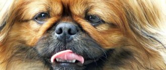
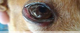
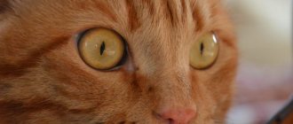
In sick dogs, the gland becomes red and thickened, resembling a small cherry formed in the corner of the eye. If one eye is affected by the disease, it is possible that similar symptoms may also occur in the other. Most often, such diseases occur in young dogs, up to one year old. In addition to redness and increase in size, clear or mucous discharge from the gland may appear. The disease is often combined with conjunctivitis.
Treatment.
Conservative treatment with antimicrobial and anti-inflammatory drugs is ineffective. The adenoma is removed surgically.
The optimal treatment is surgical fixation of the gland in its place on the inside of the third eyelid. After this iron; will perform its functions, and the risk of dry eye disease will also be prevented. As a rule, such operations are successful.
Another treatment option is to remove this gland, which may result in insufficient hydration of the eye. If you ignore a third eyelid adenoma, there is a risk that after two or three weeks of illness, further complications will follow. If stitching does not produce results, then you need to resort to a radical method (remove promptly, but you will have to instill eye medications into your eyes for quite a long time to moisturize the cornea)
Prevention.
Timely treatment of general and eye diseases is necessary.
Causes and prevention of the disease
The exact causes of inflammation of the third eyelid in dogs have not been well studied to date. The chain of lacrimal glands located around the cartilaginous junctions of the nictitating membrane provides the main source of tear film and ocular lubrication, thereby experiencing high physiological stress. The fibrous tissues that hold the third eyelid in the correct anatomical position weaken at some point, and the glandular tissues begin to bulge above the inner surface of the eyelid, which provokes their inflammation. The final result is swelling of the nictitating membrane, its prolapse and diffuse inflammation.
Third eyelid "adenoma" in dogs can develop in one or both eyes. More often, the disease is observed among representatives of brachiocephalic breeds, which proves a strong genetic component, expressed in the form of a weak connection of the fibrous tissue of the third eyelid. In addition, traumatic inflammation, as a localized protective response to damage to the tissues of the third eyelid, as well as hypertrophy - an increase in the size of the organ solely due to the expansion of existing cells, and not through new cellular growth, may also play a role in the development of "cherry eye" in dogs.
So, the main causes, or more precisely, risk factors for inflammation of the third eyelid in dogs are hereditary factors and trauma. In addition, in practical veterinary medicine the following conditions are encountered that can provoke the development of this disease:
- True Gardner's lacrimal gland adenoma.
- Hyperplasia of the lacrimal glands of the third eyelid due to leukemia.
- Eversion of the cartilage of the nictitating membrane, which is often observed in active puppies during play or when scratching.
Most often, “cherry eye” affects young dogs under the age of one year, when the body is actively growing. Of the brachiocephalic breeds, the disease often affects English and French bulldogs, pugs, and also – with large heads and loose constitutions – Cane Corsos, mastiffs, Newfoundlands, and Great Danes. Also, inflammation of the third eyelid is not uncommon among Spaniels, Chow Chows, Shar Peis, Basset Hounds, Beagles and Bloodhounds.
Prevention
As mentioned above, the exact cause of inflammation of the third eyelid in dogs is not well understood, but requires professional treatment. However, since the pathology appears most often among certain breeds, it is believed to be characterized by hereditary factors. Thus, one of the most effective ways to prevent the disease is a detailed study of the medical history of the puppy’s parents when purchasing it.
Special Notes
Cherry eye in dogs is a condition that usually appears unexpectedly and for no apparent reason. In a typical case, the animal looks normal, but literally within a couple of minutes, a dense mass of red, inflamed tissue may appear in the inner corners of one or both eyes.
Worth about the third eyelid in dogs is not a life-threatening condition for the animal, and also does not require quick treatment. However, at the first appearance of symptoms, you should not delay the provision of assistance - over the next few days, related pathological conditions will begin to develop, starting with conjunctivitis, which can lead to irreversible consequences, including loss of quality of vision.
Some dogs are born with a visible third eyelid, which is more common on both sides. Externally, this condition manifests itself in the form of a not completely hidden third eyelid with the upper and lower ones open. This disorder is more of a cosmetic problem that does not require serious intervention, but puppies of valuable breeds will never be calibrated.
What is this?
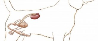
The third eyelid is a semilunar fold located on the inner corner of the eye. Needed for auxiliary protection of the eyes: when they are touched, or if the dog tilts its head, the third eyelid closes the cornea from possible damage.
However, the third eyelid itself, much more often than we would like, becomes the cause of various pathological conditions.
Animals at risk
The risk group includes animals that are most vulnerable to the listed disorders. These include:
- brachycephals with bulging eyes (Japanese chins, Shih Tzus, Pekingese, pugs);
- breeds with folded skin (chow-chow, sharpei, English bulldogs);
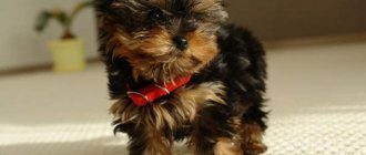
- pets with long hair (Yorkshire terriers, Afghan hounds, lapdogs).
In the first case, vulnerability to external factors is explained by the visual organ being too large. In the second - skin folds on the face, increasing the likelihood of inflammatory processes due to accumulating dirt and dust. In the third - long hair, fraught with frequent irritation and injury.
Adenoma
Adenoma of the third eyelid (or hyperplasia, “cherry eye”) is the official name for prolapse of the lacrimal gland of the 3rd eyelid. In fact, this is an enlargement of Gardner's lacrimal gland (up to 15 mm in diameter), which occurs with swelling and accumulation of secretions.
The gland of the third eyelid is located inside the stroma of the third eyelid and is visualized without additional devices and instruments. The tear film in dogs is produced by three glands, all of which are equally important in protecting the eye, but the disease only affects one.
The reasons for the appearance of adenoma are related to the developmental characteristics of the animal and external factors. So, the causes and treatment of third eyelid adenoma:
- Weak ligaments are not able to support the gland in its normal position;
- The cartilage of the eyelid is everted;
- Hyperplasia;
- Injury;
- Heredity.
You should know that any, even the smallest inflammation or prolapse of the third eyelid requires treatment. Examination of the animal will allow the doctor to select individual treatment. The most common method used is to remove a pink formation in a dog's eye. Preliminary therapeutic treatment with antibiotics is carried out in order to relieve inflammation and slightly reduce the volume of the gland.
After surgery, the four-legged patient is recommended to wear a protective collar for 2 weeks. As recommended by your veterinarian, use eye drops and ointments. A caring attitude towards your pet and following all recommendations will allow you to avoid complications in the form of conjunctivitis, and forget about the disease in a month.
This most often occurs in puppies and young dogs under one year of age. Next, the gland strengthens and becomes more elastic.
Signs and symptoms
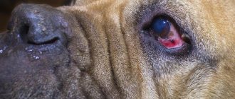
- Actually, the prolapsed gland itself: it is red and very clearly protrudes forward. Its color and size are due to the fact that when the eyelids move, it is constantly exposed to mechanical stress, swells, swells and rubs the cornea.
- The animal becomes very restless and constantly scratches its eyes, which further injure the gland.
- Often, adenoma of the third eyelid is accompanied by purulent discharge and conjunctivitis. Because when you come into contact with dirty paws, you come into contact with pathogenic microflora, which develops very quickly and causes all sorts of problems.
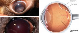
Keratoconjunctivitis or dry eye
When dogs develop a condition called keratoconjunctivitis or dry eye, their tear glands produce less fluid than normal.
Tears perform important functions:
- Removal of potentially damaging material from the surface of the eye.
- Nutrition of corneal tissue.
Not surprisingly, a lack of tears can cause big problems, including:
- Corneal ulcer;
- Chronic outflow of mucus from the eyes;
- Pain.
Most cases of vision loss in dogs begin with the development of keratoconjunctivitis.
The cause of the disease may be the canine distemper virus, pathology of the third eyelid, lacrimal gland, chemical poisoning, or injury.
Treatment is carried out mainly with medication, but surgical intervention is not excluded. Applicable:
- Anti-inflammatory eye drops;
- Eye moisturizers “artificial tears”;
- Broad-spectrum antibiotics;
- Antiallergic drugs.
Inflammation and swelling (eversion)
Causes conjunctivitis, volvulus of Gardner's gland or adenoma resulting from:
- injuries or exposure to foreign bodies, dust, smoke in the eye;
- inversion or inversion of the eyelid;
- prolonged exposure of the eyes to direct ultraviolet rays;
- long-term use of medications;
- a disease of the plague that affects the lymphatic and blood vessels.
The disease manifests itself as redness of the eyes and the appearance on the surface of the conjunctiva and third eyelid of lymphatic follicles enlarged to the size of millet grains. Inflammation may be accompanied by mucous discharge from the eyes.
It will be easier for a specialist to help the animal if no more than 6 hours have passed since the onset of ectropion. In this case, the prolapsed third gland is repositioned. After this time, the need for surgery increases.
There is a high probability of ectropion occurring in puppies up to a year old, while the gland has not yet strengthened. Subsequently, its elasticity increases and problems do not arise.
Inflammation of the third eyelid
Owners often come to veterinary clinics with dogs whose third eyelid is inflamed. This happens due to the fact that the membrane is affected by pathogenic microflora. The conditions for its occurrence are created by the following factors:
- Any, even the most minor, eye injury. Dog owners should be especially attentive after walking in the forest, since an ordinary branch that hits a pet in the eye can lead to a painful reaction.
- Exposure of the eye to active chemicals, smoke, as well as the entry of foreign particles into the conjunctival cavity.
- Long-term drug treatment can also lead to inflammatory processes in such a delicate structure as the third eyelid.
- The disease canine distemper can also be expressed in such a symptom as inflammation of the membrane.
As for the symptoms, the inflammatory process manifests itself in the fact that the third eyelid swells and turns red in the corner of the eye. At the very beginning, its size reaches a grain of wheat, becoming larger over time. Treatment of the third eyelid in a dog in this case must begin with a diagnosis. It is strictly not recommended to do this on your own; you must seek the help of a specialist.
The veterinarian prescribes the animal to take medications that can stop inflammatory processes, for example, corticosteroids. Along with this, various antiseptic lotions are used that will disinfect the dog’s eye and prevent the development of infection. Tetracycline ointment and Korneregel are excellent and affordable means for such treatment. It is also acceptable to use folk remedies, for example, the eyes can be washed with decoctions of oak bark or chamomile.
If inflammation progresses against the background of some other disease, then serious treatment is needed, including antibiotics, fungicides and drugs that can fight viral infection. In dogs, treating this disease is always a long and painstaking process that requires a lot of time and attention from the dog breeder.
Prolapse (loss)
Hyperplasia of the third eyelid is a kind of “loss” of the nictitating membrane, when it “falls out” from the corner of the eye. The eyeball is half covered with reddish membrane tissue. The cause of the pathology is that the ligament holding the third eyelid is weakened.
It differs from inflammation: in color - crimson-red and enlarged third eyelid, which noticeably extends beyond the edge of the eye, and also in the fact that this is not a disease - this loss appears as the body’s response to some negative stress/intervention and when the main cause of the disease is eliminated, the third eyelid gradually restores its normal position.
Entropion of the third century
This type of third eyelid prolapse most often occurs in the youngest dogs. The appearance of the pathology is associated with an increase in the diameter of the eyeball as the dog grows older. This provokes the process of lengthening the cartilage stalk, which is attached to the ligamentous apparatus of the organ. If the leg lengthens excessively, the cartilage breaks over time, causing it to lose its flexibility and cease to perform supporting functions. As a result, the eyelid begins to bulge, disturbing the animal, and attempts to insert it yourself are doomed to failure.
A cartilage fracture interferes with the normal functioning of the eyeball, which becomes extremely vulnerable to the harmful influence of microbes; infection can easily get there. This will inevitably lead to inflammatory processes. Dog breeds such as Newfoundland and Great Dane are at risk. Unfortunately, the animal cannot be cured without surgery. Only a qualified veterinarian will be able to properly suturing the cartilage so that the eyelid returns to its place.
Removal
There is no drug treatment program for problems with the third eyelid. The only way out is surgery to remove the third eyelid.
Purpose of surgery:
- do not disturb the anatomical structure of the third eyelid,
- preserve the mobility of the eyelid as much as possible;
- exclude the development of the disease in order to preserve the animal’s vision.
What does the eyelid look like after surgery?
The specialist will inform you about the need to remove cartilage or part of the lacrimal gland only as a last resort, if the consequence of maintaining the tumor may be loss of vision.
The operation is performed under general or local anesthesia, is not complex, and does not require a long recovery period.
After the operation, to prevent the dog from injuring himself with his paw, he needs to wear a protective collar . During the first 7-10 days after surgery, you should prevent the development of bacteria by using antibacterial medications for the eyes.
Suturing and reduction
Reduction of the third eyelid gland is as necessary as removal of tumors. The likelihood of developing diseases that can deprive the dog of his vision becomes the basis for making a decision on surgical treatment.
The photo shows a dog before and 3 days after surgery.
It is possible to realign the gland without surgery at the initial stage of the disease, in the first 12 hours. If more than 12-24 hours have passed, then surgical intervention is recommended.
There are many surgical options for treating third eyelid gland prolapse. Regardless of which method is used, the gland tissue must be completely preserved .
In cases of prolonged illness, surgery is inevitable. Operations for suturing and reducing the third eyelid are performed under an operating microscope. In order not to disturb the animal, general anesthesia is used, but deep sleep is not required, since the doctor will spend very little time - 15 minutes - to restore one eye.
The surgeon uses special ophthalmic threads, which eliminate the possibility of causing microtrauma to the eye and help the surgeon place the prolapsed gland in the originally intended place, avoiding the formation of postoperative scars. Modern microsurgical instruments in the hands of an experienced doctor guarantee the absence of kerato-conjunctivitis in the postoperative period.
Dangers and complications
Timely therapy allows you to avoid many complications: atrophy, eye loss, partial or complete blindness. Due to the variability of possible causes, in addition to vision loss, it is worth considering other dangers associated with the original pathology or associated infection.
The possibility of disruption of internal organs and systems, which could lead to the death of the pet, should not be ruled out. It is much safer to try to prevent the disease by avoiding its occurrence.
How to help before seeing a doctor
We hope that you have already understood that a doctor specializing in ophthalmology can significantly help your dog. However, it is not yet possible to do this; it is necessary to provide first aid and at least somehow relieve inflammation, reduce pain and anxiety of the animal.
In this case the following is possible:
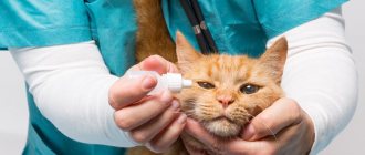
- Apply Dexamethasone . Eye drops based on corticosteroids with anti-inflammatory and antiallergic properties. For a dog, 1-2 drops in the third eyelid area 2 times a day is enough. The active substance quickly penetrates the tissues, but is contraindicated in purulent processes. After instillation, tears may leak.
- Rinse the eye with Tsiprovet . The active ingredient is the antibiotic ciprofloxacin. Possessing a broad-spectrum antimicrobial effect, it quickly relieves inflammation of an infectious nature and has an anti-inflammatory effect. When instilled, 1-2 drops cause a burning sensation, the animal may worry, but it quickly passes.
- Put a special protective collar on the animal so that it does not damage its eye even more. IT IS NECESSARY!
What are the forecasts?
In most cases, the glands return to normal function within a few weeks after surgery. In approximately five to twenty percent of cases, prolapse of the third eyelid gland may recur and further surgery will be required. Many animals that experience prolapse in one eye can eventually develop in the other. Surgery is always necessary due to the risk of developing dry eye when the gland prolapses. In severe or chronic cases, it is necessary to completely remove the gland, especially if the function is severely weakened or absent.
Sources:
https://bulldog-fill.com/vet2.html https://sobakus.com/zdorove/glaza/bolezni-tretego-veka.html https://yaroshenko.vet/articles/cherry-eye/
Corneal wounds
The surface of the eye is covered by a clear, skin-like tissue called the cornea. Just like skin, the cornea can be damaged, and lacerations (cuts), punctures and ulcers are quite common in dogs. Trauma is often to blame, such as when a dog runs through long grass and gets hit in the eye. In other cases, problems with the eyes themselves (such as poor tearing or abnormal anatomy) can put dogs at risk for corneal damage. A pet with a corneal wound will frequently rub the affected eye and squint due to pain. The eye may also be red.
Treatment of corneal wounds includes:
- Fight infections with antibiotic eye drops or ointments;
- Elimination of pain;
- Allowing time for the cornea to heal.
In severe cases, surgery or other treatment may be required to protect or repair the cornea and promote healing.