Why can a dog hurt his eye?
The causes of eye injuries are very simple:
- a dog fight or a skirmish with a cat;
- injury due to negligence (injured by a firecracker in a domestic dog, hit by a speck when a pet leans out of a car window, injured by a twig or thorn of a plant in a hunting dog);
- head injury due to a blow or an accident;
- contact with irritating substances - acids, alkalis, etc.;
- thermal burn;
- foreign objects getting into the eyes of detection dogs.
If the eye is closed and the dog is squinting to protect it, or the eyelid is not functioning properly, then this is a sign that he needs medical attention. If you see a foreign object, do not try to remove it or treat your dog without a professional examination by a specialist. Inaccurate movement sometimes damages the organs of vision even more and causes dense scarring or blindness.
Predisposing factors
Most often, eye injuries occur due to the following predisposing factors:
- If the dog was chasing cats
through the bushes. Protruding branches pose a serious danger to the eyes (and not only in the case of dogs).
- that have suffered from firecracker explosions
are often admitted Very unlucky dogs receive fireworks directly in the eye, which in many cases ends in blindness.
- Hunting and service dogs
are always at risk, since while working, branches, dry grass stubble, leaves, sand, etc. often get into their eyes. It is especially hard on the ages of these animals: scratches and small wounds appear there all the time (especially during the hunting season). Please note that a simple tetracycline ointment in this case can only slightly muffle the symptoms. If you don't go to the clinic, your pet risks becoming a one-eyed dog!
- Young, excitable and overly nervous dogs often injure their own eyes during rough play or “scuffles” with other animals.
- Of course, dogs of fighting breeds
, as well as dogs “oriented” to protection, also have an increased chance of getting eye injuries.
Types of damage
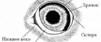
Damage is divided into simple and complex. Simple ones include injury to the cornea (the clear outer layer at the front of the eyeball) or sclera (the white area of the eyeball). The wound may extend into or through these areas.
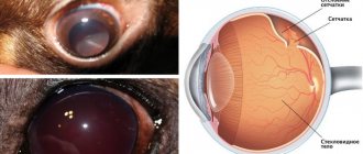
Complicated injury, along with the cornea and sclera, includes damage to other structures of the dog's eye:
- iris;
- retina;
- lens;
- century.
Did you know? Have you ever wondered why your dog can't see the treat that's right in front of him? It turns out that her nose is too large, which blocks part of the visible range, forming a “blind spot” in front of her nose.
Injuries to the cornea and sclera
The injury to the cornea or sclera in a dog may not be visible to the naked eye. Your pet can get it while playing, interacting with other animals, or running through thick grass.
But the symptoms will be noticeable:
- the dog squints;
- fluid accumulates in the eye tissues (edema);
- bleeding is observed;
- you can see a foreign body in the eye.
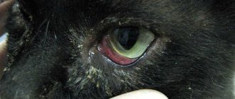
Lacerated wounds quickly harden. As a result, a blood-filled thickening may be observed under the moist tissue of the eyeball (subconjunctival hematoma). Defects in the iris, distortion of the pupil, blood in the anterior chamber of the eye, cataracts, detachment of the back of the eye (retina) from the eyeball, and protrusion of the eyeball (exophthalmia) may also be noticeable.
Risk factors:
- existing visual impairments;
- puppyhood - very excitable and inattentive during games;
- professional hunting;
- fight.
Treatment depends on the severity of the injury.
The animal is treated at home if the eyeball is safe. In other cases, hospitalization will be required. Important! Do not use alcohol-based products to treat your vision. This will cause a chemical burn.
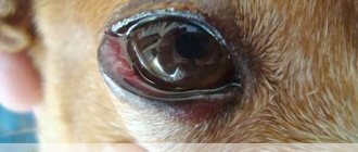
Damage to the eyelids
Damage to the eyelid is accompanied by bleeding. The eyeball is not affected.
The causes of injury may be the following:
- blow with a blunt object;
- scratch;
- laceration, cut or puncture wound;
- tear.
Such damage may be accompanied by complications - infection, inflammation. To cure them, a long period of therapy will be required - up to 1 month.
Eye contusion (concussion)
The typical cause of a bruise is a blow from something blunt or against something.
Bruises are divided into 4 groups:
- 1 - with minor hemorrhages;
- 2 — with minor damage to the sclera;
- 3 - with damage to the eyeball;
- 4 - with lesions of several adjacent tissues.
In addition to the main symptoms inherent in a particular type of bruise, it is accompanied by acute or aching pain, photophobia, swelling, and lacrimation.
Did you know? If the eye is bruised, it is recommended to immediately apply ice to it.
do this cooling several times on the first day after getting a bruise Exposure to temperatures allows you to regulate blood flow to the bruised area.
Eye loss
An “eye prolapse” is technically a dislocation of the eyeball. This happens during a fight with certain breeds of dogs. Pekingese, Pugs, Japanese Chins, Chihuahuas and Bulldogs have a narrow orbital socket and a large eye that is supported by eyelids and a small eye socket. Another reason for protrusion of the organ of vision is increased intracranial pressure. Under its influence, the eye is “squeezed out” from the inside.
Symptoms of pathology:
- unnatural appearance of the organ of vision;
- bloody discharge from the orbit;
- swelling of the mucous membrane;
- increased tearfulness;
- frequent blinking;
- photophobia.
There is nothing the owners can do to help their pet at home other than providing him with first aid. Therefore, the dog must be taken to a veterinary ophthalmologist immediately.
Types of damage
In veterinary practice, the following types of eye injuries in pets are distinguished:
- Damage to the eyelids. Such injuries can be through or non-through. Mechanical damage to the lower or upper eyelid is often accompanied by bleeding. The optical system of the eye is not impaired in such pathologies. The animal experiences profuse lacrimation immediately after injury.
- Corneal injury . Damage can be non-penetrating or penetrating. The greatest danger is posed by deep injuries to the cornea, often leading to loss of visual function. The introduction of sharp objects into the cornea is fraught with the penetration of pathogenic microorganisms into the eye tissue and the development of an inflammatory reaction.
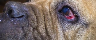
Eye injury from a cat's claw
- Eye contusion . Similar injuries occur when kicked, hit by a stick, or when the animal bumps into a hard object while running. The damage is accompanied by an increase in intraocular pressure, displacement and rupture of the internal structures of the organs of vision. In severe cases, the eyeball falls out of the socket. Contusion can lead to damage to the lens, which can lead to blindness in your pet.
Veterinary ophthalmologists distinguish between simple, complex and complicated injuries. The owner of the animal should understand that even seemingly mild ophthalmological damage can cause vision loss.
Signs of an eye injury in a dog
Common signs of eye injuries in dogs:
- squinting;
- rapid blinking;
- inability to open the eye;
- tearfulness;
- reddened mucous membranes;
- bloody sclera;
- photophobia;
- the pet tries to hide;
- restless behavior.
Symptoms of injury may include bleeding, swelling of the eyelid, or swelling of the tissue around the eyes. Please note that it is not necessary to hurt the eye for symptoms to appear. This could also be the result of a blow to the head.
What is a corneal ulcer?
The corneal layer is the front part of the eye, consisting of several layers:
- Upper epithelial. Protective shell of the organ.
- Stroma. Base of the cornea.
- Descemet's membrane. Posterior border wall.
- Posterior epithelial layer. Corneal endothelium, which maintains mild dehydration of the eyeball.
Normally, the surface of the cornea is transparent and smooth, with no roughness. The vessels of the circulatory system are not visible in the layers. Due to the large number of nerve endings, this area is highly sensitive.
A corneal ulcer affects the top layer. It is similar to a scratch on the skin, but is considered much more dangerous. The condition is complicated by severe pain. Because of this, the dog loses its appetite and has trouble sleeping. As a result, she quickly becomes physically and nervously exhausted.
Degradation of the epithelial layer of the organ is soon observed. The eye loses protection against infectious agents. A corneal ulcer is a serious disease that often results in infection of the organ by bacteria and blindness.
First aid
Only a specialist can provide professional assistance to a dog after an injury. But the owners can also provide first aid:
- The injured eye should be rinsed with saline solution, clean boiled water, or the “Natural Tear” drug, which is sold at the pharmacy.
- Then a cold compress can be applied to the swollen area after the bruise for 10 minutes; for other types of injuries this is not necessary.
- Apply chloramphenicol drops to the eye (2-3 drops).
- Apply a sterile bandage.
- Deliver the pet to the clinic.
When applying a bandage, do not fix it too tightly so as not to aggravate the situation.
Before going to the doctor, the dog needs to be kept quiet and shaded. Important! Take your pet to the hospital for evaluation and treatment within 24 hours of the injury to prevent complications.
How is the diagnosis carried out?
To help your veterinarian make a diagnosis, be sure to provide a detailed report of the incident, including:
- when did symptoms start?
- whether they are getting better or worse;
- what incidents may have caused the injury.
In the case of a foreign object or a visible wound (i.e., a scratch), diagnosis will be easy. However, if there is no apparent cause, the veterinarian will perform a thorough eye examination:
- the dog’s reactions to visual stimuli (light and objects close to the eye);
- pupil size, shape, symmetry and reflexes.
All this will help determine how deep the dog’s eye injury or bruise is. A blood test will be done if there are signs of an infectious disease. If damage to the skull is suspected, an x-ray will be taken. Based on the results of all examinations, the doctor will make a conclusion about the disease and prescribe a course of treatment.
Diagnosis of the condition of the cornea and other parts of the eye
If even slight damage to the pet’s organ of vision is detected, the owner should not hesitate to visit a specialized institution. In addition to the physical examination, the ophthalmologist will conduct instrumental diagnostic methods - ophthalmoscopy, ultrasound.
In some cases, the veterinarian resorts to a test sample with a fluorescent substance to identify the location and extent of damage to the cornea. In some cases, intraocular pressure is measured.
Special means of ophthalmological examination are carried out after preliminary sedation of the animal. Special research methods make it possible to identify deep damage to the ocular structures - prolapse of the iris, dislocations and subluxations of the lens, disruption of the hydrodynamics of the eye.
If the organs of vision are damaged, it is best to show the animal to a specialist - a veterinary ophthalmologist. The doctor will not only conduct special research methods, but also, if necessary, provide surgical treatment for the problem that has arisen.
Treatment of eye injuries in dogs
The treatment regimen depends on the type of injury:
- For penetrating wounds without gaping wound edges, an antibiotic or atropine ophthalmic solution is injected into the dog's eye. An Elizabethan collar is also worn to prevent damage to the eyeball tissue if the dog decides to scratch.
- If the wound has minor damage to the edge, then a soft contact lens is added to the above elements. Your doctor may suggest it to create a barrier between the healing cornea and the environment.
Important! If 2 different liquid medications are prescribed for eye drops, you should wait about 10 minutes between each use so that each medication can take effect.
You need to limit your walking time. Use belts instead of a collar to ensure that nothing interferes with blood flow and oxygen supply to the tissues of the head. It also reduces the risk of increased intraocular pressure. Monitor the condition of the wound and remember all the changes that occur. This may be important for the veterinarian to clarify the treatment regimen.
With the help of drugs
The medications described in this section are intended to provide general information about possible treatment. The doctor will prescribe it for a specific dog based on the established diagnosis.
Main groups of drugs:
- Antibiotics are used for lacerations to prevent the development of bacterial infection and inflammation of the eyeball tissue. The main drugs - "Neomycin", "Polymyxin B" and "Bacitracin", "Gentamicin solution" - are instilled into the eye. In case of extensive damage, injections of Ciprofloxacin, Cefazolin or Gentamicin are prescribed.
- Anti-inflammatory drugs are 1% Prednisolone Acetate or 0.1% Dexamethasone solution. They are used after the wound is sutured and it is in the healing stage. Prednisolone can be administered by injection or orally.
- Pupil dilatation medications, known as mydriatics, are intended to relieve eye pain. This is a 1% “Atropine Solution”.
- For general anesthesia, Aspirin, Butorphanol (for mild pain), Oxymorphone (for acute pain) and Tramadol (for severe pain) are used.
When is surgery necessary?
Injuries to the dog's eye that require surgery include the following:
- cuts;
- wounds;
- tissue rupture;
- other lesions with a moderately gaping edge, occupying more than 2/3 of the cornea.
Such defects require sutures and the use of painkillers and antibacterial drugs.
Treatment
Any treatment should be carried out only after examination by a veterinarian, examination and a clear diagnosis. In particular, if a dog has an inflamed eye, the doctor will tell you how to treat it; you should not self-medicate.
Examination by a specialist
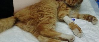
The first thing to do is to carefully transport the animal, if possible, to a clinic that you can trust with your dog.
Ideally, show the dog to an ophthalmologist. However, such specialists of a narrow profile do not work in all clinics, so in some situations you can turn to surgeons or general practitioners.
The examination may include full general tests to detect infections and a mandatory examination. You may need to measure intraocular pressure and even prescribe surgery.
It is important to identify the problem based on the results of tests and examination and make the correct diagnosis. The owner must strictly follow all recommendations received.
Eye drops
Sometimes it is enough to instill eye drops in case of an eye injury in a pet; this also applies to inflammatory diseases such as conjunctivitis.
Owners can get confused because they don’t always know how to put drops in their dog’s eyes. You need to follow some simple recommendations.
First, you need to wash your hands thoroughly to avoid infection, which will aggravate the inflammation process. Then carefully clean off pus and secretions, and also rinse the area around the eyes.
As a rule, dogs do not like such procedures and can behave very nervously, but under no circumstances should you scold them or raise your voice. Calming intonations, some encouragement, and affection will help you cope with the animal and make the procedure as painless and comfortable as possible from a psychological point of view. Although large dogs should wear a muzzle to avoid injury to the owner.
Drops for eye injuries are instilled in strict accordance with the prescribed dosage and doctor's prescription.
Caring for a dog after an eye injury
The doctor will determine how often you need to show the dog to him to monitor the progress of the wound healing. Deep or long penetrating lesions that have not been sutured should be inspected every 24 to 48 hours to ensure the integrity of the eyeball, monitor for infection, and control inflammation. Superficial penetrating wounds are usually rechecked at intervals of 3 to 5 days until healing occurs.
Did you know? The pupils tend to dilate if the pet is hunting or playing. This is the effect of adrenaline being pumped into the blood. As a result, his eyes become better able to detect the slightest movement and succeed in catching his lunch.
Symptoms and diagnosis
The main sign of mechanical damage to the eye is the suddenness of its appearance. His eyelids swell and the dog constantly puts his paw to his eyes.
There are often cases when blood begins to “leak” from there.
In some cases (subconjunctival hematoma), it remains in the tissues of the eyeball, causing the latter to swell and sharply increase in volume. If a foreign body is present, it can often be detected visually.
If this “body” sticks out of the eye at all (grass stubble stuck in), then it can be very difficult to miss it.