What is retinal detachment in dogs?
Detachment of the retina in a higher animal, be it a dog, a cat or a human, essentially has the same etiology, but in order to understand it, it is necessary to remember general information about the structure of the organs of vision. It is thanks to the retina that the light energy perceived by the eye , is processed and transformed into neural stimulation, a kind of signal in which visual data sent to the brain is encrypted.
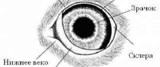
Important! The retina is a thin layer of ectodermal tissue, consisting of nerve cells and located on the back wall of the eyeball on the inside. The main function of this neural layer is to convert into nerve impulses and subsequently transmit to the brain the image projected by the lens and cornea.
In a healthy state, the retina should fit very tightly to the choroid of the eye; its connection with the eyeball along the perimeter of the optic nerve circumference is especially strong. It is in this area that the thickness of the neural layer is maximum. If this attachment weakens or is broken, it is said to be retinal detachment.
Why does retinal detachment occur?
Retinal detachment is not a disease, but rather a symptom that can be caused by a variety of reasons.
In dogs, in particular, a similar pathology can occur as a result of:
- pathologies of the structure of the visual organs that are hereditary in nature - progressive atrophy or dysplasia of the retina, primary hyperplastic persistent vitreous syndrome, Collie Eye Anomaly, etc.;
- suffered an injury resulting in hemorrhage in the eye or retinal rupture;
- serious acute inflammatory processes in the organs of vision associated with a significant accumulation of blood or exudate between the pigment epithelium and the neural layer (so-called choriotetinitis);
- tumors and neoplasms in the back of the eye, lymphomas, myelomas, etc.;
- stretching of the membrane of the eyeball, which, in turn, can occur as a complication of glaucoma;
- age-related retinal degeneration - dysplasia, dystrophy, etc.;
- damage to the vascular bed (this happens as a side symptom in diabetes mellitus, hormonal disorders, increased blood viscosity, as well as a constant state of high blood pressure).
Depending on what pathology caused the problem, retinal detachment is usually divided into several types:
| A type of disruption of the connection between the neural layer of cells and the choroid of the eye | Nature and features of development |
| Serous | Develops against the background of accumulation of inflammatory fluid or blood in the subretinal space. In the first case, the pathology is called exudative, in the second - hemorrhagic. Exudative detachment, as a rule, is the result of a severe infectious disease (bacterial, viral or fungal), while hemorrhagic detachment is usually associated with non-invasive problems - high blood pressure, lack of platelets in the blood, impaired clotting, etc. |
| Traction | Caused by tension of the neural layer on the part of the vitreous body, for example, as a result of displacement of the lens or iridolenticular diaphragm, inflammation of the uvial tract, retinal degeneration and other pathologies |
| Rhegmatogenous | Occurs due to thinning or tearing of the retina, usually of an age-related nature |
| Traumatic | It can manifest itself as a direct result of damage to the eye (penetrating wound, contusion, etc.) or develop gradually, as a complication after an injury, including due to the inflammatory process that arises after this |
| Iatrogenic | It develops as a complication after unsuccessful manipulation of the eye, for example, microsurgical removal of the vitreous body (vitrectomy), removal of cataracts using the ultrasound method (phacoemulsification), etc. |
| Mixed | Pathology occurs against the background of the synchronous action of several causes |
Based on the area of distribution, local, widespread, subtotal and total retinal detachment are distinguished. In general, all dogs are susceptible to pathology, regardless of breed, size and lifestyle. But there are a number of factors that repeatedly increase the risk of disruption of the tightness of the neuroepithelium to the choroid.
These include, in particular:
- hereditary predisposition;
- elderly age;
- characteristics of the breed (for example, congenital anomaly of the eye is very common in Scottish, Shetland and Australian shepherds, border collies, Nova Scotia retrievers, long-haired whippets, as well as Lancashire heelers; vitreous degeneration - in livery dogs and shih tzus; retinal dysplasia - in Labradors, retrievers, American cocker spaniels, English springer spaniels, etc.);
- hypertension (high blood pressure);
- progressive cataract;
- lens displacement;
- eye surgeries.
Did you know? In humans, the axes of the eyes are parallel, but in dogs they are in the form of rays diverging at an angle of 20 degrees.
Thanks to this feature, the animal, without turning its head, covers a space of 240–250 degrees with its gaze, while a person only 150.
Forum KOTODOM
Many cat owners do not even suspect that their proud pet is gradually losing his sight. Blindness in cats can be reversible or irreversible, partial or complete. What can cause blindness in cats?
1. Cataract. Cats can also develop cataracts. This is a condition in which the lens of the eye becomes cloudy. Cataracts usually develop due to eye injury, diabetes, or a genetic problem. Cataracts are treated surgically.
2. Glaucoma. A tumor, injury or any other hereditary disease can lead to excess pressure in the eyes, which can result in glaucoma. This is a common cause of blindness in cats. Glaucoma is treated by lowering intraocular pressure.
3. Tumors. Tumors of the eye often lead to blindness. In many cases, the eye has to be removed surgically.
4. Progressive retinal atrophy. This is an incurable disease and is most likely hereditary. This is a very slowly developing disease, under the influence of which the cat gradually completely loses vision. Because the disease progresses slowly, cats learn to cope with the gradual loss of vision and can live with vision loss.
5. Hypertension. Cats with kidney disease and diabetes are predisposed to hypertension. Hypertension can contribute to retinal detachment due to an overactive thyroid gland or kidney disease. There is no specific treatment for hypertension, but the cat should be kept on a salt-free diet.
6. Conjunctivitis. This infection causes the inner eyelid to become red, swollen and itchy. Although conjunctivitis does not cause complete blindness, repeated infection can cause blurred vision. Treatment may be in the form of eye drops or oral medications depending on the cause of the infection. There are other causes of blindness in cats, including: trauma, retinal degeneration, neoplasia, hypoxia, congenital underdevelopment of the optic nerves, and encephalitis. It is very important to make a correct diagnosis, since treatment will be determined by the diagnosis. In many cases, blindness is the result of hidden disorders or diseases that have a significant impact on the cat's health. Sudden blindness in cats is usually provoked by hypertension, thyroid disease, and kidney disease.
Signs of blindness in cats: 1. The cat constantly bumps into objects. 2. The cat has poor control over the coordination of paw movements. 3. Cloudiness of the eye or dilation of the pupil even in bright light. 4. Noticeable clumsiness of the cat. 5. The cat cannot find a cup of food or water. 6. The cat sleeps a lot and is inattentive. 7. The cat gets scared easily and seems timid. 8. The cat refuses to play and does not hunt.
Caring for a blind cat, of course, falls entirely on the owner. The first thing to do is take your cat to the vet as soon as you suspect vision problems. If your cat partially or completely loses its vision, do not abandon the poor animal under any circumstances. After all, this is cruel. Treat her the same way you would treat a blind person. A blind cat is quite adaptable and uses other senses to compensate for the loss of vision. Make sure your cat doesn't wander outside alone for long periods of time. Let your cat outside only under supervision. You can put a collar and leash on your cat and take her for a walk on a leash. Label the collar with your name and address in case your cat gets lost. A blind cat can walk around the house on its own. Cats have scent glands on their paws that leave a trail that cats can easily follow. You should not carry your cat around the house in your arms. Let your cat be independent. Limit your cat's movement near hot water, balconies, and stairs, as an accident may occur with a blind cat near these dangerous places. When approaching the cat, talk to it so that it does not get scared. If the cat is blind in one eye, approach it from the side of the sighted eye. Do not move furniture, tray, or cup of food unless absolutely necessary. Unexpected changes can disorient a blind cat. When playing with your cat, use toys that make sounds, this will help your cat maintain physical fitness and enjoy life.
Blindness can cause a feeling of helplessness in both the animal and the owner. But cats have a strong will and quickly adapt to this condition. This requires support, attention and care from the owner. A blind cat is still your cat and should be cared for like a healthy cat.
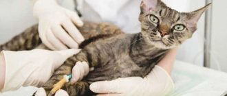
CONSULT YOUR VETERINARIAN
Cats can lose their vision for a variety of reasons. Vision often weakens with age, but due to injury or disease, even a kitten can go blind. What to do if your cat is blind? How to help?
Firstly, you need to take a healthy approach to the situation. You should not try on the loss of vision on yourself and transfer the terrible human experiences about this to your cat. Cats themselves are unlikely to be prone to getting emotional and causing tragedy; Blind domestic cats can lead normal lives and feel happy. A blind cat needs help, but not pity.
Secondly, you should definitely consult a good veterinarian. The doctor will be able to correctly diagnose, prescribe the necessary treatment and recommend special care for a blind cat. What if, under certain conditions, vision can be restored? Or maybe the loss of vision is partial, and proper care will prevent complete blindness? In any case, a blind cat should be regularly shown to a veterinarian to monitor its eyes and general health.
Thirdly, it depends mainly on the owners themselves how comfortable and fulfilling their pet’s life will be day after day.
Forecast and possible consequences
It is possible to successfully treat retinal detachment in a dog, but only in the early stages of the development of the pathology. In addition, the likelihood of a favorable prognosis directly depends on what exactly caused the problem (which of the above types it belongs to). In any case, there can be no talk of any self-medication here, just like the hope that the animal’s condition will stabilize on its own.
If the onset of retinal detachment is ignored, the dangerous condition will progress and can lead to such systemic disorders in the organs of vision as:
- destabilization of metabolic processes in eye tissues;
- interruptions in the supply of retinol, trace elements and other vital substances from the pigment epithelium to the retina;
- difficulty in cellular nutrition, leading to disruption of the structure and functions of tissues as a whole;
- problems with blood circulation in the eye, accompanied by the release of a special signaling protein - the so-called vascular endothelial growth factor;
- increased intraocular pressure;
- hemorrhages into the vitreous body (hemophthalmos).
As a result of all these pathological processes, the dog often develops glaucoma, and then irreversible retinal atrophy. Due to retinal atrophy, visual acuity and clarity initially suffer, and then gradually complete blindness occurs. If atrophy affects both eyes, it is called circular.
Important! Retinal atrophy is a decrease in the volume of cells in the neuroepithelium, primarily in its central part - the macula or macula. The macula is the organ of central vision in humans and other vertebrates.
Symptoms and signs of PRA
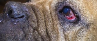
The condition usually begins to develop and show signs in adult dogs, usually between three and eight years of age. The first sign for many owners is that the dog begins to lose its night vision and has trouble navigating obstacles or finding its way in low light and dark conditions. As the condition progresses, the dog's pupils may appear dilated and take on the distinct appearance that characterizes the condition. Cataracts can also develop at the same time as the condition, resulting in a milky or clouded appearance of the eye lens in some cases.
PRA develops relatively gradually and can take months or even years to progress to complete blindness, meaning dog owners are often unaware of the condition until it is quite advanced. Many dogs adjust to their progressive loss of vision very naturally and do not experience major difficulties or become able to accommodate their progressive blindness by using their other senses, which again can make it difficult to identify the condition with ease.
[custom_ads_shortcode3]
How does the disease manifest itself?
Due to the seriousness of the consequences and the direct relationship between their severity and the timeliness of seeking emergency veterinary help, it is very important to detect signs of retinal detachment in a dog as early as possible. Although an animal, unlike a person, cannot report the onset of vision problems, an attentive owner is able to notice them by the changing behavior of his pet.
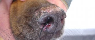
In particular, the following signs indicate a violation of the normal attachment of the neuroepithelium and choroid:
- impaired coordination of movements;
- disorientation in space (the animal may bump into different objects, behave uncertainly, or “get lost” in an unfamiliar place);
- increased tearfulness;
- the appearance of floating spots on the mucous membrane of the eyes, clouding of the lens;
- reduced or absent reaction of the pupil to light (the pupil remains dilated at all times);
- hemorrhages in the eye;
- deterioration of the pet’s general condition and mood - loss of playfulness, apathy, refusal to go for walks, etc.
Since retinal detachment is often a symptom of an internal disease, both infectious and systemic, other signs characteristic of a particular disease may be added to the overall clinical picture. For example, hypertension (high blood pressure), which causes complications in the eyes, manifests itself in dogs with such signs as:
- unsteady gait;
- pendulum-like movement of the eyes;
- rapid pulse;
- labored breathing;
- swelling of the limbs;
- restless behavior;
- sudden attacks of vomiting.
Having noticed such symptoms, the animal must be immediately shown to a specialist.
Did you know? It has been proven that domestic dogs see much worse than their wild relatives. It turns out that visual acuity, like muscles, can gradually atrophy in the absence of constant training.
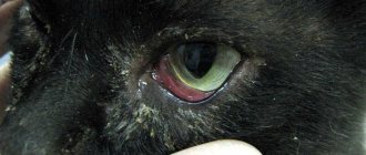
Signs of blindness
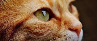
A cat's blind eye is often covered with a veil, or the pupil remains dilated even in bright light.
The animal will not be able to say that it sees poorly. However, a person can determine by certain signs that a cat has lost its sight:
- the animal stops moving a lot and spends more time dozing;
- walks only on the floor, jumps less or not at all;
- walks around the house carefully;
- The blind cat's whiskers are broken off. This is due to the fact that the whiskers are the main sensory organ of a blind cat. With them she feels the world, catches vibrations in the air;
- eyes do not glow in the dark;
- while moving, it crashes into objects that are not located in their usual places.
A cat that is blind in one eye may lash out in self-defense if you make sudden movements on its blind side.
How is the diagnosis carried out?
Retinal detachment is a condition that cannot be visually diagnosed. Therefore, a good veterinarian, after a conversation with the dog owner and an initial examination of the animal, should refer the “patient” to an ophthalmologist, who, in turn, conducts a comprehensive examination by:
- studying the fundus of the eye under a directed beam of light using a special device, for example, a fundus lens (ophthalmoscopy);
- examination of the condition of the anterior chamber, internal tissues of the eye, iris and lens under a slit lamp (biomicroscopy);
- checking pupillary reactions and the functional state of tissues due to exposure of the eye to a light stimulus (electroretinography);
- Ultrasound B-scan of the eye.
All these procedures together make it possible to detect the presence and localization of areas of detachment (they manifest themselves as mobile segments of a dirty gray color on the iris), violations of the integrity of the neuroepithelium and their configuration, the presence of blood or exudate in the subretinal space, as well as other concomitant pathologies of the visual organs . Ultrasound scanning, in particular, reveals a detachment in the form of a visually noticeable film on the vitreous, in contact with the dentate line and always moving during pupil movement, as well as a characteristic configuration of the optic nerve head, reminiscent of the Latin letter “V”.
Important! Ophthalmoscopy and other non-contact procedures are an informative diagnostic method if the light-refracting tissues of the eye remain transparent. In the presence of concomitant pathologies such as corneal edema, hemorrhages or clouding of the lens, retinal detachment can only be determined by contact - using ultrasound.
In addition to narrowly targeted forms of examination, to establish an accurate clinical picture and select the optimal treatment tactics, the doctor may prescribe other diagnostic procedures and tests - laboratory blood examination (including to identify possible inflammatory processes and infections), cardiogram, ultrasound of internal organs, measurement or blood pressure monitoring, etc.
Treatment of retinal detachment
Due to the lack of close contact between the neuroepithelial tissue and the choroid of the eye, serious pathological disorders begin to develop in the dog’s body. However, with timely intervention, this process can not only be stopped, but even reversed and the animal can see normally again.
Treatment of retinal detachment is possible in one or several stages, both conservatively and surgically, sometimes a combination of both of these methods is used. The nature and sequence of assistance to the animal is determined individually in each specific case, depending on:
- causes of pathology;
- nature (scale of distribution), size, configuration of detachment;
- the time elapsed from the onset of the destructive process to the visit to the doctor;
- the presence and number of retinal breaks, their location;
- the presence of signs of neuroepithelial atrophy;
- age and general condition of the animal, etc.
Find out how to treat a dog if its eyes are purulent, the reasons for the appearance of purulent discharge.
With the help of drugs
It is necessary to clearly understand that medical (conservative) vision correction for retinal detachment is generally impossible. However, this method sometimes allows you to eliminate the cause that caused the complication on the organs of vision. In this case, if treatment is started in a timely manner, its result can be an almost complete recovery of the animal.
For example, as a therapy for identified pathology that has given rise to a violation of the tightness of the neuroepithelium to the choroid of the eye, the doctor may prescribe the following pharmaceuticals:
| Group of drugs | Indications for use |
| Vasodilator drugs (Amlodipine, Vetmedin, Cardalis, etc.) | Aimed at symptomatic lowering of blood pressure |
| Angiotensin-converting enzyme inhibitors (Captopril, Enalapril, etc.) | Normalize cardiovascular activity, restore metabolic processes in tissues |
| Diuretics (Furosemide, Torasemide, etc.) | Removes excess salts and fluid from the body, reduces swelling, thereby improving blood flow through the vessels, which leads to normalization of pressure |
| Corticosteroids (Dexaford, Kela Dexa, Prednisolone) | Have anti-edematous and anti-inflammatory effects |
| Antibiotics (Gentamicin, Amoxicillin, Brovaseptol, Metronidazole, Tromexin, etc.) | Used when there is a confirmed presence of a severe bacterial infection in the dog’s body. |
Surgical intervention
If retinal detachment as a process is identified and progresses, but the veterinarian believes that the animal’s vision can still be restored, and the age and general condition of the dog gives reason to consider the use of serious anesthesia possible, a decision is made to return the neuroepithelium to its original position through surgery - so-called retinopexy. Surgical treatment of retinal detachment in dogs is carried out in several possible ways.
For example, during an operation, a doctor can attach the neuroepithelium to the choroid using a kind of “welding” method, forming a scar suture at the junction, which holds the tissue in a given position and blocks the further development of pathology. Such an operation, in turn, is possible by exposing tissue to extremely low or, conversely, high temperatures. In the first case, the procedure is called cryopexy (from the Greek “kryos” - cold and “pexis” - fastening), in the second - diathermy or laser photocoagulation, i.e. using a narrow beam of light. Sometimes this method of treatment is also called local filling, since it is used in cases where the violation of the integrity of the connection occupies a small area.
A more complex method of surgical correction of retinal detachment is called vitrectomy. Its essence is to remove the deformed vitreous body and affected areas of tissue from the surface of the neuroepithelium in order to eliminate the increased tension of the neural layer and, accordingly, prevent further detachment. In this case, the neural layer of cells must be straightened, and a special composition must be introduced into the eye cavity - a special physiological solution, a silicone substitute or a so-called perfluoroorganic compound, which ensures a stable adhesion of the tissues to each other at the anatomical and physiological level.
Did you know? For humans, the main organ for understanding the world around us is vision, but for dogs, hearing and smell are much more important. It is also interesting that a dog’s eyes are designed in such a way that at close range the animal does not see objects sharply: in order to focus, the animal needs to move at least 35 cm away from the object being examined.
It is not surprising that such an operation requires filigree precision from the surgeon, is expensive and can only be performed in large veterinary clinics that have the necessary modern equipment. It happens that in order to completely restore an animal’s vision, it is necessary to perform not just one, but a whole complex of operations, for example, a combination of laser photocoagulation and vitrectomy with replacement.