Mitral valve endocardiosis (MVA) in dogs is a cardiac pathology in which the valve thickens and loses its shape, and its leaflets lose the ability to close completely.
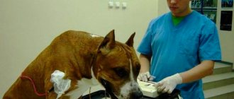
Suspicion of mitral valve endocardiosis in a dog
Causes of endocardiosis in dogs
The only established cause of the development of pathology is considered to be a hereditary factor acting in conjunction with age. The presence of genes in which EMC is encoded does not mean that the dog will certainly get sick. This is confirmed by the fact that not all pets that are at risk by breed have heart problems. Inflammatory diseases, chronic emotional and physical overload, as well as other factors that have so far been little studied can trigger the “launch” of EMC.
That is, the conditions under which the dog is kept and its diet do not affect the development of EMC in any way. Another thing is that a sick animal requires a special lifestyle and diet, but this is a separate topic.
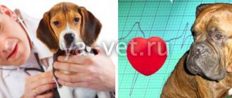
Dogs with heart problems can live long lives
What etiological factors are at work?
Mitral endocardiosis in dogs (MEC) is characterized by thickening of the mitral valve and loss of its elasticity, as a result of which the valves do not close completely.
Valvular endocardiosis develops as a result of the action of two main factors on the body: genetics and age. In veterinary practice, this is considered the most adequate cause of the development of the disease and is considered first when the animal is admitted. But this does not mean that all dogs without exception that have a “crooked” gene will necessarily have heart problems from birth.
The “triggering” of endocardiosis can provoke:
- inflammatory processes in the body, systematic hypothermia, chronicity of the process with any localization;
- constant stress, improper maintenance, emotional overload, worries;
- physical activity, long-term forced training, long-distance running.
Age is considered a factor in the progression of the disease, this is explained by long-term observation of dogs susceptible to EMC. It was found that endocardiosis is rare in young and young individuals, even if they are at risk. The onset of the disease is noted in middle-aged dogs, the peak of pathology occurs at 8-11 years.
Important! Endocardial disease in dogs is more common in small and medium-sized breeds. Predisposed: Schnauzers, Pekingese, Toy and Small Poodles, Chihuahuas, Spitz, Spaniels. Their large dogs the mitral valve is the weakest point in Dalmatians, Germans.
Which dogs are at risk?
Most often these are representatives of small and medium-sized breeds:
- Chihuahuas and toy terriers;
- Spitz and dachshunds;
- Pekingese and poodles;
- Yorkshire terriers and French bulldogs.
Large pets (shepherds) also occasionally get sick, but the process is easier for them. Veterinarians note the highest predisposition to EMC, as well as to its earliest manifestation, in Cavalier King Charles spaniels. The relationship between the weight of the animal and the risk of EMC is clearly expressed in miniature dogs: the lighter the dog, the higher the risk of pathology and the more severe its course.
Miniature breed dogs are more likely to suffer from mitral valve endocardiosis.
Mitral valve endocardiosis in dogs, treatment regimen
Mitral valve endocardiosis in dogs - what is important for owners to know. The heart of a dog is a symbol of devotion and fidelity at all times. But despite their energy and endurance, our faithful friends, especially with age, begin to suffer from cardiovascular diseases, and this often takes owners by surprise. To spot signs of heart failure in time, you need to know very little and be attentive to any changes in your dog's behavior. Then, as a result of a timely diagnosis and adequate therapy, your beloved dog will warm your life with its presence for a long time.
What to do if your beloved dog is at risk
Calm down and understand that the risk of getting sick is not a disease, but it is important to understand that such a pet needs special attention. The owner should:
- periodically examine the dog to have an idea of the state of its health;
- control your pet’s breathing, and you will have to learn to determine its frequency at rest. Normally, it should not exceed 27 breaths/min. If it turns out to be higher, this is a cause for concern and consultation with a veterinarian;
- feed the dog in a balanced and correct manner so that it does not become overgrown with fat. Excess weight in itself will not cause ECH, but it greatly increases the load on the heart
As long as the dog is active, eats normally, breathes freely and looks good, he is healthy.
A pet prone to heart disease is a subject of special concern for the owner
General information about the disease
It is believed that EMC is a pathology exclusively in mature animals, since it develops and makes itself felt only in the second half of life. To understand the nature of endocardiosis, you will have to remember how the heart works. It is, in fact, a muscle biopump that ensures constant blood movement through the veins and arteries. Dogs, like humans, have a four-chambered heart. Between its sections there are valves that allow blood flow in one direction. Affected by the disease, over time they become deformed, become thicker and stop closing as tightly as is physiologically necessary, so the blood begins to flow back, and the pressure in the heart increases. Because of this, stagnation forms and pressure increases in the vessels adjacent to the heart.
Structure of a dog's heart
Problems with the left valve cause pulmonary edema, a fatal condition, and when the right valve is affected, fluid accumulates in the abdominal cavity (effusion). Statistics show that of all the heart valves, the mitral (left) is affected more often than others, therefore:
- in 70% of cases, endocardiosis develops on the left side;
- in 5% - on the right;
- in 25% of cases there is bilateral damage.
This explains the symptoms of EMC, the most typical of which are shortness of breath, fainting and signs of extreme fatigue.
Endocardiosis: what kind of disease is it?
EMC cannot be considered solely a “disease of old age,” although it develops in the 2nd half of a dog’s life. The heart is a four-chamber biopump that ensures blood movement through vessels of different sizes. Between its sections there are “plug” valves that allow blood to move only in a certain direction. With endocardiosis, the mitral valve ceases to close tightly, a “leak” occurs, and blood flows back, which creates increased pressure in one of the sections.
A failure in one mechanism always entails an imbalance in other anatomical structures. As a result of a critical accumulation of blood volume, it stagnates and the pressure in the vessels increases.
Important! Deformation of the left valve entails the development of pulmonary edema; if the right valve malfunctions, effusion (liquid fraction) begins to accumulate in the abdominal cavity.
According to statistics from cardiologists at the Ros-Vet EC, pathology is most often detected:
- in the left side of the heart (70%);
- in the right (5%);
- bilateral involvement (25%).
It is precisely this pathogenesis of the development of the disease that is responsible for the typical symptoms of endocardiosis in dogs - fainting, shortness of breath, high fatigue. If they appear, the owner must immediately take the pet to a veterinary clinic for a full examination.
Symptoms
EMC in dogs can develop asymptomatically for years. Even chronic heart failure (CHF), a typical symptom, does not appear immediately in some cases. It is worth special mention in the context of the pathology under consideration.
CHF is an irreversible phenomenon. The veterinarian has to take this into account when asked about the prognosis. The dynamics of cardiac failure in dogs are classified according to the stage of the underlying disease and accompanying signs:
1. Asymptomatic period. During this time, the animal coughs occasionally, may breathe heavily and show fatigue only when:
- great mental stress;
- physical stress.
It would seem that everything is within the normal range.
2. The appearance of symptoms. During this period, the dog quickly gets tired and exhibits breathing problems even under moderate, habitual loads, and it is possible:
- Increasing symptoms. The dog becomes less mobile, cannot catch its breath for a long time with minimal exertion, and sometimes even at rest;
- Noticeable deterioration in condition. The dog increasingly looks lethargic, has difficulty breathing, does not ask to go for walks, preferring to lie on his favorite bedding. Periodically, a protracted and debilitating cough bothers you, and your limbs may swell.
Dogs with heart problems get tired quickly
The presence of signs of CHF is not an unambiguous confirmation of mitral valve (MV) endocardiosis - cardiac failure can also appear for other reasons, but it is always a reason for prompt consultation with a doctor.
In addition, it is worth knowing that edema due to heart problems in dogs is rare, as it means an extremely severe degree of damage to the right ventricle.
Shortness of breath in a dog is one of the signs of EMC
You can also suspect the development of EMC in a pet in cases where:
- the pulse, which can be determined by placing your hand on the femoral artery of the pet, is weak and incomplete;
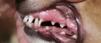
- the dog has hoarse breathing and a cyanotic tongue;
- mucous membranes become pale (a sign of anemia);
- the dog eats little and drinks a lot;
- the animal breathes with an open mouth even at home, at rest;
- pink foam appeared in the nostrils and nasopharynx (one of the symptoms of pulmonary edema);
- ascites (dropsy) develops;
- veins in the neck are swollen;
- The animal often jumps up in its sleep, as it suffocates while lying down.
Problems with the mitral valve can cause your dog to faint during coughing or physical activity.
Symptoms of canine endocardiosis
The most common symptoms of endocarditis in dogs are coughing (in some cases with white foam, which the dog swallows back), shortness of breath and exercise intolerance. Sometimes the dog becomes restless at night due to difficulty breathing when lying down. Fainting also occurs in some cases during physical activity or anxiety, during coughing (so-called cough fainting) or associated with supraventricular tachyarrhythmia.
An increase in coughing attacks is observed after drinking and physical activity. Sustained diffuse pulmonary edema develops, leading to moist rales. Over time, damage develops not only to the left, but also to the right side of the heart, this entails dilation of peripheral veins, ascites, and enlargement of the liver. Due to myocardial degeneration and stretching of the atria, their premature contraction often occurs - paroxysmal tachycardia.
The peculiarity of this disease is that it occurs without symptoms for the first few years.
Holosystolic murmur when listening to the heart is more pronounced in the upper left part (between the 4th and 6th left ribs) and is typical for those patients who have mitral regurgitation. This noise can travel in all directions. Mild regurgitation is often inaudible or is heard exclusively in early systole (in this case, a protosystolic murmur occurs).
Physical activity or emotional arousal often leads to an increase in the intensity of soft noises during mitral regurgitation. At further stages of endocardiosis, a more pronounced murmur is observed. In dogs that have massive regurgitation and severe heart failure, the murmur may be soft or inaudible. In some cases it resembles a musical tone.
Some animals with chronic mitral valve endocardiosis have a mid-late systolic clicking sound, with or without a murmur. A galloping sound is sometimes heard in the upper left side of the heart in dogs with advanced disease. Tricuspid regurgitation usually causes a holosystolic murmur, which is heard more clearly in the left upper part of the heart.
The pulsation of the jugular vein, vibration on the right in the area symmetrical to the location of the heart on the left, as well as features of the noise heard in the valve projection help to differentiate the radiating noise of mitral regurgitation from the noise of tricuspid insufficiency in the right half of the chest.
Pulmonary sounds when listening can be both normal and pathological. Harsh, intense breathing and crepitation sounds heard at the end of inspiration (most clearly heard in the central fields) occur with pulmonary edema. Due to rapidly developing pulmonary edema, expiratory and inspiratory wheezing and shortness of breath increase.
Some dogs with mitral regurgitation have abnormal pulmonary sounds, which are caused largely not by heart failure itself, but by concomitant respiratory disease. Sinus tachycardia is common in dogs diagnosed with congestive heart failure. Dogs with chronic pulmonary disease often have sinus arrhythmia with a normal heart rate. Due to pleural effusion, lung sounds are weakened.
Clinical examination
During auscultation of a dog that is not currently showing any clinical signs, the following is revealed:
- systolic click (at an early stage): high pitch, the presence of a sharp sound between the heart sounds S1 and S2; this sound is often mistaken for an additional heart tone (which causes the appearance of a gallop rhythm);
- systolic apical murmur of the tricuspid or mitral valve;
- early or late soft holosystolic murmur, corresponding to moderate or severe regurgitation.
A complete examination of the dog reveals:
- loud heart murmur (levels 4-6/6);
- weakened 1st tone;
- ventricular arrhythmias, in particular atrial fibrillation, indicating a severe course of the disease and a poor prognosis;
- weak and rare pulse observed in the femoral artery;
- pallor of the mucous membranes;
- tachypnea, orthopnea, respiratory distress;
- wheezing, pulmonary edema;
- pink foam in the nostrils, as well as in the nasopharynx in the presence of acute and severe pulmonary edema;
- ascites, swelling of the neck veins (in case of right-sided heart failure).
Diagnostics
In addition to visual examination, including auscultation (listening) and palpation (palpation), the following methods are used as diagnostic methods for EMC in dogs:
Chest X-ray
The image allows you to see the degree of enlargement of the atrium and ventricle on the left, as well as assess the dynamics of the process (on a repeat image, a month later). It is noteworthy that the increase in this part of the heart to a critical size can occur without signs of heart failure.
Dog in the x-ray room
The image of the right atrium is also carefully analyzed, although its enlargement may be masked by abnormal volume of the left atrium. Among other things, radiography allows you to assess the condition of the lungs - their size and structure, which is also extremely important. Based on the results of radiography, it is possible to determine the stage of EMC and the degree of influence of the process on the lungs.
Electrocardiography (ECG)
It reveals pathological changes in the heart muscle by the rhythm of its beating, which, depending on the condition, is defined as:
- sinus, as well as paroxysmal or constant supraventricular tachycardia;
- ventricular extrasystoles and atrial fibrillation;
- extrasystole of the supraventricular part.
Among other parameters, an ECG allows you to evaluate the functionality of the mitral valve.
Four-legged patient on ECG
Echocardiography (Echo CG)
It can also be used to determine the size, structure and functionality of the heart. In case of insufficiency of the atrioventricular valves, which includes the MV, effusion (fluid) is often detected in the pericardium (the sac around the heart), as a result of congestive heart failure. Echo CG for endocardiosis in dogs is carried out in Doppler mode to obtain comprehensive information about the anatomical changes and hemodynamics (the speed of blood flow through the chambers and vessels) of the heart.
Pet on Echo KG
Ultrasound of the heart
This type of research is widely used for diagnosing cardiac and other pathologies. Ultrasound allows you to see the organ being examined on the screen, evaluate its function, tissue structure, and also determine the degree of blood supply and other indicators.
Dog on ultrasound
Video - Ultrasound - examination of a dog's heart
Blood and urine tests
They are uninformative. With EHR, indicators of laboratory blood tests (general and biochemical), as well as urine, may be normal or show deviations due to heart failure, as well as pathologies of non-cardiogenic origin:
- pharyngitis and pneumonia;
- tracheal collapse and dirofilariasis;
- chronic bronchitis and bacterial endocarditis,
- pulmonary fibrosis;
- other conditions.
Blood and urine samples for analysis
When receiving referrals for different types of pet examinations, some may feel like there are too many of them. It's a delusion. None of the methods can be singled out as the main one or, conversely, unnecessary because only based on the totality of their results, a specialist is able to objectively assess the health of a four-legged patient and the degree of development of pathology, as well as plan therapy and predict its results.
Dog blood tests
Chronic heart failure in dogs
Chronic heart failure (CHF)
is not an independent separate disease, it is a complex of disorders in the cardiovascular system (impaired pumping function of the heart, congestion in the pulmonary and systemic circulation, etc.) that accompany the primary disease.
The development of the disease leads to changes in hemodynamics and the structure of the heart itself as an organ. The more pronounced the changes in the heart, the less important it is what exactly served as the primary trigger of the disease - CHF becomes the main problem. In most cases, it is impossible to eliminate the cause, so treatment of already developed heart failure is carried out.
Symptoms of CHF in dogs
At the initial stage, symptoms are not pronounced, but as the disease progresses, clinical signs become more noticeable.
Symptoms such as:
CHF is classified according to the degree of development of symptoms. There are four functional classes of CHF.
- 1 functional class.
Symptoms are absent or appear during severe psycho-emotional and physical stress. - 2 functional class.
Symptoms may appear with moderate exercise, but do not appear at rest. - 3 functional class.
Symptoms occur with moderate exercise and rarely at rest. - 4 functional class.
Symptoms appear at rest, and moderate physical activity leads to a sharp increase in symptoms.
Endocardiosis
is a chronic degenerative valve disease in which the valve thickens and becomes deformed. This pathology almost never occurs in cats. The most commonly affected valves are the mitral (left) and tricuspid (right) valves in dogs. Isolated endocardiosis of the tricuspid valve is extremely rare.
Mitral valve endocardiosis may be hereditary. As a rule, it occurs in older dogs of small and medium breeds. Vulnerable breeds include dachshunds, poodles, cocker spaniels, and Yorkshire terriers. Degenerative collagen nodules form on the valve leaflets, which deform the leaflets.
The latter do not close tightly and allow reverse blood flow (so-called regurgitation
) from the ventricle to the atrium. The atrium stretches and increases in size. This stage of compensatory increase in atrial volume allows most patients to remain without symptoms of CHF for quite a long time.
With further progression of the disease, clinical signs of CHF appear: cough, exercise intolerance, shortness of breath, the risk of developing pulmonary edema - a condition that poses a serious threat to life. Various arrhythmias can complicate the disease.
With severe endocardiosis, sudden rupture of the chords and even rupture of the atrium wall can occur, leading to death within a very short time.
According to the severity of regurgitation and the development of symptoms, 4 stages of mitral valve endocardiosis (MV) are divided:
- Stage 1.
The MV is deformed, but the atrium is not enlarged. The animal has no clinical symptoms. - Stage 2.
The atrium and ventricle are slightly enlarged. There are no symptoms. - Stage 3.
Increased pressure in the left atrium. Moderate congestion in the lungs. The leading symptom is cough. - Stage 4.
Decreased pumping function of the heart. High risk of pulmonary edema. An increase in the size of the liver is often observed, and there may be ascites (free fluid in the abdominal cavity).
How dangerous is the disease?
To accurately determine the animal’s condition, it is necessary to undergo a comprehensive cardiological examination. It includes anamnesis, physical examination, chest x-ray, echocardiography, and electrocardiogram.
The prognosis for life in dogs is quite variable and can range from several months to several years, depending on the stage of the disease, response to therapy and the owner’s willingness to carry out long-term therapeutic treatment. Treatment of CHF usually consists of strictly following an individually selected regimen of medications (pills).
Source: https://zoostatus.ru/lechenie/bolezni-sobak/khronicheskaya-serdechnaya-nedostatochnost-u-sobak-endocardioz/
Stages of the disease
The course of EHR occurs in 4 stages, each of which is characterized by certain changes in the heart.
Table 1. Stages of EHR development and processes accompanying them.
| Stage | Changes that occur in a dog's heart |
| I | There are no changes yet on the part of the left atrium and ventricle, but the tissues of the mitral valve are already susceptible to degenerative processes that lead to the formation of nodular defects. |
| II | The area of damage increases and they merge. The process involves the valve chords. |
| III | Growths appear on the urinary tract, its chords thicken, and their tissues become coarser. The thickness of the valve itself also becomes larger, and its flexibility deteriorates. In the basal part there is a high risk of microbleeding and the formation of calcifications. |
| IV | The valve tissues rapidly degrade. It becomes deformed, the edges curl up. In especially severe cases, the chordae rupture due to loss of elasticity. As the muscles of the ventricle contract, the valve functions increasingly worse. |
Four-legged patient with MV endocardiosis
Echocardiographic diagnosis of heart valve endocardiosis in dogs
Using echocardiography in dogs with endocardiosis
a number of pathological changes are revealed:
- the leaflets (especially the septal ones) of the mitral and/or tricuspid valves are deformed, thickened, shortened, with nodes at their end parts
- increase in the anteroposterior size of the left atrium and the ratio of the diameter of the left atrium to the size of the aortic root
- an increase in ESR in the initial stage of the disease; subsequently, ESR and the mitral-septal separation index also increase (EPSS indicator, which correlates with the severity of systolic dysfunction)
- hyperkinesis of the free wall of the left ventricle and interventricular septum, with their normal size or slight hypertrophy
- at the initial stage there is an increase in the shortening fraction and ejection fraction of the left ventricle, at the later stage there is a decrease in them
- systolic prolapse with parachute of the leaflet into the atrium cavity
- sometimes it is possible to detect a rupture of the chordae tendineae
- regurgitant jet with turbulence using color Doppler mapping
Conservative treatment
It includes the use of medications prescribed by a doctor. Tablets and mixtures chosen according to your own judgment, especially if they once helped neighbor Murka, can cause harm. The physiology of pets is such that many “human” drugs do not work on them or work differently. It is noteworthy that even certain cat medications are not suitable for dogs, and vice versa. As for traditional treatment methods, it will not be of any use to dogs with EHR. Infusions and decoctions of herbs are powerless against mitral valve degeneration.
Medications to stimulate heart function are the basis of treatment for EMC in dogs.
When following the recommendations of a veterinarian, you should understand that conservative therapy does not promise complete healing, since such a result cannot be achieved with medications. Her goals:
- control and relief of symptoms of heart failure;
- maximum support for heart functionality;
- correction of neurohormonal hyperactivation, stimulating the development of pathology.
Treatment with drugs is indicated at stages when the symptoms of the disease are obvious. Doctors believe that in the early period, when the EHR reveals itself exclusively as a heart murmur detected on the ECG, and the dog is cheerful and cheerful, there is no need to give it medication. Later:
- To prevent and eliminate edema, diuretics (diuretics) are prescribed;
- Angiotensin-converting enzyme (ACE) inhibitors are used to stimulate cardiac activity. As for more powerful pacemakers, it is not recommended to use them while heart failure remains minimally expressed;
- to combat increasing arrhythmia, beta blockers and calcium channel blockers are used;
- To stop coughing, the dog is given vasoconstrictors and diuretics. Why? Because with EMC, the cause of cough is the accumulation of fluid (effusion) in the bronchi. Chronic cough over a long period of time inevitably forms bronchitis (inflammation of the bronchi), which is treated with corticosteroids.
Table 2. Approximate list of medications that form the basis of EMC therapy in dogs.
| Drugs | Expected Result |
| ACE inhibitors or pimobendan, digoxidine and others | Relieving symptoms of heart failure, stimulating heart function |
| Furasemide, spironolactone, hydrochlorothiazide, nitroglycerin and others | Improving urine flow, normalizing blood pressure |
| Hydrocodone, butorphanol, theophylline and others | Cough elimination |
| Kupentil and others | Slowing down blood clotting processes |
| According to indications, other medications are also prescribed | |
The veterinarian determines which medications and in what doses to treat the dog. When choosing methods and methods of therapy, he will be guided by objective data about the condition and well-being of the animal.
Dangerous condition of the pet - endocardiosis
Canine endocardiosis is a progressive myxomatous degeneration of the atrioventricular valves. The disease is characterized by the progressive deposition of polysaccharides in the heart valves, which over time disrupts their normal functioning and leads to the development of congestive heart failure.
Endocardiosis is the most common heart disease in dogs. In the veterinary literature, it has many synonyms, such as myxomatous mitral valve disease, chronic degenerative valvular disease, chronic valvular fibrosis, etc.
The heart of an animal, like a human, is conventionally divided into two halves (right and left), each of which consists of an atrium and a ventricle between which there are valves; in the left section the valve is called the mitral valve, in the right section - the tricuspid. These valves ensure normal blood flow during contraction of the heart muscle.
The exact causes of the disease have not been established; the suspected cause is a hereditary defect of connective tissue.
Clinical signs
Older dogs of small and medium breeds, such as dachshunds, chihuahuas, Pekingese and others, are predisposed to the disease. In large breed dogs, this pathology is not clinically significant.
With refractory form
When congestive heart failure becomes refractory (that is, unresponsive or difficult to respond to drug treatment), therapy is intensified or revised, taking into account the individual characteristics of the patient:
- To combat recurring pulmonary edema, the dosage of diuretics is increased and the animal’s mobility is limited for several days. As the condition improves, the volume of drugs may be reduced to the original level or remain slightly higher.
- To stimulate cardiac activity, ACE inhibitors are given to the maximum permissible amount. As for stronger drugs (for example, Digoxidine), their volume remains standard if the concentration of the drug in the blood does not become subtherapeutic, that is, insufficient for a therapeutic effect.
During the therapy, it is important to monitor the four-legged patient’s kidney function, as well as the level of electrolytes in his blood. Salt is excluded from the dog’s diet during this period. In some cases, when the animal is receiving maximum doses of ACE inhibitor and diuretics, the veterinarian will add a minimum of hydralazine or amlodipine, with constant monitoring of blood pressure.
Refractory form of EMC requires intensive therapy
Periodic jumps in heart rate can provoke decompensated congestive hypofunction (insufficient work) of the dog’s heart, as well as sudden weakness or fainting. Despite periodic relapses of symptoms, many animals suffering from chronic atrioventricular valve regurgitation remain able to lead normal lives for several years after the earliest signs of heart failure appear.
Treatment and prognosis for endocardiosis
Drug treatment for canine endocardiosis is aimed at controlling the symptoms of congestive heart failure, supporting heart function, and correcting excessive neurohormonal activity that contributes to the development of the disease. Drugs that reduce the size of the left heart ventricle (diuretics) reduce the volume of regurgitation and fibrous mitral annulus. Drugs that promote arterial vasodilation increase cardiac activity and reduce the amount of regurgitation by reducing blood pressure.
The progression of the disease leads to the need for regular assessment of the dog’s condition, as well as periodic adjustments to treatment. In many dogs with severe mitral regurgitation, compensation is maintained for several years with proper treatment. Heart failure develops slowly in a significant proportion of affected dogs, while in other animals severe pulmonary edema occurs.
Alternating episodes of decompensation in animals undergoing long-term treatment for heart failure can often be successfully reversed. When treating, it is necessary to take into account the clinical status of the animal and the factors that in a particular case complicate the course of the disease. Surgeries such as mitral annulus repair and other valve replacement and restoration techniques may be used in some cases, but are not widely available.
Currently reading:
- What to do if your dog has heart failure
- Thyroid dysfunction in dogs (hypothyroidism)
- Prognosis and treatment of kidney failure in dogs
- Compositions, dosages of Hills food for all dogs
Surgery
The possibilities of modern veterinary medicine provide a chance to completely rid dogs of EHR problems through surgery. The surgery involves replacing the mitral valve, using a purse-string suture technique to shrink the holes. Despite the fact that the experience of such operations in Russia is still small, the country has veterinary cardiac surgeons capable of performing them at a high level.
The indication for surgery is congestive heart failure, which cannot be eliminated with medication. EHR is just such a case. However, there is one important clarification: surgical intervention can be counted on only when the animal has not yet developed secondary cardiomyopathy (impairments in the structure and function of the heart that arise as a result of the primary disease). This is another reason to remind pet owners that a sick pet should see a doctor as soon as possible.
Surgery in a veterinary clinic
Patient monitoring:
In asymptomatic dogs, x-rays are performed when the first heart murmur is detected and then every 6 to 12 months thereafter to detect progressive cardiomegaly.
The frequency of re-evaluation of animals receiving treatment for heart failure depends on the severity of the disease and the presence of complicating factors.
Patients with newly diagnosed or decompensated congestive heart failure should be evaluated more frequently. After an episode of congestive heart failure - once a week during the first month of treatment; The chest x-ray and ECG may be repeated at the first weekly check and at subsequent visits if any changes are observed on the general physical examination.
Dogs with chronic heart failure whose symptoms are well controlled may be evaluated less frequently, usually several times a year.
Caring for a sick dog
A sick dog requires special treatment and control. First of all, you should take care of a visit to the animal clinic. It is hardly worth calling a veterinarian to your home, since without an examination he will not be able to make an accurate diagnosis and prescribe treatment.
Before visiting the clinic and upon returning home, you should:
- provide your pet with complete rest. This means that family members, especially children, should be told about the dog's condition and warned not to harass it. It is advisable to reduce the volume of receivers in the apartment to a minimum;
- organize a flow of fresh air into the room where the dog is lying. To do this, open the window, and in the warm season, open a window or balcony. The floor where the dog lies on the bedding must be clean, so wet cleaning is needed daily;
- Make sure your pet has free access to a bowl of fresh, clean, cool water. You shouldn't force your dog to eat when it doesn't want to. Salt and other foods that provoke severe thirst should be eliminated from the diet as much as possible;
- If the dog has stopped caring for himself, the owner will have to take on this mission - wipe his eyes daily with cotton swabs soaked in cooled strong tea. Using cotton wool or a piece of soft cloth soaked in petroleum jelly, carefully remove crusts from the nose and its passages so that the dog can breathe easier.
The pet is sick
After a visit to the doctor, an attentive, responsible and caring owner strictly follows all recommendations for caring for the animal, feeding and treating it.
Complications
Possible complications of EHR in dogs include:
- persistent heart rhythm disturbance;
- phenomena of decompensation affecting the pulmonary circulation.
The most severe consequences of MV endocardiosis are:
- chord separation;
- rupture of the atrium on the left side, leading to lightning shock and death of the pet.
Dog with severe endocardiosis
Forecast
The outcome of the disease depends on the stage it was at the time of its detection, as well as on how timely and complete the therapy was. Unfortunately, more often a dog ends up in a veterinary clinic when severe health problems are already evident. In this case, it is difficult to save the animal, and if this is possible, then its life expectancy is unlikely to be long.
Owners should know that:
- dogs with mild, less often moderate, mitral valve insufficiency can live long and happily if the disease does not develop;
- animals with obvious and clinically confirmed symptoms of EMC who have undergone therapy can live for at least another year.
EHR detected at an early stage is not a death sentence
Treatment
Before starting treatment, differential diagnosis should be carried out so as not to be confused with pathologies such as: mitral valve dysplasia, regurgitation cardiomyopathy, infective endocarditis, systemic hypertension. In addition, the doctor’s prescriptions will depend on the stages of the disease. Stages of pathology:
- asymptomatic;
- stage of heart failure;
- chronic relapsing stage.
Before treatment, differential diagnosis is carried out.
Asymptomatic stage
- At the asymptomatic stage, therapy consists mainly of monitoring the animal’s condition.
- Regularly monitor blood pressure, take x-rays of the sternum every year, and maintain your pet’s optimal weight.
- But you also need constant moderate physical activity.
- Avoid excessive overvoltage.
- Reduce or completely eliminate your intake of salty foods.
During the asymptomatic stage, you need to monitor the dog's weight.
For heart failure
- For heart failure, furosemide, amlodipine, ACE inhibitors along with pimobendan, digoxin, spironolactone, hypothiazide are prescribed.
- Antiarrhythmic therapy is recommended if necessary.
- Avoid physical activity, monitor your heart rate and breathing rate. If necessary, thoracentesis is used to eliminate pleural effusion.
The drug Furosemide is used for heart failure.
Chronic form
The chronic form requires an increase in the dose of furosemide and inhibitors.
And also increase the dosage or frequency of diuretics. If secondary pulmonary hypertension occurs, sildenafil is prescribed. Use antitussives or bronchodilators.
For secondary pulmonary hypertension, the drug Sildenafil is prescribed.
conclusions
Endocardial disease in dogs progresses slowly. If the dog’s condition changes negatively, one day it may cause its death. In most cases, this is due to the fault of the owners who did not find the time and opportunity to visit a doctor or to unconditionally follow his recommendations. Only those dogs who are lucky with an intelligent and caring owner have a chance to live.
A dog's happiness and health depend on its owner