Each growth on a dog's eyelid can be fraught with danger in the form of a malignant tumor, which is life-threatening if the process spreads. Therefore, if any formation is detected in this part of the eye, it is recommended to show it to a veterinarian/oncologist. If the doctor has doubts about the benignity of the neoplasm, they will conduct a histological examination and make an accurate diagnosis. Early detection of even the most dangerous forms of cancer can save your pet's life.
Possible reasons
The following will list the pathologies that most often cause growths to appear in dogs of different ages:
- Eosinophilic granuloma. A common problem among decorative breeds prone to allergic reactions. Often found on the eyelids of dogs in the form of a small pea. May be single or multiple. The reason may lie in poor diet, insect bites or taking antibiotics. Single nodules tend to resolve on their own without any treatment. For multiple granulomas, glucocorticosteroid therapy may be prescribed. In all cases, the prognosis is favorable.
- Warts. There are breeds that have a tendency to develop warts all over their body. They are often found in pugs, mastiffs, bulldogs and terriers. They do not pose any danger, but if they are localized in problem areas, they will require removal using coagulation or cryodestruction. If a dog has a small formation on his eyelid that looks like a wart, then under no circumstances should you try to remove it yourself, as there is a risk of infection and the onset of an inflammatory process, which can seriously harm the eye.
- Papilloma. Formations that are very similar in appearance to warts, but differ from them in that they are prone to a malignant course. First, a small growth the size of a grain appears on the eyelid, which in a few months can grow to the size of a large pea. Most oncologists are inclined to believe that all papillomas must be removed followed by histological examination. This can be done using electrocoagulation or laser. For calm pets, local anesthesia will be sufficient. Sedation is used in cases where there is a risk that the animal will behave aggressively, which may affect the quality of the operation, given that it is performed on the eye. There is one problem with papillomas - they often appear again in the same place. Most often, English and French bulldogs suffer from them, since their entire muzzle has been covered with them since childhood.
- Melanoma. Sounds like a death sentence for humans, but is not so dangerous for animals. It occurs very often, but does not have a tendency to relapse and metastasize, therefore it is considered a conditionally malignant type of cancer. It may appear as a small black growth on the eyelid, which does not bother the dog in any way until it begins to ulcerate. The diagnosis can only be confirmed using biopsy and histology. In all cases, it must be completely removed surgically within healthy tissue. The prognosis is favorable provided that it is removed in a timely manner.
- Demodecosis. If localized in the muzzle area, it can cause small growths to appear on the dog’s eyelids. Hair loss and flaking are also observed in this area. In puppies, this phenomenon is called juvenile demodicosis. Everything goes away within 2-3 weeks. In adult dogs, the activity of this type of mite can provoke a sharp decrease in immunity or cancer, so it is recommended not to delay identifying the cause. Scraping or plucking the affected area will confirm the presence of a mite.
- Sebaceous gland cyst of the eyelid. Hard lumps the size of a grain of rice. May appear in both eyes. They do not bother the dog at all (no itching or pain). There is a tendency towards inflammation. Without cytology it is impossible to confirm the diagnosis. Large cysts must be surgically excised.
- Barley. A common occurrence among all breeds. The appearance can be triggered by hypothermia, a bacterial infection or severe stress. A round pink growth appears on the eyelid. As the process progresses, purulent contents begin to be released from it. Barley can be internal or external. May affect the lower or upper eyelid. In severe cases, the eye may even become swollen. For treatment, eye anti-inflammatory drugs (tetracycline and erythromycin ointments) are used.
- Sebaceous gland carcinoma. A malignant formation on the eyelid that must be immediately removed. Such tumors can ulcerate, grow to large sizes and metastasize. An effective method of removal is to influence the formation with liquid nitrogen, which leads to the destruction of cells and the disintegration of the tumor itself. There is a chance of relapse (in 10-15% of cases during the first year after surgery), so the dog is advised to regularly examine the eyelid area.
Well, don’t forget that there are hair follicles on the eyelids that can periodically become inflamed (due to hypothermia). This process is accompanied by the appearance of a small red nodule on the eyelid, which disappears within a couple of days if the course is not complicated by a bacterial infection.
Loading…
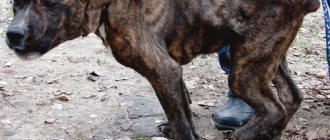
Dogs' eyes are very sensitive and are often subject to various injuries and damage. If a dog has a swollen eyelid, this is an alarming symptom that requires increased attention. There are several reasons for this phenomenon; characteristic symptoms will help you understand them.
[custom_ads_shortcode1]
Pathological anatomy
Sebaceous gland carcinoma develops as a result of malignant proliferation of sebocytes, from which the tumor developed, difficult due to the diffuse or multicentric origin of the tumor.
Although there are several ways to classify sebaceous gland carcinoma, most authors recognize four histological types: lobular, comedocarcinoma, papillary, and mixed (9). Histologically, sebaceous gland carcinoma can be further subdivided into well-, moderately and poorly differentiated variants. The more common lobular type simulates the normal structure of the sebaceous gland: poorly differentiated cells are located on the periphery, and more highly differentiated lipid-producing cells are localized centrally. In the comedocarcinomatous type, the lobule has a large necrotic central core surrounded by living tumor cells. In the papillary type, papillary processes and zones of sebocyte differentiation are observed. With a mixed type, any combination of the other three types is observed. When a tumor develops from the Zeis glands and is limited to their border, the affected glands are microscopically detected near the edge of the eyelid with an intact cartilaginous plate.
A widely known and often mentioned feature of sebaceous gland carcinoma is the ability for intraepithelial (pagetoid) spread into the epidermis of the eyelid and the epithelium of the conjunctiva. According to various estimates, such an increase is observed in 44-80% of cases (4.9). In an analysis of a group of 25 patients, Chao et al. (4) found that pagetoid tumor growth was associated with a significantly higher risk of exenteration.
Why do my eyelids swell?
As a rule, the main causes of swelling are inflammation and injury. This symptom is also a sign of:
- allergies;
- conjunctivitis;
- blepharitis;
- barley;
- insect bite.
Specific symptoms and the mechanism of development of each of the listed diseases will help you understand the approximate causes of edema. At the same time, it is recommended not to self-medicate, but to entrust the health of the animal to a specialist.
[custom_ads_shortcode2]
Allergic reactions in dogs
If your dog's upper eyelid is swollen, it may be due to an allergic reaction. There are:
- Seasonal allergies.
- Contact dermatitis of the eyelids.
- Food intolerance.
Seasonal allergies in dogs are extremely rare. This disorder is characterized by intolerance to the pollen of certain plants. Eye irritation develops due to pollen, which is accompanied by swelling, severe itching, and redness of the entire eye.
Food allergies also rarely cause swelling of the eyelids. In the vast majority of cases, intolerance to food or new food is manifested by damage to the skin around the mouth and ears; the eyes are much less often involved in the pathological process. If this happens, the symptoms spread to the eyes and eyelids, the skin of the ears, the epidermis on the body and the area around the mouth. Symptoms of food allergies: swelling of the epidermis, itching, redness of the eyes, lacrimation, sneezing.
Most often, swelling is associated with contact or allergic dermatitis. This is a specific form of intolerance that develops when the epidermis is irritated by an allergen. Allergies on the eyelids appear due to the ingress of household chemicals, dog bath products or medications.
Another separate type of pathological reaction of the body is intolerance to the poison of certain insects. In this case, swelling and redness develop directly at the site of the bite. This reaction appears in response to a bee or wasp sting, or, less commonly, a mosquito bite.
Recognizing an allergy is quite simple, since its symptoms appear a short time after exposure to the irritant. Signs of a food allergy appear 10-60 minutes after eating, a contact allergy is noticeable after half an hour, a reaction to insect bites - after a few minutes.
Treatment: taking antihistamines, antiallergic eye drops, a cool compress on the eyelid to reduce swelling.
[custom_ads_shortcode3]
Are warts dangerous for dogs?
The neoplasms vary greatly in appearance but are caused by a single “agent.” Warts can appear on the dog's body or on mucous membranes, most often in the mouth. Skin lesions begin in areas covered with delicate skin - on the paw (elbows), inner thigh, groin area, armpits.
The appearance of warts on the mucous membranes is considered a more dangerous manifestation of the disease. A wart located on a dog’s lip, palate or inside cheek can be torn off when chewing food, which can lead to a secondary complication – infection. Veterinarians note that in advanced stages, papillomas spread from the oral cavity and appear on the dog’s face, more precisely, on the outside of the mouth.
The ears and ear canals of animals are covered with thin skin, and a small layer of subcutaneous tissue is literally penetrated by nerve endings and capillaries. Warts on the ear most often cause itching and discomfort, prompting the animal to scratch. And again the danger of scratching and complications due to infection.
Let's briefly look at the types of papillomas. Externally, the new growths have some similarities with cauliflower, but can vary in:
Size – from 0.1 to 5 mm. It is possible that the size of warts may go beyond the boundaries of the “classical gradation”.
Colors - flesh, brown, gray, yellow and combinations of shades.
Structure – dense, hard, soft.
The method of “implementation” into the skin is ingrown, attached to a stalk.
Papillomas, which are attached to the dog’s body or mucous membrane with the help of a leathery stalk (see photo), literally dangle, which increases the risk of damage. Ingrown warts are dangerous due to possible transformation into a malignant tumor - squamous cell carcinoma. The risk of degeneration does not depend on the color, size and structure of the neoplasm.
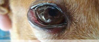
The dog's eyelid is swollen photo
This disease is manifested by inflammation of the mucous membrane of the eye. As a rule, conjunctivitis is bacterial in nature. The pathology is accompanied by severe redness of the eyes, profuse lacrimation, and sticking of the eyelids. In severe bacterial infections, purulent exudate oozes from the eyes, irritating the eyelids and causing them to swell.
For conjunctivitis, treatment is carried out using antiseptic and antibacterial drops. The treatment regimen is selected by the doctor, depending on the dog’s age and general health, since conjunctivitis in animals can be a secondary symptom of serious diseases.
[custom_ads_shortcode1]
What to do when pigment spots appear on a dog?
You need to show the dog to a veterinarian so that he can determine whether the pet has good or bad pigmentation. Any changes in the skin indicate pathologies that are asymptomatic or have just begun to progress.
The doctor will scrape the skin to check for bacteria or fungi. The dog will have to donate blood, urine and fluid from the joints, and undergo an ultrasound of the thyroid gland, kidneys and adrenal glands. Based on the results, the veterinarian will prescribe a course of antibiotics, immunosuppressive medications and ointments.
Important! The veterinarian carries out differential diagnosis to exclude serious diseases that have similar symptoms. These include skin cancer and lupus
Blepharitis
If your dog's lower eyelid is swollen, blepharitis should be ruled out. This pathology is characterized by inflammation and severe swelling of the eyelid, both lower and upper. Blepharitis most often acts as a complication of other diseases.
It can be bacterial, viral, fungal or allergic in nature. Often inflammation develops due to eyelid injuries and infection entering the wound.
Blepharitis is dangerous due to the spread of inflammation to the conjunctiva and cornea. Characteristic signs: swelling of the eyelids, redness of the entire eye, small pustular rash. With blepharitis, severe itching occurs, so the dog may scratch the skin until wounds and crusts form.
Treatment: warm soothing compresses on the eye, antiseptic treatment, antibacterial drops.
[custom_ads_shortcode2]
Melanoma in dogs: symptoms of the ocular form
This type of tumor in animals can form on:
- century;
- cornea and sclera;
- retina and iris;
- conjunctiva;
- choroid.
The main symptoms of the disease in this case are:
- swelling of the eyelids and surrounding areas;
- redness of the conjunctiva;
- corneal clouding;
- frequent tears;
- decreased visual acuity;
- ulcers on the eyelids.
Sometimes sick dogs also experience clouding of the cornea. Often eye melanoma in dogs is accompanied by ulcers on the eyelids. Sometimes, among other things, dark spots form on the animal’s eyeball.
Stye on the eye
Styes in animals appear due to mechanical damage to the eyelid. This disease is bacterial in nature and is very recognizable, as it is a large subcutaneous pimple on the eyelid. The disease develops due to blockage of the sebaceous gland ducts, followed by infection.
Treatment consists of antiseptic treatment of the eyelid and the use of antibacterial agents. In this case, eye ointments with an antibiotic (Tetracycline ointment, Levomecitin) are effective; therapy can be supplemented with Albucid drops, but they cause a strong burning sensation, so it is difficult to apply eye drops to your pet.
Allergic swelling in the eyes successfully disappears a few hours after the irritant stops and antihistamines are taken. If the disease is bacterial in nature, a short course of treatment is necessary. As a rule, the duration of therapy does not exceed 4-5 days.
If a dog's eyelid is swollen so much that it touches the cornea, then it is highly likely that the pet will walk squinting or with one eye closed. This condition is dangerous because if something injures the cornea, then there is a risk of developing an inflammatory process, due to which the lens becomes cloudy and the whites turn red. It is even more difficult if purulent discharge appears, due to which the eyelashes stick together and the hair under the eyes constantly gets wet.
[custom_ads_shortcode3]
Causes of skin lesions
The disease is considered idiopathic (for unknown reasons), and is more often diagnosed in poodles and dogs, Akita Inu, Dachshund, Samoyed, Vizsla, etc., both purebred representatives and their crosses. Presumably, this is a genetically inherited pathology that is transmitted to puppies in an autosomal recessive manner.
Clumping of hair on the face, typical of keratinization defects.
The pathogenesis of the disease has not been identified; it is believed that it can manifest itself in the case of
- congenital defects of the sebaceous and sweat glands. The secretion “leaks”, and the animal’s body reacts to it as a foreign body, with inflammation;
- changes in lipid metabolism (lipids negatively affect the formation of sebum);
- disruption of the process of primary keratinization, here atrophy of the sebaceous glands and an inflammatory process around them occurs.
There is a hypothesis that the disease is of cell-mediated autoimmune origin. When diagnosed, this is confirmed by a decrease in the concentration of macrophages and T-lymphocytes if cyclosporine is used. Secondary destruction of the sebaceous glands is expressed by signs characteristic of hyperkeratosis. It often develops with extensive demodicosis, leishmaniasis and other skin diseases.
Analysis of the most common reasons
Unfortunately, it is not always possible to notice in time that a dog’s eyelid is swollen. This is especially true in cases with animals that have a thick undercoat (Newfoundland, Chow Chow, some types of shepherd dogs and poodles). Or the owner simply noticed the problem already when accompanying symptoms appeared in the form of a non-opening or sour eye.
Making a diagnosis is of great importance, since it is necessary not only to cope with the inflammatory process, but also to exclude its root cause. Therefore, first of all, pay attention to the following options:
- Insect bites. Particularly dangerous are the bites of wasps and bees, which occur mainly in the autumn and spring periods. After being bitten by stinging insects, the dog's eyelid swells and his eye becomes swollen. This is especially true for bites on the brow ridges, the temple area and the eyelid itself. It is important to immediately examine the lesion, as a sting may remain in it. It must be removed without breaking off. In most cases, the swelling subsides within a few hours. Only those cases are dangerous when bites occur in the area of the nose and neck, as there is a risk of asphyxia. Allergy sufferers after stinging insect bites can be given an antihistamine, which will not relieve swelling, but will definitely help with itching.
- Injury. A dog can get injured in the eye area while walking through the forest, bushes or tall grass. You can get stuck on a branch, weed thorns or coniferous tree needles. You can also get injured in a fight or during active play. Therefore, if a dog’s eyelid is swollen and red after a blow, then the option of a bruise and hematoma should be considered. If there are no open wounds, the swelling will subside within a few days; redness usually lasts about a week. If there is a deep scratch (from claws or grass), then this place should be treated with an antiseptic solution (chlorhexidine, miramistin).
- Blepharitis. An inflammatory disease that most often causes a dog's eyelids to swell. It can be triggered by trauma, fungal, bacterial infection, demodicosis and entropion. Along with swelling, there is always souring, eyelashes stuck together from pus, and redness of the eyelid mucosa. In advanced cases, clouding of the cornea may develop and lacrimation may increase. But the most unpleasant thing about blepharitis is the itching, due to which the animal tears its skin until it hurts. For successful treatment, it is necessary to take control of the root cause (if you cannot get rid of it completely). Subsequently, symptomatic treatment is carried out, depending on the clinical picture. In most cases, Tobrex drops are immediately prescribed (1-2 drops 3 times a day). If itching is present, Lecrolin without a preservative is prescribed. In case of bacterial infection, Floxal or Colbiocin ointments are added.
- Allergy. It can also appear on the face in the form of redness, swelling and rashes. Therefore, if a dog’s eyelids are swollen after changing food or introducing new foods to the diet, then the possibility of a food allergy should be ruled out. Characteristic symptoms include itching and increased lacrimation. With allergies, not everything is so simple, because along with food, such a reaction can be provoked by insect bites, taking medications and contact with chemicals. For any type of allergy, eliminating the allergen is considered an important condition for eliminating symptoms. In some cases, this is impossible to do (when the allergens are dust, lint, pollen, etc.), so only symptomatic treatment is carried out (a combination of antihistamines and anti-inflammatory drugs) during an exacerbation.
- Turn of the century. A congenital problem in some breeds with a large number of folds in the muzzle area (mastiffs, bulldogs, chow-chows, sharpeis, etc.). Even having surgery at an early age does not guarantee that the problem will not appear in the future. Therefore, if a dog has a swollen eyelid and his eyes cannot open, then the possibility of bloat, blepharitis and keratitis should be excluded. If the eyelid reaches the cornea and injures it, then blepharoplasty is urgently necessary, as there is a risk of developing ulcerative keratitis and partial or complete loss of vision in the affected organ.
When swelling of the eyelids in dogs also excludes the possibility of a foreign body (plant seeds, thorns, needles from cacti or coniferous trees), a corneal ulcer, which is accompanied by severe itching (because of this, the pet can rub the affected eye with its paw, thereby injuring the eyelid and conjunctiva ), and bacterial infection (very often occurs against the background of demodicosis and allergies).
Loading…
The third eyelid in a dog is a small semilunar fold of mucous membrane, necessary to protect the cornea from damage when tilting the head. Normally, it is almost invisible and has a pale pink color, but at the same time, this part of the eye is vulnerable to injury and infection. Inflammation of the third eyelid, regardless of the cause, is a common reason for your dog to visit the veterinarian.
[custom_ads_shortcode1]
Neoplasms of the eyes in animals
Eye tumors mostly occur in older animals, but there are exceptions. In dogs, primary intraocular neoplasms more often occur; in cats, slightly less than half of such tumors are metastases or a manifestation of a systemic oncological disease. Primary lesions are always unilateral, except for melanocytosis in dogs; secondary neoplasms can affect both eyes.
In terms of frequency of occurrence in dogs and cats, melanocytic neoplasms of the choroid are in first place. Melanoma is a primary tumor that most often affects one eye and manifests as diffuse hyperpigmentation of the iris or localized pigmentation. However, there are also non-pigmented ones, i.e. unstained melanomas. In addition to changes in the color of the iris, during a clinical examination, you can see a change in the shape of the pupil, the shape of the iris, blood in the anterior chamber of the eye, signs of uveitis or endophthalmitis with tumor necrosis, a decrease in intraocular pressure (with damage and destruction of the ciliary body), or an increase in pressure if the outflow is disrupted intraocular fluid and the development of glaucoma. Unlike humans, in dogs and cats, ocular melanoma occurs more often in the iris and ciliary body than in the choroid, and has a good prognosis for survival if the eye is enucleated (removed) before the tumor invades the sclera or metastasizes. Metastases can be found in regional lymph nodes, lungs and liver.
Prognosis and treatment planning often depend on the location of melanoma. In this article we consider intraocular tumors, but we cannot fail to mention other locations of ocular melanoma.
Melanocytic tumors of the eyelid are predominantly benign in dogs, but can metastasize in cats, especially if the conjunctiva is involved
Limbal melanomas often do not exhibit aggressive behavior in all animal species, although dogs under 4 years of age tend to actively grow, while dogs over 8 years of age progress more slowly and may not require therapy for a long time.
There are directly opposite data from studies on the frequency of metastasis of diffuse iris melanoma in cats. Some of them showed a higher incidence of metastasis than nodular anterior uveal melanoma in dogs [4]. Other studies suggest that although the tumor is potentially malignant, the risk of metastasis appears to be relatively low in most cats [1]. In contrast, posterior uveal tumors are rare in both dogs and cats and have a benign course.
The second most common eye tumor is lymphoma.
This is a secondary tumor that can appear in one or both eyes. When tumor cells spread hematogenously, the choroid is a favorite place for lymphatic cells to settle inside the eye. Clinical symptoms of this neoplasm may include a change in the color of the iris, its swelling and thickening, hyphema (blood in the anterior chamber of the eye), precipitates on the corneal endothelium, synechiae, miosis, hypotonia, and the Tyndall effect. That is, signs of uveitis. Changes in pupil shape, loss of vision, corneal edema with vascularization, and intrastromal corneal hemorrhage may also be observed. Due to obstruction of the iridocorneal angle by tumor cells or inflammatory products, secondary glaucoma often develops. When examining the fundus of the eye, it is sometimes possible to detect hemorrhages (bleeding), an increase in the diameter of blood vessels, and retinal detachment. In advanced cases, dogs with intraocular lymphoma may present with neurological signs. More often, intraocular lymphomas have a B-cell phenotype and respond better to chemotherapy than T-cell lymphomas.
Adenomas and adenocarcinomas of the ciliary epithelium are sometimes found in dogs
Such tumors can be seen as a mass protruding from behind the iris into the pupil. Depending on which ciliary epithelium the tumor originated from, it may be pigmented or not. This formation must be differentiated from melanoma or melanosis of the same area.
Ocular sarcomas in cats, also called post-traumatic sarcomas, most often occur months or years after severe eye trauma. Mostly after 6-7 years. The vast majority of cats with this pathology are middle-aged and older and have a history of penetrating trauma to damage to intraocular structures (lens, vitreous, iris). But several cases have been described in which there is no history of trauma, surgery, or inflammation. Primary ocular sarcomas in cats are extremely aggressive tumors. In addition to clinical signs consistent with trauma, signs of ocular sarcoma include chronic, relatively refractory uveitis, glaucoma with buphthalmos, and a previously damaged eye that is now enlarged. The tumor usually spreads through the optic nerve into the orbit and affects it. Often even before the disease is suspected. After enucleation, relapses or the occurrence of distant metastases are often observed. The prognosis is unfavorable.
Other secondary ocular tumors are rare. Although any metastatic tumor has the potential to involve the eye, the most common tumors include breast cancer, hemangiosarcoma, thyroid, pancreatic, and renal cancers, cutaneous malignant melanoma, seminoma, and rhabdomyosarcoma.
Treatment
The main treatment method for intraocular tumor process is surgery.
For localized and small melanomas, adenomas, adenocarcinomas, methods such as partial surgical removal or laser destruction of the tumor and adjacent ciliary body (iridectomy or iridocyclectomy or laser photocoagulation) can be used. But often, by the time clinical signs appear, many tumors are too large for such treatment (affecting more than a quarter of the eye circumference or affecting other structures of the eye). In addition, there is a risk of developing complications from the procedure itself, which often decides the issue in favor of enucleation of the eye. Also, glaucoma, uveitis or hyphema that cannot be corrected against the background of an eye tumor forces the removal of the affected eyeball. The survival prognosis is good provided that the tumor has not spread beyond the eyeball. It is recommended to conduct frequent postoperative examinations (every 3 months for a year, then annually), paying special attention to the submandibular, retropharyngeal and bronchial lymph nodes. Additional chemotherapy or radiation therapy may be used, although the effectiveness of these treatments is unclear.
Diffuse iris melanoma seen in cats often progresses slowly. However, in some cats the tumor develops rapidly and metastasizes. Cats with this disorder should be examined regularly by a veterinary ophthalmologist to determine when enucleation should be performed. Because the disease is slowly progressive, the clinical conflict is to simply observe the patient or to enucleate a functional eye if retaining such an eye would endanger the animal's life through metastasis. However, many affected cats, even those with metastatic disease, can live for a long period of time with few harmful effects.
The treatment for ocular sarcoma is early enucleation. The prognosis is usually unfavorable due to early metastasis or invasion of surrounding tissues. Some researchers suggest removing all injured cat eyes as a preventive measure for the development of this tumor. But the value of this approach remains to be determined.
Ocular lymphoma can be treated quite successfully with chemotherapy. There is a possibility of complete or partial restoration of vision in animals if the disease does not cause serious complications.
After removal of the tumor-affected eye, the animals easily adapt and live well. However, careful attention to the remaining eye is required and if there is the slightest problem, an examination by an ophthalmologist is recommended.
Khodyakova L.Yu., veterinary ophthalmologist at the Krasnogorsk branch of the VK “Svoy Doctor”
List of used literature:
1. Slatter. Fundamentals of veterinary ophthalmology. 5th edition. 2013
2. R. Riis. Ophthalmology of small animals. 2006
3. J Feline Med Surg. 2021 Sep; Feline Ocular Post-Traumatic Sarcomas: Current Understanding, Treatment and Monitoring/Carrissa Wood 1, Erin M Scott 2
4. Melanocytic Ophthalmic Neoplasms of the Domestic Veterinary Species: A Review. Wang AL, Kern T.
Causes of inflammation of the third eyelid
A healthy 3rd eyelid in a dog does not protrude beyond the eye and is practically invisible. A small fold of mucous membrane is located in the inner corner of the eye and is secured by small cartilages. The third eyelid swells, taking on an unhealthy red hue when the lymphatic follicles in the inner part become inflamed. This happens for several reasons:
- eye injury;
- weak ligaments that do not allow to hold the glands;
- heredity;
- exposure to dust, dirt, smoke or chemical components in the eyes;
- injury to blood and lymphatic vessels;
- reaction to a long course of treatment with certain drugs.
Dogs that have previously suffered from plague are at risk. Purulent inflammation, uveitis and tuberculosis can also cause inflammation of the third eyelid in a pet. The most common variants of the disease are:
- adenoma;
- hyperplasia;
- turn of the century.
It is worth considering that some dog breeds have a predisposition to one form or another of the disease. The anatomical features of the eye structure in bulldogs, pugs and Shar-Peis often cause hyperplasia. Shepherd dogs, Great Danes and Dobermans suffer from entropion of the third eyelid, and decorative and hunting dogs are prone to adenoma.
Important! Before treating inflammation of the third eyelid, it is necessary to establish its cause and the severity of the disease. This will allow you to select the most effective therapy. In extreme cases, surgery will be required.
[custom_ads_shortcode2]
Treatment
Most often, surgical treatment is required: the tumor is removed using a laser or scalpel. For benign neoplasms, total removal of the tumor leads to recovery.
When removing large tumors, eyelid surgery may be required. Maintaining the ability of the eyelids to completely cover the surface of the gas and the mobility of the upper eyelid is the key to maintaining eye health. Such operations are carried out in stages.
Small mastocytomas and lymphosarcoma are treated therapeutically (corticosteroids, chemotherapy drugs).
Some tumors respond well to cryotherapy (freezing). This therapy may be considered for benign meibomian gland tumors, melanocytomas, histiocytomas, some small mast cell tumors, squamous cell carcinoma, and some other tumors.
Sometimes, to remove a tumor, it is necessary to remove the entire eyeball - this happens when the tumor penetrates the tissues of the eye and orbit.
Read reviews about our veterinary center. Call the number and schedule a consultation right now or request a call back. (c) Veterinary center for the treatment and rehabilitation of animals “Zoostatus”. Varshavskoe highway, 125 building 1.
Adenoma of the third century
Adenoma of the eye in a dog is not an entirely accurate term for defining this disease, since it is not a tumor. (The presence of a benign neoplasm is determined using a biopsy.) This is the name for prolapse of the lacrimal gland, which occurs due to weakened ligaments.
Most often, adenoma occurs as a result of injury, but the disease can also be caused by careless play, scratching with a paw, or an accidental blow to the eye. Other reasons:
- heredity;
- hyperplasia;
- eversion of cartilage.
Read Causes of hematobartonellosis in dogs: signs and methods of treatment Often eyelid adenoma in dogs occurs before the age of one year, when the ligaments are not yet strong enough. As you get older, they become stronger and more elastic, and the risk of prolapse of the gland decreases. In some breeds, due to the structure of the eyes, adenoma appears more often, regardless of age (pugs and bulldogs). This occurs because the gland is not held tightly enough between the orbit and the eyeball.
Signs of gland prolapse:
- The third eyelid clearly protrudes forward, it is red in color, and due to the constant movement of the eye, the gland swells and becomes more inflamed.
- The dog behaves restlessly, tries to scratch the sore eye, as a result of which the gland is more injured.
- Purulent discharge is a frequent companion of adenoma. Bacteria that get into the eye from dirty paws cause purulent conjunctivitis. If the disease is not treated in time, it will lead to tissue death.
Even minor inflammation requires mandatory treatment. In most cases, the adenoma is removed after examination by a doctor, after first treating the dog with a course of antibiotics to eliminate the inflammatory process.
[custom_ads_shortcode3]
Diagnosis of an animal
Considering the impossibility of independently determining the type of tumor process and the risk of rapid development of metastases in the animal’s body, the owner should immediately contact a specialized institution if there is the slightest change in the pet’s visual organs.
First of all, the ophthalmologist will examine the eyelids using a binocular loupe. In order to determine the degree of lacrimation, the specialist will perform a Schirmer test using test strips. If a violation of the integrity of the cornea is suspected, the veterinarian usually resorts to a fluorescein test. It is mandatory to examine the fundus of the eye using an ophthalmoscope and determine intraocular pressure.
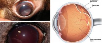
In order to clarify the density of the tumor and carry out a differential diagnosis, an ultrasound examination of the eye is performed in a specialized institution.
Ultrasound of the eyeball in a dog
In some cases, to exclude the inflammatory nature of the pathology, the animal may be prescribed bacteriological, virological and mycological methods for studying biological tissue. A general and biochemical blood test will help assess the severity of the disease and adjust therapeutic measures.
If metastases are suspected, a veterinarian may perform an X-ray examination of the orbit of the eye and chest. It is in the lung tissue that metastatic changes in ocular cancer tumors are most often found.
The main method for diagnosing third eyelid tumors in dogs in veterinary oncology is histological examination. For this purpose, in a specialized institution, a biopsy is taken from a sick individual and the tissue is examined. Based on the results of the cytological analysis, the veterinary oncologist makes a final diagnosis and selects a treatment method.
Third eyelid hyperplasia
Hyperplasia (prolapse) is often a complication of conjunctivitis or leukemia. If the disease is not treated, it can develop into an adenoma. Externally, the pathology looks like a swollen third eyelid, covering half of the eye.
The color of the mucous membrane ranges from reddish to deep crimson. In fact, hyperplasia is not a disease; it is a consequence of certain pathological processes in the animal’s body. When the underlying disease is eliminated, the third eyelid returns to normal.
- genetic predisposition;
- worms;
- hidden viral infections;
- stress;
- eye injuries.
In case of hyperplasia, surgical removal of the third eyelid is not resorted to. To eliminate symptoms, treatment of the underlying disease is necessary.
[custom_ads_shortcode1]
Prevention
In most cases, papillomatosis in dogs is mild, and pets recover without medical intervention. An animal that has once had papillomatosis acquires immunity. She does not develop new warts, but the virus remains in the body forever, and the dog can infect animals with reduced immunity. The likelihood of catching the virus while walking is very high, so it makes sense to vaccinate your dog against papillomatosis.
In addition, you need to take care of your pet’s immunity. You can increase the body's resistance to viruses if you follow all the rules for keeping a dog and create a balanced diet for it.
Benign skin tumors in dogs are a disease that in most cases does not require treatment. However, any skin problems are a reason to consult a veterinarian.
What does melanoma look like in dogs? This is a malignant tumor based on melanocytes (cells growing in the deep layers of the skin, which protect them from exposure to light and give it pigment). Localization of the tumor: mouth, limbs, eyes. Males are more predisposed to the disease than females. The most common breeds affected by melanoma are setters, retrievers, and cocker spaniels. Neoplasms of this type often penetrate deep into tissues and metastases develop.
Entropion of the third century
Often occurs in young dogs when the growth of the eyeball is not yet complete. Entropion occurs when cartilage loses flexibility due to refraction. As a result, the shape and position of the third eyelid changes; hyperplasia is a common consequence of the disease. As a result of the inversion of the third eyelid, the organ cannot function normally and protect the cornea from pathogenic flora.
Volvulus can be easily identified by redness of the mucous membrane and the appearance of enlarged lymph follicles. The inflammatory process may be accompanied by mucus secretion. It is possible to straighten the eyelid without resorting to surgery if more than 6 hours have not passed since the prolapse of the gland.
[custom_ads_shortcode2]
Why do papillomas appear?
As already mentioned, papilloma is a benign neoplasm that can be either very small or quite large. Papillomas have a variety of forms: pimple, cone, multilayer wart, etc. All such tumors are penetrated by a large number of blood vessels, and when injured, they bleed heavily.
Papillomatosis is a viral disease that appears in a dog due to infection with microorganisms from the Papillomaviridae family. The routes of infection by these viruses are different:
Direct contact between animals. Dogs in kennels are usually infected this way.
Injection with a non-sterile syringe, which has already been used to inject an infected dog.
Active communication of dogs in places of mass walking, during which they come into contact with each other, play, and fight.
Using the same grooming items for a large number of dogs.
As soon as the virus enters the upper layer of the skin through microcracks, it immediately begins to supply special proteins to the epithelial cells, causing them to actively divide, forming a tumor. In the case of the eyes, papillomas grow from the epithelial tissue located on the eyelid.
From the moment of infection to the appearance of papilloma on the eye, it can take about 2 months. The virus can also be constantly present in the blood of some dogs, but does not manifest itself in any way as long as their immunity works well, and will begin to develop if:
the dogs experienced severe stress;
the animal was treated with strong antibiotics, which weakened the body’s resistance;
immunity “failed” for some other reason.
Methods of treating a dog
If symptoms of inflammation of the third eyelid appear in a dog, regardless of the reasons, the pet needs first aid before the doctor arrives. This will help relieve pain and alleviate symptoms. What eye products can you use on your own before being examined by a veterinarian:
- Dexamethasone. The drug cannot be used if there is purulent inflammation. Dexamethasone should be dripped into the third eyelid area twice a day, 2 drops.
- Tsiprovet. An antibiotic that helps relieve inflammation and kill pathogenic bacteria. Place a drop in the inner eyelid 4 times a day.
Read: Causes of alopecia in Spitz: effective ways to eliminate the problem. It is important to understand that drops will only relieve symptoms; inflammation of the third eyelid should only be eliminated by a doctor. First of all, the veterinarian must determine the cause of the disease, after which he will draw up a treatment plan. Drug therapy for the disease includes:
- antiseptics for washing eyes;
- anti-inflammatory drugs;
- corticosteroids;
- tetracycline.
To wash your pet's eyes, you can use decoctions of medicinal herbs (chamomile, oak bark, St. John's wort, etc.). Drug treatment is an addition to the main treatment - surgery. The procedure is carried out under general anesthesia, suturing and fixing the prolapsed gland. The operation must meet the following criteria:
- preservation of the gland;
- preservation of the third century;
- normalizing eyelid mobility.
For adenoma, surgery is not required if the tumor is small, does not interfere with vision and does not cause inconvenience to the animal. In this case, regular monitoring by a doctor is necessary. If the tumor begins to grow in size, surgery will be required.
After a successful operation, relapse is practically excluded. To recover, the dog will need care - treating the stitches with antiseptic drops and ointments. To prevent your pet from accidentally scratching its eyes, it is necessary to wear a protective collar.
A dog's second eyelid is completely removed in exceptional cases when it is impossible to preserve the gland. In this case, the animal requires regular instillation of moisturizing drops, since the activity of the lacrimal glands is disrupted. Surgeons warn that removing the third eyelid will negatively affect the animal's future vision.
During treatment and recovery, it is advisable to completely avoid walking, especially in cold weather. It is worth paying attention to nutrition. Experts recommend switching your pet to dietary food to avoid allergies and possible complications.
In addition to the main therapy, it is worth giving the dog vitamins and immunostimulating drugs. Proper care during treatment and recovery after surgery will help your dog recover faster.
[custom_ads_shortcode3]
What does papilloma look like on the eye of dogs?
At the beginning of the disease, papillomas in dogs look like pimples around the eyes or small formations on the edge of the eyelid. After some time, they increase in size and take on a cone-shaped or “cauliflower” appearance.
Papillomas around the eyes do not bother the dog at all and are completely safe for it. It’s another matter if the tumor “settles” on the lower or upper eyelid at the border of eyelash growth. This arrangement annoys the dog and brings it inconvenience:
When blinking, the tumor irritates the conjunctival membrane of the eye, which can cause inflammation.
The tumor bothers the dog, and it tries to remove the irritant with its paw, injuring the papilloma.
A damaged papilloma bleeds, and if contamination gets into it, it can become inflamed.
The tumor causes constant lacrimation in the dog.
Very rarely, some of the papillomas, such as squamous cell papillomas, can become cancerous.