Doctors diagnose congenital ptosis of the upper eyelid in the maternity hospital. As a last resort - in the first year of a child’s life. The disease is not only a cosmetic defect. Often it becomes a consequence or symptom of another pathology. Find out more about this disease, its causes, diagnosis and treatment methods from this article.
In this article
- What is upper eyelid ptosis?
- Why does ptosis of the upper eyelid occur?
- The main causes of ptosis of the upper eyelid
- Symptoms of congenital ptosis of the upper eyelid
- What are the degrees of ptosis?
What is upper eyelid ptosis?
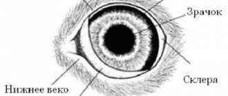
The eyelids should cover the iris by no more than 1.5 - 2 mm. If the functioning of the muscular system is impaired, the muscles that control the rise and fall of the eyelid are weak, innervated, and do not work correctly, doctors talk about ptosis - an ophthalmological disorder. It leads to partial or complete blocking of the iris of the eye. In severe forms of the disease, it is almost impossible to raise the eyelid; it completely covers the palpebral fissure.
The name pathology was first used by the ancient Greeks. Translated from ancient Greek, ptosis means “fall.” An abnormally low position of the eyelid in relation to the eyeball often occurs in old age. It is associated with loss of mobility of the eye muscles. There is also congenital ptosis of the upper eyelid. In the acquired form of the disease, drooping of one eyelid usually occurs. With congenital, the eyelids of both eyes droop.
Methods of treating a dog
If symptoms of inflammation of the third eyelid appear in a dog, regardless of the reasons, the pet needs first aid before the doctor arrives. This will help relieve pain and alleviate symptoms. What eye products can you use on your own before being examined by a veterinarian:
- Dexamethasone. The drug cannot be used if there is purulent inflammation. Dexamethasone should be dripped into the third eyelid area twice a day, 2 drops.
- Tsiprovet. An antibiotic that helps relieve inflammation and kill pathogenic bacteria. Place a drop in the inner eyelid 4 times a day.
Read Causes of abscess in dogs, signs and methods of treatment
It is important to understand that drops will only relieve symptoms; inflammation of the third eyelid should only be eliminated by a doctor. First of all, the veterinarian must determine the cause of the disease, after which he will draw up a treatment plan. Drug therapy for the disease includes:
- antiseptics for washing eyes;
- anti-inflammatory drugs;
- corticosteroids;
- tetracycline.
To wash your pet's eyes, you can use decoctions of medicinal herbs (chamomile, oak bark, St. John's wort, etc.). Drug treatment is an addition to the main treatment - surgery. The procedure is carried out under general anesthesia, suturing and fixing the prolapsed gland. The operation must meet the following criteria:
- preservation of the gland;
- preservation of the third century;
- normalizing eyelid mobility.
For adenoma, surgery is not required if the tumor is small, does not interfere with vision and does not cause inconvenience to the animal. In this case, regular monitoring by a doctor is necessary. If the tumor begins to grow in size, surgery will be required.
After a successful operation, relapse is practically excluded. To recover, the dog will need care - treating the stitches with antiseptic drops and ointments. To prevent your pet from accidentally scratching its eyes, it is necessary to wear a protective collar.
A dog's second eyelid is completely removed in exceptional cases when it is impossible to preserve the gland. In this case, the animal requires regular instillation of moisturizing drops, since the activity of the lacrimal glands is disrupted. Surgeons warn that removing the third eyelid will negatively affect the animal's future vision.
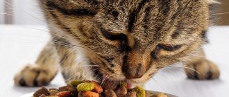
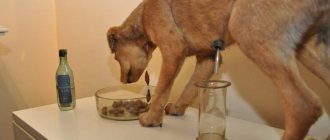
During treatment and recovery, it is advisable to completely avoid walking, especially in cold weather. It is worth paying attention to nutrition. Experts recommend switching your pet to dietary food to avoid allergies and possible complications. In addition to the main therapy, it is worth giving the dog vitamins and immunostimulating drugs. Proper care during treatment and recovery after surgery will help your dog recover faster.
Why does ptosis of the upper eyelid occur?
The levator muscle, controlled by the oculomotor nerve, is responsible for raising the eyelid. It is attached to the upper edge of the tarsal cartilage in the region of the periosteum. The muscle then passes along the walls of the orbit, approaching its upper edge. Then it passes into the tendon, the anterior fibers of which are attached to the tarsal cartilage and directed to the palpebral bundle of the main orbicularis oculi muscle. With a normal structure, the levator muscle provides lifting of the eyelid and its components: skin, cartilage and conjunctiva of the transitional upper fold.
The work of the muscle that lifts the eyelid depends on the coordinated movement of the eyelids, which is provided by cartilage, skin and the conjunctival superior fornix. The connection between the levator and the central nervous system is provided by the sympathetic and oculomotor nerves. In front of the muscle that lifts the eyelid, there is a thin layer of fatty tissue surrounded by the superior orbital artery, frontal and trochlear nerves. Their main function is to separate the orbit from the levator. When positioned correctly, it is in good shape. This promotes proper functioning of the oculomotor muscles.
The normal functioning of the levator muscle depends on Whitnell's ligament, a dense fibrous tissue that supports the eyeball. Externally, Whitnell's ligament connects the fibrous capsule of the lacrimal gland and the periosteum of the frontal bone. The ligament performs an important function - it limits the tension of the levator muscle on the posterior side. This allows you to support the upper eyelid. If the ligament undergoes pathological changes, the levator muscle becomes denser, which leads to the development of ptosis of the superior ptosis.
Third eyelid in a cat: treatment
Therapy begins when the cause of the disease is established. It is prohibited to inject medications or give pills on your own. Medicines can distort the symptoms of the disease, and this will affect the quality of diagnosis and complicate the care of the animal.
Drug treatment: drops, ointments, solutions
If there is discharge from the eye, it is necessary to regularly rinse the visual organ.
To do this, use solutions that clean the conjunctiva of the eye well and do not cause:
- saline;
- water-based furatsilin or chlorhexidine;
- eye lotions (Rosinka, Beafar Oftal).
When rinsing, first remove dirt from the eyelids with a napkin soaked in the solution. Then, using a syringe without a needle, the ocular surface is irrigated.
Cats don't like this procedure. If the animal feels satisfactory, it begins to break free. To ensure proper rinsing, it is recommended to wrap the cat in a towel.
Important: use a new wipe for each eye.
After the eye surface is washed, you need to wait 2-5 minutes, then instill drops . The time interval is necessary to avoid interaction between the instilled agent and the components of the rinse solution.
The following types of drops can be used to treat the third eyelid:
- Levomycetinaceous;
- Gentamicin;
- Ciprofloxacin;
- Decta-2 (a combination drug consisting of dexamethasone and gentamicin);
- Leopard (contains chloramphenicol and furatsilin).
The choice of drug depends on the main diagnosis . Antibiotic drops are needed only if conjunctivitis, blepharitis and other infectious inflammations of the eye are detected. If the cause is helminthic infestations or dysfunction of internal organs, then preventive therapy is prescribed for the eye to avoid secondary infections.
Be sure to read:
Otitis media in cats: symptoms and treatment, description, treatment with medications and folk remedies
The medicine is instilled by bending back the edge of the lower eyelid. After this, carefully close the eye and lightly massage the skin of the upper eyelid to evenly distribute the drug.
Eye ointments , unlike drops, remain in the conjunctival sac longer, so they are placed in the cat’s eye less often. Veterinarians often recommend using ointments at night. Like the drops, they are placed under the lower eyelid and then gently massaged into the upper eyelid to distribute the medication evenly.
When is surgery needed?
The operation is performed relatively rarely due to the fact that a frequent complication of surgical treatment is disruption of the lacrimal gland and the development of dry keratoconjunctivitis. A serious complication that causes dry eye and increases the risk of secondary infections.
Indications for surgery:
- Severe prolapse with prolapse of the lacrimal gland from the conjunctival sac.
- Adenoma. Here, surgery is necessary if the cat experiences severe discomfort and constantly rubs its face. When the neoplasm does not cause discomfort in the animal, conservative treatment is limited.
- Deep traumatic injuries. An operation is needed when a foreign body is “settled” deep in the eyeball and cannot be removed by rinsing with solutions.
Surgical treatment is a last resort and the intervention is often accompanied by complications. Veterinarians perform operations only if there are serious indications.
Folk remedies
Instead of medicinal solutions for washing cat eyes, you can use folk remedies:
- chamomile;
- calendula;
- thyme.
Herbal infusions cleanse well and soothe the mucous membranes. But it is important to ensure that there are no impurities in the liquid - small particles of plants will increase irritation.
The main causes of ptosis of the upper eyelid
The congenital form of eyelid ptosis usually occurs due to underdevelopment of the levator muscle. The disease can also develop due to aplasia - the complete absence of muscle. Due to pathological processes that can occur during pregnancy, the levator muscle is sometimes connected to the superior rectus muscle. Peripheral innervation—the supply of organs and tissues with nerves—may also be disrupted. In more rare cases, congenital ptosis becomes a consequence of aplasia of the oculomotor nuclei.
Incomplete development of nerves often leads to the development of ptosis. This occurs in the prenatal period and is usually associated with endocrine or neurological pathologies. The disease develops when the expectant mother takes certain medications that are prohibited during pregnancy. The cause of eyelid ptosis is often disturbances in the birth process, for example, trauma to the newborn. Sometimes ophthalmologists diagnose a baby with congenital myasthenia gravis, an autoimmune neuromuscular disease characterized by rapid fatigue of the primary cavity muscles. Less commonly, the cause of ptosis is paresis of the third pair of cranial nerves.
Congenital ptosis is very often one of the concomitant conditions of serious diseases. Almost all of them are characterized by a violation of the innervation of the levator muscle, which can occur for various reasons. Typically, drooping upper eyelids are a consequence of pathologies such as:
- Horner's syndrome is a symptom complex that occurs due to damage to the sympathetic nervous system against the background of damage or abnormal formation of the chest and neck. With this disease, congenital ptosis is combined with dilation of the conjunctival vessels, narrowing of the pupil, and a deeper location of the eyeball;
- Duane's syndrome is a congenital ophthalmological pathology in which the movement of the eyeballs is impaired. This condition develops due to improper functioning of the nerves that control the movements of the visual organs. Duane's syndrome is often called "atypical strabismus";
- Marcus-Gunn syndrome is a pathology in which the eyelid is not constantly drooping, but rises when opening the mouth, yawning or chewing. This syndrome is a consequence of a congenital pathological connection between the nucleus of the oculomotor and trigeminal nerves.
Ptosis of the upper eyelid is very rarely an independent disease. Firstly, it occurs against the background of other pathologies. Secondly, it is often accompanied by various ophthalmological disorders. Most often, ophthalmologists diagnose, in addition to ptosis, strabismus - strabismus or amblyopia - “lazy eye”.
Sometimes drooping eyelids are accompanied by anisometropia. This is a disease of the visual organs in which the refraction of one eye differs significantly from the parameters of the other.
Causes of inflammation of the third eyelid
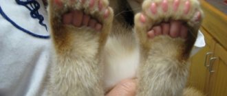
A healthy 3rd eyelid in a dog does not protrude beyond the eye and is practically invisible. A small fold of mucous membrane is located in the inner corner of the eye and is secured by small cartilages. The third eyelid swells, taking on an unhealthy red hue when the lymphatic follicles in the inner part become inflamed. This happens for several reasons:
- eye injury;
- weak ligaments that do not allow to hold the glands;
- heredity;
- exposure to dust, dirt, smoke or chemical components in the eyes;
- injury to blood and lymphatic vessels;
- reaction to a long course of treatment with certain drugs.
Dogs that have previously suffered from plague are at risk. Purulent inflammation, uveitis and tuberculosis can also cause inflammation of the third eyelid in a pet. The most common variants of the disease are:
- adenoma;
- hyperplasia;
- turn of the century.
It is worth considering that some dog breeds have a predisposition to one form or another of the disease. The anatomical features of the eye structure in bulldogs, pugs and Shar-Peis often cause hyperplasia. Shepherd dogs, Great Danes and Dobermans suffer from entropion of the third eyelid, and decorative and hunting dogs are prone to adenoma.
Important! Before treating inflammation of the third eyelid, it is necessary to establish its cause and the severity of the disease. This will allow you to select the most effective therapy. In extreme cases, surgery will be required.
Symptoms of congenital ptosis of the upper eyelid
Signs of damage to the levator muscle are difficult to miss. In most cases, the eyelid droops partially; in rarer cases, it droops completely, covering the eyeball. A person constantly looks tired or sleep-deprived. People with congenital ptosis of the upper eyelid are forced to constantly tense their forehead, arch their eyebrows, and raise their chin. They do all this in order to improve visibility. Doctors often say about children who have congenital ptosis that they have the astrologer pose. She is characterized by an upward-pointing face. This position allows you to compensate for the reduced field of view.
The main symptom of ptosis is drooping of the upper eyelid. Ophthalmologists also identify a number of signs that allow doctors to make a final diagnosis. Typically, patients have the following manifestations of this disease:
- raised eyebrows;
- increased eye fatigue;
- difficulty with eyelid drooping;
- development of strabismus or amblyopia;
- impaired sensitivity of the cornea;
- formation of epicanthus;
- decreased visual acuity;
- sleepy facial expression;
- thrown back head;
- double vision;
- burning and itching;
- exophthalmos;
- enophthalmos.
Due to disruption of the levator muscle, blinking movements cause discomfort to a person. The eyes get tired quickly, burning and itching occurs. Some patients complain of headaches. Improper functioning of the levator palpebrae superioris muscle may be due to various factors. Therefore, before starting treatment for ptosis, the doctor needs to determine what led to the drooping eyelid. Typically, making a diagnosis requires a preliminary examination of the patient by an ophthalmologist and neurologist.
Causes of third eyelid prolapse
global $ads_google; //data-ad-slot=”2475549904″ $ads_google = empty($ads_google) ? false : true; ?> if ($ads_google == false) {?>
$ads_google = true; ?> } ?>
With a quick glance at the cat, it is difficult to immediately determine the cause of the swelling of the thin fold. It can be associated with both external influences and internal factors. Veterinarians name a lot of reasons for the loss of the third eyelid:
- Infection of a bacterial, fungal or viral nature;
- Impaired functioning of the liver, intestines, kidneys;
- Hormonal imbalance in the body;
- Severe exhaustion, dehydration of the body;
- Weakening of the ligaments and muscles of the eye;
- Inflammatory process in the auditory analyzer;
- Introduction of foreign bodies into the mucous membrane of the eye;
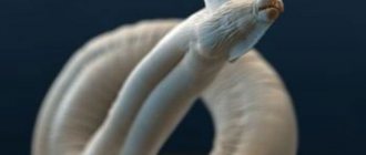
- Infection with parasitic worms;
- Mechanical damage to the eye area;
- Development of allergic conjunctivitis;
- Benign or malignant formation;
- Malnutrition of the eyeball (atrophy);
- Deformation of the elastic cartilage of the third eyelid;
- Eye size that is too small (microphthalmos).
Problems with the conjunctival fold can occur in cats of any breed, age, or gender. However, it has been noted that the Persians and the British are the most sensitive in this regard. Genetic predisposition increases the risk of third eyelid prolapse in cats of these breeds, regardless of the cause.
ON A NOTE
:
In rare cases, the nictitating membrane becomes noticeable in an animal after castration and under severe nervous stress.
What are the degrees of ptosis?
There are 3 degrees of congenital ptosis. They differ in the number of millimeters between the edge of the eyelid and the central part of the pupil. In order for the doctor to determine the degree of ptosis of the upper eyelid, the patient must relax and take a natural position. Highlight:
- 1st degree ptosis - the difference is from +2 to +5 mm;
- ptosis 2 degrees - the difference is from +2 to -2 mm;
- ptosis of the 3rd degree - the difference is from -2 to -5 mm.
If a person has grade 1 ptosis of the upper eyelid, then the eyeball is closed by approximately one third. This form of the disease is often called partial ptosis. It is characterized by the formation of folds on the upper eyelid and bags under the eyes. The eyebrows rise and the nasolabial triangle becomes more defined. With partial ptosis, the patient notes that it is harder for him to blink, and there is a sensation of a foreign body in the eye.
The second degree of ptosis is characterized by the formation of deep folds between the eyebrows. Fine wrinkles begin to form around the eyes, and the eyelid drops to the level of the eyelashes. Ophthalmologists often call this form of the disease semiptosis. Headaches occur due to muscle tension. Many people complain of feeling dry. Some people experience conjunctivitis. Treatment for this condition is necessary.
With grade 3 (severe) ptosis, the symptoms of the disease intensify. The eyelids almost completely cover the eyeball. Doctors call this condition complete ptosis. The skin loses its former appearance and becomes more wrinkled. In severe cases, the mucous border of the eyelid is everted. The size of the pupil changes, the eyeball looks more recessed into the orbit or, conversely, convex.
Third eyelid in a cat: when treatment is not required
When a mucous membrane covers part of a cat's eye, it is not always a sign of a serious pathological condition .
Be sure to read:
What to do if your cat has bald spots on its fur?
In some cases, you can do without the use of drugs:
- severe weight loss or dehydration;
- recovery after a serious illness;
- decreased immunity;
- mild form of cat flu.
In case of these disorders, you need to carefully monitor the pet’s condition and make sure that he receives a sufficient amount of vitamins and other useful substances from his food. If necessary, you need to regularly remove discharge from the nose and eyes.