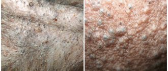
What are comedones
Surely everyone knows about the existence of so-called “black” dots. The latter are considered one of the main cosmetic problems among people. So here it is. Comendons are almost complete analogues of “black dots”. The problem is that they can be dangerous to the animal's health.
In dogs, due to the structural features of their sebaceous glands and hair follicles, comedones in many cases are a manifestation of purulent folliculitis or even furunculosis. In this case, the bases of the hair follicles become inflamed. Since the inflammatory process is often purulent in nature, the health and even life of the dog can be in serious danger.
Hyperpigmentation in dogs
Lentigo
Lentigo is a macular melanosis that is intensely black and usually occurs as multiple lesions, most commonly located on the ventral trunk. Lentigo occurs in adult dogs. Foci of hypermentation in dogs with lentigo increase in size and number over several months; subsequently, they become static and remain unchanged throughout the dog’s life. The lesions are sometimes clustered and may spread further diffusely to the ventral surface of the body. These sharply demarcated macules do not itch and do not cause discomfort to the patient. They should not have a rough surface and should not be palpable thickened. When a hyperkeratotic or thickened surface is present, other diseases such as epidermal nevus or papilloma-induced lesions should be considered.
A hereditary form of lentigo, called lentigo profuse, has been reported in pugs and is believed to have an autosomal dominant pattern of inheritance. Many of the lesions described in these dogs were clinically and histologically hyperplastic, however, it is likely that the authors were, in fact, describing pigmented epidermal nevus or papilloma-induced lesions. Similarly, generalized lentigo, probably an epidermal nevus, has been described in the cat.
Histologically, lentigo lesions in the early stage are characterized by a sharply localized increase in the number of melanocytes and melanosomes. Epidermal pigmentation is greatly increased, since almost every keratinocyte contains melanosomes. Usually no structural changes or only mild structural changes are observed in the epidermis. As the lesion progresses, the skin may thicken slightly and mild orthokeratotic hyperkeratosis may be observed.
It is a cosmetic disorder with no known significance in dogs and cats as there are no reports that it can become malignant. The only significance is that the lesion may need to be differentiated from pigmented tumors, especially melanomas, papilloma-induced lesions (which may have malignant potential) and pigmented nevus.
Epidermal nevus
Nevi are defects in skin development and, in some cases, are hereditary. They may be related to dermatomes or peripheral nerves and may have a linear shape. Most epidermal and all melanocytic nevi are associated with increased pigmentation, but the degree of pigmentation varies among epidermal nevi. Nevus keratosis pilaris is often pigmented but appears as speckled macules or plaques.
These lesions are often cosmetic, although secondary infection may occur and some may be accompanied by itching or inflammation. Treatment is surgical removal or laser surgery. Etretinate is useful in some cases of epidermal nevus or nevus keratosis pilaris where surgical removal is not possible.
Canine acanthosis nigricans
This disease is most commonly seen in dachshunds and has led to the belief that this primary (idiopathic) form is hereditary in nature. Similar lesions can be acquired and occur secondary to a variety of chronic inflammatory diseases. Therefore, this term is also described as a type of reaction associated with phenomena such as friction, hypersensitivity diseases and seborrhea with Malassesia.
Post-inflammatory hyperpigmentation in dogs
The most common form of hyperpigmentation in dogs is post-inflammatory. Many dogs and some cats produce more pigment in or around areas of inflammation. Hyperpigmentation may appear in many diseases characterized by chronic erythematous papular lesions. Often, this type of post-inflammatory hyperpigmentation has a lattice appearance and is most commonly seen in superficial bacterial pyoderma. Many other chronic inflammatory diseases also lead to hyperpigmentation; examples include chronic or convalescent lesions from canine demodicosis, canine mange, and the central zone of skin following convalescence from rounded dermatophytosis. Sometimes the area of pigmented comedones appears as a hyperpigmented area or plaque. These areas or plaques may be dark blue to gray in color and should raise suspicion for demodicosis, hyperadrenocorticism, or nevus keratosis pilaris.
More diffuse hyperpigmentation in dogs may arise from chronic diffuse inflammation, which may be associated with events such as ultraviolet exposure to skin that has lost hair or from chronic irritation due to skin friction. Diseases with severe itching may be suspected because both friction and inflammation can lead to hyperpigmentation. Often hyperpigmentation in dogs is observed in hypersensitivity diseases.
Melanotrichia (hair hyperpigmentation) may also be seen in areas recovering from deep inflammation (eg panniculitis, vaccine reaction). It is seen more frequently in certain breeds such as the Yorkshire Terrier, Silky Terrier, Bedlington Terrier, Old English Sheepdog and Poodle. Some dogs, especially poodles with sebaceous adenitis, may have multifocal areas of melanotrichia. Since inflammatory lesions in adenitis of the sebaceous glands are observed even in areas without melanotrichia, it is possible that this disorder is not observed due to inflammation, but is observed as a result of other pathological mechanisms. Multifocal areas of melanotrichia are observed in poodles after attacks of intervertebral disc disease.
The exact mechanism of hyperpigmentation in dogs due to these inflammatory diseases is unknown. Research suggests that keratinocytes may be able to locally stimulate melanogenesis by releasing melanocyte-stimulating factors. It is possible that these factors are present in low quantities in the normal epidermis, but their levels and activity increase in response to stimulation or stress of keratinocytes. Focal post-inflammatory melanotrichia seen in adult dogs with silver or gray coats may result from a reversion to a puppy coat caused by genes at the graying locus (G). If melanotrichia is observed due to the influence of the G locus, the hair returns to its normal adult color at the next molt. In other dogs, melanotrichia may occur due to the influence of melanocyte-stimulating factors such as LTB4.
Hormone-associated hyperpigmentation in dogs
Diffuse hyperpigmentation can also occur in dogs due to metabolic or hormonal diseases such as hyperadrenocorticism, hypothyroidism, and sex hormone-related dermatoses. The mechanism is unknown, although it has been suggested that hyperpigmentation may occur due to the direct effects of hormonal changes on melanocytes. Adrenocorticotropic hormone and other lipotropic hormones of the pituitary gland stimulate melanogenesis and this mechanism may explain the occurrence of hyperpigmentation in some cases with hyperadrenocorticism and changes in the amount of adrenal sex hormones. However, the direct effect of other hormones on melanocytes has not been well established. In some cases, it has also been suggested that ultraviolet radiation may play a role. When alopecia precedes hyperpigmentation, the skin is exposed to light, which can contribute to pigmentary changes. Protecting the skin reduces the likelihood of hyperpigmentation in some animals.
Melanotrichia may also occur due to resolution of metabolic or hormonal diseases; it is often observed in dogs with hyperadrenocorticism treated with mitotane. The mechanism is unclear, but the influence of the G locus is likely to be important in dogs with silver coats.
Drug-induced hyperpigmentation in dogs
Hyperpigmentation due to prescription drugs is rare. Treatment with mitotane may be associated with hypermelanosis and melanotrichia. Because the changes are usually temporary, even with continued mitotane therapy, hyperpigmentation is probably caused by hormonal changes and not by the drug. In an experiment, the drug minocycline caused hyperpigmentation in dogs and this effect was presumably caused by iron deposition. In humans, a variety of other metallic substances can cause changes in skin pigment; for example, changes may occur due to parenteral or local absorption of metals such as silver, gold and mercury.
Papillomavirus-associated hyperpigmentation in dogs
Canine papillomavirus induces at least two syndromes that may present as pigmented lesions. Cutaneous exophytic papillomas are usually seen in older dogs and are more common in male dogs. They appear as cauliflower-like nodules or keratin plaques, mainly on the head, eyelids and paws. They can be flesh-colored, pale, brown or black. These lesions may regress on their own. Papillomavirus-associated pigmented plaques have been compared to epidermodysplasia verruciformis. This condition is seen primarily in young Pugs, Miniature Schnauzers and Shar Peis and may have a genetic basis. These lesions often appear as irregular, black, pigmented macules or plaques. They are observed mainly in the armpits, abdomen, ventral chest and neck. They do not spontaneously regress and often progress slowly. There are reports that they transform into squamous cell carcinoma.
Pigmented tumors
Many tumors have different pigmentation from the surrounding skin. The color difference may reflect the type of fabric and associated color or other pigments. Vascular tumors may have a red or red-blue color. Histiocytic, lymphocytic, and plasmacytic tumors often appear pale, red, or purple. Common hyperpigmented (black) tumors are melanocytoma and melanoma; however, a variety of tumors can exhibit hyperpigmentation. Basal cell tumors, trichoblastomas, fibromas, epidermal nevus, and epithelial nevus may also frequently present as hyperpigmented lesions.
Causes of comedones on the skin of dogs
Experts today believe that the reasons for the appearance of comedones on the skin of dogs are extremely numerous:
- Injuries. The skin surface is inevitably damaged and contaminated with pathogenic/conditionally pathogenic microflora. As a rule, inflammation of staphylococcal or streptococcal etiology develops.
- Dermatitis of flea origin (as well as lice-eating and lice-eating). When there are a lot of parasites and they constantly bite the dog, it begins to itch all the time. The result is the same as in the previous case. Demodicosis and other tick-borne pathologies lead to a similar outcome.
- Autoimmune diseases, which in this case manifest themselves in the form of eosinophilic inflammation of the hair follicles and histiocytosis (as for the latter, its true causes are still unclear).
- Various fungal pathologies.
- Allergies (especially in chronic forms).
- Poor nutrition of the animal.
Types of allergic skin reactions in puppies
Sizes of spots in the abdominal area show different sizes - tiny, small, medium and large. Sometimes a number of varieties occur simultaneously, depending on the degree of exposure to the allergen. Pekingese often suffer from allergies. Here are the types of dermatitis found in Pekingese puppies.
Types of dermatitis that cause an allergic reaction:
- Dermatitis caused by flea bites. Appears due to parasite bites. An allergic reaction occurs when insect saliva enters the animal's body. Sometimes red spots appear, which then turn brown. Before treating such dermatitis, fleas must be eliminated.
- Atopic form of dermatitis. Appears in animals prone to atopy from childhood. Reddish spots appear on the animal's skin. The disease can be caused by rodents or pollen from selected plants during flowering.
- Quincke's edema is a dangerous form of dermatitis. The disease occurs when a large amount of allergen enters the body: it enters the puppy’s stomach along with food, when bitten by insects with saliva, with fragrant pollen when inhaled. This is the most dangerous form of dermatitis, which can sometimes lead to the death of an animal, especially a small puppy. At the first stage of the disease, the skin on the abdomen and mucous membranes turn red. Local swelling appears. In such a situation, you need to urgently contact a veterinary clinic. Ignoring the situation and delaying visiting a doctor is dangerous for the animal’s life.
- Contact dermatitis. Red spots appear on the stomach and in places of contact with the allergen. Shampoo sometimes causes an allergic reaction. Use the product as directed. Three-month-old puppies are bathed exclusively with shampoo intended for puppies.
- Dermatitis caused by eating poor quality food. If the puppy consumes natural products, milk, the dog may become allergic to the ingredients of the product. A similar reaction often occurs to meat products. To eliminate the cause of the red spots, it is necessary to temporarily switch the puppy to a special top-class food. You will need to contact a veterinarian who will help you create a diet appropriate for the puppy’s age.
Primary and secondary comedones
Veterinarians distinguish between primary and secondary comedones. Let's say right away that the secondary variety occurs in almost 100% of cases. In this case, the pathology develops against the background of some other disease. For example, flea dermatitis is primary, and comedones are secondary. Accordingly, all the cases described above (with the exception of neoplasms of an autoimmune nature) refer specifically to the secondary variety.
Accordingly, the primary type is much less common. These include commandons of the following origin:
- Acne is of genetic origin. Veterinarians believe that bulldogs, boxers, Dobermans and Rottweilers are inherently predisposed to this pathology. In addition, other skin diseases are very common among them. Finally, many schnauzers have a pathology that is called “comedone syndrome.”
- Comendons of autoimmune etiology.
- Endocrine acne. They develop, as the name implies, due to disruption of the endocrine secretion glands.
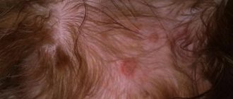
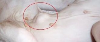
The dog has a black belly: Reasons for the appearance of “blackness”
- The most common reason is pigmentation . With age, during the period of growing up, when hormones take their toll, various kinds of dark spots may appear on the body. There is no need to be afraid of them.
- Another no less banal reason is dust and dirt . If a dog walks on the street, or simply climbs on balconies or under sofas at home, then small dark specks often appear on the belly, which are not so easy to wash off with water. These are pores clogged with dust. In this case, wet the belly, soap it and leave the animal in the bath for 5-10 minutes. The dirt should come off, but it is not always possible to get rid of it 100% on the first try.
- Lentigo . This is a fairly common occurrence among dogs. It cannot be called a disease, it is only a cosmetic defect of the skin, although many veterinarians consider it a manifestation of metabolic and immune disorders of the dog. Pugs and French bulldogs are most susceptible to this disorder, and, unfortunately, no treatment has yet been found.
- Dermatitis . With this disease, the color of the spots can vary from light pink to black. In addition, dermatitis can be accompanied by itching, ulcers and other skin problems. For correct diagnosis and treatment, a diagnosis by a veterinary dermatologist is required.
- Dermatophytoses . These are various fungal diseases, including lichen. Some varieties of fungi can cause spots on the skin, but in addition to spots, there may be other accompanying symptoms such as hair loss, itching, scabs, and weeping spots.
- Comedones. Simply put, blackheads. They occur not only in people, but also in dogs. The reason for their appearance is the same as in humans – blockage of the sebaceous glands. If comedones appear, you should wipe the skin with a bandage soaked in chlorhexidine. It is important to choose a bandage and not a cotton pad, since the rough fabric of the bandage will also cause light peeling. Also, areas where blackheads accumulate can be wiped with an alcohol solution of salicylic acid. But it is worth paying attention to the fact that some breeds, for example, miniature schnauzers, do not need to get rid of black spots, since this cosmetic defect is allowed by the breed standard.
- Demodicosis . This is an infection of a dog with subcutaneous mites; in fact, everyone has demodex, even people. It is not dangerous as long as the body does not have problems with the immune system. But as soon as the immune system weakens, Demodex begins to actively multiply, black crusts appear on the skin, it becomes rough, hair falls out and peeling appears. At the moment, demodicosis, if confirmed by a veterinarian, is treated by taking Bravecto tablets according to the treatment regimen.
Read the article “How to properly trim a dog’s nails and ear hair”:
Watch the video: Skin diseases in dogs - A veterinarian explains
Predisposing factors
The following predisposing factors contribute to the appearance of neoplasms:
- Incorrect, unbalanced diet.
- Poor pet hygiene. Greasy and matted wool is an excellent “reason” for the development of inflammation of the hair follicles.
- Poor living conditions. They contribute to both the deterioration of the coat and the appearance of parasites in the animal’s fur.
- Helminthic infestations. Worms themselves consume a lot of vitamins and nutrients, which causes the animal’s general condition to deteriorate, the immune system weakens, and the risk of developing autoimmune pathologies and other dangerous diseases increases.
List of diseases accompanied by comedones
As we have said many times, dog acne rarely grows on its own. This happens much more often if the pet is already sick with something. Here is an approximate list of diseases accompanied by comedones:
- Pathologies of the thyroid gland . In this case, primary type comedones develop.
- Comedones syndrome , or “schnauzer comedones syndrome,” which we already mentioned above.
- Sebadenitis. Hereditary autoimmune disease. We have already written that pathologies of this type can cause the growth of tumors, but in this case they appear with a 100% probability.
Changes in skin pigmentation
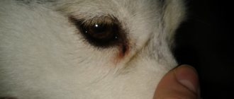
I have 4 Yorkshire terriers, the oldest is 5 years old, the girl suddenly developed dark pigmentation on her stomach and they are starting to spread. There are no other signs or symptoms - scratching, itching, peeling, flaking. Only dark bluish pigmentation. Her veterinarian was unable to determine the cause or make a diagnosis, but only gave me a list of possible causes - fungus, hormones (the dog was spayed), thyroid, stress. My three other Yorkies do not have this pigmentation and their bellies are a healthy pink color. What should I do, undergo another examination?
Read advice from Dr. Christy Conn:
The condition you are describing is called skin hyperpigmentation or simply darkening of the skin. Hyperpigmentation is usually a symptom or sign of an underlying disease. The diseases that hyperpigmentation can cause have been described by your veterinarian. With such a large list of possible diseases and various diagnoses that need to be ruled out, some pet owners are simply lost in such an incomprehensible situation. In this case, it is best to start eliminating the most dangerous diseases and the most common conditions. Skin diseases are rarely life-threatening, but there is one disease that should be checked for immediately. The described bluish/purple skin may indicate possible bruising. A blood clotting problem can cause bruising and, if left untreated, can threaten your dog's health. Therefore, do a blood test on your dog, check for the presence of blood cells, platelets and blood clotting rate. This test will also help determine the presence of other common causes of hyperpigmentation, described below. If, in addition to spots on the dog’s skin, no other signs are found that indicate any disease, most likely it may be hypercortisolism syndrome, hypothyroidism, acanthosis, or a chronic inflammatory reaction. It is worth knowing that sterilization does not remove all hormones, but only those produced by the ovaries. Hypercortisolism syndrome and hypothyroidism are caused by an excess of adrenal hormones or a lack of thyroid hormones. Both diseases can be detected by a blood test, so this should be done first when spots are detected on the dog’s skin. Hyperpigmentation can also be caused by excess melanin deposition due to chronic irritation. Friction of the skin on the carpet is a common problem in overweight dogs, in such cases hyperpigmentation appears in the area you described. By losing weight, your dog's hyperpigmentation may improve. Allergies caused by contact can also cause hyperpigmentation. Acanthosis is darkening of the skin due to genetic causes that cannot be treated, or secondary factors, such as allergies or friction, as already described above. A thorough physical examination and medical history of the dog will help determine the most likely cause and determine the best course of action. For example, if there is a medical history of allergies suffered by the dog, it is worth doing an allergy test. A good relationship between the veterinarian and the owner is the key to a successful search for the source of the disease problem. If you notice that your veterinarian is not properly explaining the likely causes or what the best steps to take are, it may be worth seeking out a different veterinarian.
Ask the Vet: Change in Dog Skin Pigmentation.
Don't lose us, subscribe to the VKontakte page vk.com/26dogs.
Treatment methods for comedones in dogs
In modern veterinary medicine, the following methods of treating comedones in dogs are used:
- The simplest and most effective option is to apply medications directly to the affected areas of the skin. But for the products to work well, it is very advisable to cut off all the hair from these areas, and also get rid of the crusts of exudate. Of course, scabs should be removed by first soaking them. This is done by applying ordinary sterile oils, or 3% hydrogen peroxide. Suitable oils (sea buckthorn, almond, or refined olive) can be purchased at any nearest pharmacy. We recommend using sea buckthorn, as it not only perfectly softens the skin, but also promotes its speedy healing.
- The animal is bathed using shampoos with a keratolytic effect.
- Before the “oil” treatment, it does not hurt to remove excess sebum from the skin.
- Retinoids and other vitamins are prescribed orally to stimulate the regeneration process of the skin epithelium.
- Anti-inflammatory corticosteroids are also prescribed to stop the inflammatory process. But! Since they “impair” already weakened immunity with prolonged use, they are allowed to be used for a maximum of a couple of weeks. In addition, for some types of autoimmune pathologies, their use is strictly prohibited.
- Since comedones are often accompanied by bacterial inflammation of the skin, the dog is simultaneously prescribed broad-spectrum antibiotics.
Other diseases The dog has black spots on its stomach
The animal body is also susceptible to the formation of various diseases that are directly related to the skin. And in this article we will talk directly about such an important topic as the formation of dark spots on a dog’s body. Owners are especially concerned when various dark-colored formations appear on the dog’s belly, which rightly cause panic on the part of humans. Let's look at the main reasons that can cause this disease, and also talk about how the problem can be cured. Main reasons In fact, it is almost impossible to single out any one reason that can become the main provocateur of black spots on the pet’s belly. The very wide range of diseases forces veterinarians to come up with specific treatment and diagnostic plans that allow owners to cure their dog. Here is a list of the most popular reasons why black spots can form on a dog’s stomach: • Lentigo is the very first disease that deserves attention from both the scientific world and ordinary owners. The fact is that lentigo is expressed in the formation of spots that are localized on the stomach. Sometimes spots can form large pigment groups, and they are purely cosmetic in nature, without causing harm to the pet’s body. But they are also evidence of disruption of the animal’s body. Pugs suffer from lentigo much more often than other breeds. There is currently no cure • Dermatitis is the second most common problem that can cause spots to form on the animal’s belly. But their color varies directly from the type of dermatitis that the animal suffers from. It must be remembered that dermatitis is also a consequence of improper functioning of the animal’s body, therefore a complete veterinary examination is the main method for the speedy eradication of the disease. Dermatitis is often accompanied by the formation of ulcers, as well as severe itching, which causes discomfort to the dog • Allergies are common companions in life in dogs. Our four-legged friends can also suffer from allergic reactions, which are expressed in different ways. The appearance of spots on the stomach is the same symptom as sneezing and red eyes. Their color can be varied, it all depends directly on the type of allergy. Treatment Naturally, any diseases associated with the skin of an animal require careful examination. The above list of sores is far from complete, but it includes the most common problems that provoke the formation of dark spots. Only an experienced veterinarian will be able to conduct a thorough examination of the dog’s body, after which he will formulate the correct and most effective treatment for the pet. Self-indulgence in such matters should be excluded, since skin problems are one of the most multifaceted and difficult to treat. You, as an owner, should carefully monitor your pet's diet, since the quality of nutrition is the main aspect that must be observed throughout the life of your pet. A lot depends on the quality of food. For example, seborrhea and dermatitis alone can be eliminated forever by feeding the animal high-quality food, as well as a huge amount of vitamins and minerals required for the normal functioning of the pet’s body. Try to carefully monitor this factor, and only then will you provide your beloved dog with a fun, healthy and carefree life.
List of drugs
The list of medications we have given is approximate, and therefore in any case needs to be adjusted to the specific case of the disease:
- Benzoyl peroxide. This substance has a powerful disinfectant effect and also perfectly washes away excess sebum. The drugs Benzoyl Plus® and OxyDex®, produced on its basis, have proven themselves well. But! The concentration of the active substance should be no more than 3%!
- Isotretinoin and pure retinol are used as retinoids.
- Despite the abundance of anti-inflammatory corticosteroids, the usual drug is dexamethasone.
- Microperl Humectant has an excellent keratolytic effect.
- Antibiotics prescribed include ciprofloxacin and enrofloxacin. In severe cases, doxycycline is administered once. Apply 3% tetracycline ointment to the acne itself.
- For autoimmune diseases, immunosuppressants are prescribed. These are very specific and dangerous medications; they should be selected and prescribed exclusively by a veterinarian.
Caring for a dog during rehabilitation
Comprehensive care for your dog during the rehabilitation period includes:
- Once every three days, the dog is bathed using the above-mentioned keratolytic agents.
- The pet's fur is regularly combed, trimming it around the comedones.
- To prevent the animal from gnawing on damaged areas, a surgical collar is put on it.
- Three times a week, boiled sea fish, rich in polyunsaturated omega-3 fatty acids, is added to your pet’s diet. This has a beneficial effect on the speed of skin regeneration.