Skeleton or skeletal system
The skeleton is the frame that holds all the internal organs, as well as the muscles of the dog. The structure of a dog’s skeleton can be easily represented in the diagram as two lines:
- axial, which includes 109 bones (skull and spine with ribs);
- peripheral, consisting of 180 limb bones.
Over the course of an animal's life, the composition of bone tissue changes. Therefore, in puppies and teenage dogs, the bones are more elastic and lighter, but in old age there is a high probability of increasing the risk of fragility and loss of strength. The condition of the bones and teeth is used to judge the general health of the dog.
Structure of the skull
The facial and cerebral parts of the skull are distinguished; both include paired and unpaired bones. In total, the skull consists of 27 bones connected to each other by cartilage tissue. With age, the cartilage becomes ossified, and mobility is maintained only in the lower jaw area so that the dog can chew food.
The figure shows both paired and unpaired bones of the skull.
Based on the type of skull, dogs are divided into representatives of dolichocephalic (prominent representatives are Italian Greyhounds and Greyhounds) and brachycephalic breeds (for example, pugs, dwarf Spitz). The biggest differences between them are noticeable in the structure of the facial part of the skull. So, brachycephals have a flattened muzzle and a protruding jaw. It is these characteristics that have been specially cultivated by breeders over the years to make the breeds recognizable. But such features are associated with certain health problems for pets.
Structure of teeth
Teeth are not only an important part of a dog's appearance. First of all, teeth are necessary for biting and grinding food, protecting the owner and, if necessary, attacking the enemy.
Puppies are born without teeth. At the age of two to three weeks, the first baby teeth emerge through the gums. Closer to 4-5 months, they begin to fall out to make room for permanent ones. Instead of 28 milk teeth, by the age of one and a half years, the jaw should have 42 permanent teeth. Deviations from the schedule are often due to unbalanced nutrition or breed characteristics.
The dental formula of an adult dog includes 42 teeth, with 20 located on top and 22 on the bottom.
The permanent set of teeth for an adult dog includes:
- Incisors – 6 on each jaw.
- Fangs – 2 on top and bottom. They are dangerous weapons in fights.
- There are 4 premolars on both branches of the jaws.
- Two molars on each branch of the upper jaw, as well as three below, for a total of 10 pieces.
Dog dental arcade
A tooth consists of a crown, neck and root. The crown protrudes significantly above the gum; each type of tooth has its own shape. Dentin is the main dental tissue; in the crown area it is covered with enamel, and in the root zone dentin is covered with cement. Inside the tooth there is a cavity divided into the coronal space and the root canal itself.
The number of teeth, their condition and bite (or occlusion) directly affect the health of the dog. The following typology of bites is distinguished:
The most common type is the first type of bite.
Spine structure
The spinal column is the axis of the skeleton. A skull is attached to it on one side, and it ends in a tail. Also on the sides, ribs and limbs are attached to it using cartilage tissue.
The structure of the spine can be represented as follows:
- Cervical spine - consists of 7 vertebrae, the first two of which (atlas and epistrophy) are especially mobile. They are responsible for head movements.
- The thoracic region includes 13 vertebrae. The ribs are attached to them, forming the chest. Dogs have 9 pairs of real and 4 pairs of false ribs.
- The lumbar region also consists of 7 vertebrae.
- The sacrum is a sacral bone fused from 3 vertebrae.
The dog's tail, which is a logical continuation of the spine, consists of 20-23 vertebrae. The most developed and mobile are the first five. Previously, representatives of certain breeds had their tails docked, but now such actions are not supported by the global canine community.
Separately, we should consider the structure of the dog’s penis, since it also includes a bone - the baculum, based on the connective tissue of the penis. The baculum is located at the front of the penis. Its upper edge is convex, and below there is a groove with a passing urogenital canal. In dogs, the penis belongs to both the reproductive system and the excretory system, since the urinary canal is also the vas deferens.
Limb structure
The limbs of dogs are characterized by the complexity of their structure. The front legs are an extension of the scapula, which is attached to the spine by developed shoulder muscles. The shoulder blade goes into the humerus, then the forearm and wrist joint. The forearm consists of the radius and ulna bones, and the metacarpus includes 5 bones. The hind legs are formed by the thigh, stifle, shin, hock, metatarsus and paw.
The structure of the paws can also be represented in this way:
- Pads that act as shock absorbers. They reduce stress on bones and joints and also help maintain balance. The pads consist of an impressive layer of fatty tissue, so dogs do not freeze in cold seasons, and heat is well retained in their paws.
- Pets' fingers have different numbers of phalanges. 4 fingers are three phalanges, and one is only two. An animal cannot move them in the same way as a person due to the limited space between the fingers. Normally, dogs have 5 toes on their front paws, and 4 on their hind paws. There are also rudimentary toes - dewclaws, located on the hind paws just above the foot. They do not carry any functional load, but in some cases they can be a sign of a high quality representative of the breed. This is true for Briards, Beaucerons or Pyrenean Mastiffs.
- Dog claws, unlike cat claws, do not retract into the pads and consist of hard, keratinized tissue and pulp, an area with a significant number of blood vessels and nerve endings. It is important to be very careful when trimming nails so as not to cause harm or pain to the dog. It is also necessary to monitor the condition and length of the claws, since there is a direct connection between them and the musculoskeletal system. Long claws do not allow a four-legged pet to walk normally; because of them, the skeleton can even begin to deform.
Sections of the spine and number of vertebrae in a dog
The vertebrae of the cervical spine are movably connected to each other, while the first two have significantly changed their shape: the atlas and epistropheus. The head moves on them. The ribs are attached to the bodies of the thoracic vertebrae. The lumbar vertebrae have powerful articular processes that provide a stronger connection between the vertebral arches, to which the heavy digestive organs are suspended. The sacral vertebrae are fused to form the sacrum. The size of the caudal vertebrae decreases with distance from the sacrum. The degree of reduction of parts depends on the function of the tail. The first 5-8 vertebrae still retain their parts - the body and the arch. In subsequent vertebrae the spinal canal is no longer present. The basis of the tail consists only of the “columns” of the vertebral bodies. In newborn puppies, the tail vertebrae have a low degree of mineralization, so certain breeds of dogs (for example, Airedale Terriers) undergo tail docking (circumcision) at an early age.
Rib cage
formed by the ribs and breastbone. The ribs are movably attached on the right and left to the vertebrae of the thoracic spinal column. They are less mobile in the front of the chest, where the scapula is attached to them. In this regard, the anterior lobes of the lungs are more often affected in lung disease. Dogs have 13 pairs of ribs. They are arched. The breast bone comes in the form of a clearly shaped stick. The chest itself is cone-shaped, with steep sides.
Peripheral skeleton or limb skeleton
Thoracic limb
presented:
• a shoulder blade attached to the body in the area of the first ribs;
• shoulder, consisting of the humerus;
• forearm, represented by the radius and ulna bones;
• a hand consisting of the wrist (7 bones), metacarpus (5 bones) and phalanges of the fingers. The dog has 5 fingers, represented by 3 phalanges, the first of the fingers is pendulous and has 2 phalanges. There is a claw ridge at the end of the fingers. Pelvic limb
comprises:
• the pelvis, each half of which is made up of an innominate bone. The ilium is located above, the pubic and ischial bones below;
• the thigh, represented by the femur and the patella, which slides along the femur trochlea;
• lower leg, consisting of the tibia and fibula;
• the foot, represented by the tarsus (7 bones), metatarsus (5 bones) and phalanges of the toes (5 toes from 3 phalanges, the first of the toes is trailing (detail) and has 2 phalanges. At the end of the toes there is a claw ridge).
CONNECTIONS
Among diseases of the organs of the movement apparatus, pathological processes at the junctions of bones, especially the joints of the limbs in animals, are more common than others. There are several types of bone connections.
Continuous.
This type of connection has great elasticity, strength and very limited mobility. Depending on the structure of the tissue connecting the bones, the following types of connection are distinguished:
• with the help of connective tissue - syndesmosis, and if elastic fibers predominate in it - synelastosis. An example of this type of connection is short fibers that firmly connect one bone to another, such as the forearm and tibia bones in dogs;
• with the help of cartilage tissue – synchondrosis. This type of connection has low mobility, but provides strength and elasticity of the connection (for example, the connection between the vertebral bodies);
• with the help of bone tissue - synostosis, which occurs, for example, between the bones of the wrist and tarsus. As animals age, synostosis spreads throughout the skeleton. It occurs at the site of syndesmosis or synchondrosis.
In pathology, this connection can occur where it normally does not exist, for example, between the bones of the sacroiliac joint due to physical inactivity, especially in old animals;
Rice. 5. Scheme of development and structure of the joint: a – fusion; b – formation of an articular cavity; c – simple joint; d – articular cavity; 1 – cartilaginous bone bookmarks; 2 – accumulation of mesenchyme; 3 – articular cavity; 4 – fibrous layer of the capsule; 5 – synovial layer of the capsule; 6 – articular hyaline cartilage; 7-cartilaginous meniscus
• with the help of muscle tissue – synsarcosis, an example of which is the connection of the scapula with the torso.
Discontinuous (synovial) type of joint or joints.
It provides a greater range of movement and is built more complexly. According to the structure, joints are simple and complex, in the direction of the axes of rotation - multiaxial, biaxial, uniaxial, combined and sliding (Fig. 5).
The joint has an articular capsule consisting of two layers; external (fused with the periosteum) and internal (synovial, which secretes synovium into the joint cavity, thanks to which the bones do not rub against each other). Most joints, except the capsule, are secured by a different number of ligaments. Ligaments often run along the surface of the joint and are attached to opposite ends of the bones, that is, where they do not interfere with the main movement in the joint (for example, the elbow joint).
Most of the bones of the skull are connected using a continuous type of connection, but there are also joints - the temporomandibular, atlanto-occipital. The vertebral bodies, with the exception of the first two, are connected to each other by intervertebral discs (cartilage), that is, synchondrosis, as well as long ligaments. The ribs are connected by intrathoracic fascia, consisting of elastic connective tissue, as well as intercostal muscles and transverse ligaments. The shoulder blade is connected to the body using the muscles of the shoulder girdle, and the pelvic bones are connected to the sacral bone, and to the first caudal vertebrae - by ligaments. The parts of the limbs are attached to each other using different types of joints, for example, the connection of the pelvic bone with the femur occurs using a multi-axial hip joint.
MUSCLES
Muscle tissue has the important property of contracting, causing movement (dynamic work) and providing tone to the muscles themselves, strengthening the joints at a certain angle of combination with a stationary body (static work), maintaining a certain posture. Only work (training) of muscles helps to increase their mass, both by increasing the diameter of muscle fibers (hypertrophy) and by increasing their number (hyperplasia). There are three types of muscle tissue depending on the type of arrangement of muscle fibers:
• smooth (vascular walls);
• striated (skeletal muscles);
• cardiac striated (in the heart).
Skeletal muscles are represented by a large number (more than 200) muscles. Each muscle has a supporting part - connective tissue stroma, and a working part - muscle parenchyma. The more static load a muscle performs, the more developed its stroma is. In the muscle stroma, continuous tendons are formed at the ends of the muscle belly, the shape of which depends on the shape of the muscles. If the tendon is cord-shaped, it is simply called a tendon. If it is flat, then it is an aponeurosis. In certain areas, the muscle includes vessels that supply it with blood and nerves that innervate it. Muscles can be light or dark, depending on their function, structure and blood supply. Each muscle, muscle group and all musculature of the body are covered with special dense fibrous membranes - fascia. In order to prevent friction of muscles, tendons or ligaments, soften their contact with other organs, and facilitate sliding during large ranges of movement, gaps are formed between the sheets of fascia, lined with a membrane that secretes mucus, or synovium, into the resulting cavity. These formations are called mucous or synovial bursae. Such bursae are located, for example, in the areas of the elbow and knee joint, and their damage threatens the joint.
Muscles can be classified according to several criteria. By form:
• lamellar (muscles of the head and body);
• long thick (on the limbs);
• sphincters (located at the edges of the openings, having neither beginning nor end, for example, the sphincter of the anus);
• combined (composed of individual bundles, for example muscles of the spinal column).
According to the internal structure:
• dynamic (muscles that perform dynamic loads; the higher a muscle is located on the body, the more dynamic it is);
• statodynamic (static function of the muscle during support, holding the joints of the animal in an extended form when standing, when under the influence of body weight the joints of the limbs tend to bend; muscles of this type are stronger than dynamic muscles);
• static (muscles bearing a static load; the lower the muscles are located on the body, the more static they are).
By action:
• flexors (flexors);
• extensors (extensors);
• adductors (adduction function);
• abductors (abduction function);
• rotators (rotation function).
The work of muscles is closely connected with the organ of balance and, to a large extent, with other sense organs. Thanks to this connection, the muscles provide balance to the body, precision of movements, and strength.
Thus, as a result of the joint action of muscles with the skeleton, certain work is performed (for example, an animal moves). During operation, heat builds up.
Therefore, in the warm season, with intense work, dogs may experience overheating of the body - heatstroke.
In cold weather, animals need to move more to avoid hypothermia.
Skin covering
The body of dogs is covered with hairy skin and organs or derivatives of the skin.
LEATHER
It protects the body from external influences and, through many nerve endings, acts as a receptor link for the skin analyzer of the external environment (tactile, pain, temperature sensitivity). Through many sweat and sebaceous glands, a number of metabolic products are released; through the mouths of the hair follicles and skin glands, the surface of the skin can absorb a small amount of solutions. The blood vessels of the skin can hold up to 10% of a dog's body's blood. The reduction and dilation of blood vessels are essential in regulating body temperature. The skin contains provitamins. Vitamin D is formed under the influence of ultraviolet light.
In the skin covered with hair, the following layers are distinguished (Fig. 6).
1. Cutaneous tissue (epidermis) –
outer layer. This layer determines the color of the skin, and keratinized cells are exfoliated, thereby removing dirt, microorganisms, etc. from the surface of the skin. Hair grows here: 3 or more guard hairs (thick and long) and 6-12 short and delicate undercoat hairs.
2. Dermis (actual skin):
• pilar layer, which contains sebaceous and sweat glands, hair roots in hair follicles, muscles that lift the hair, many blood and lymphatic vessels and nerve endings;
• mesh layer, consisting of a plexus of collagen and a small amount of elastic fibers.
The dermis contains scent glands that produce a characteristic odor for each breed. In hairless areas (nose, paw pads, scrotum in males and nipples in females), the skin forms patterns that are strictly individual for each pet.
3. Subcutaneous base (subcutaneous layer),
represented by loose connective and adipose tissue.
This layer is attached to the superficial fascia that covers the dog's body.
It stores reserve nutrients in the form of fat.
Rice. 6. Diagram of the structure of skin with hair: 1 – epidermis; 2 – dermis; 3 – subcutaneous layer; 4 – sebaceous glands; 5 – sweat gland; 6 – hair shaft; 7 – hair root; 8 – hair follicle; 9 – hair papilla; 10 – hair follicle
SKIN DERIVATIVES
Derivatives of the skin include the milk, sweat and sebaceous glands, claws, crumbs, hair, and nasal tract of dogs.
Sebaceous glands.
Their ducts open into the mouths of the hair follicles. The sebaceous glands secrete a sebaceous secretion, which, by lubricating the skin and hair, gives it softness and elasticity.
Sweat glands.
Their excretory ducts open to the surface of the epidermis, through which a liquid secretion is released - sweat. Dogs have few sweat glands. They are located mainly in the area of the crumbs on the paws and on the tongue. The dog does not sweat with its entire body; only rapid breathing through an open mouth and the evaporation of fluid from the oral cavity regulate its body temperature.
Mammary gland.
They are multiple and located in two rows on the lower chest and abdominal wall, with 4-6 pairs of hills in each row. Each colliculus contains several gland lobes that open into nipple canals at the tip of the nipple. There are 6-20 nipple canals in each nipple.
Hair.
These are spindle-shaped filaments of stratified keratinized and keratinized epithelium. The part of the hair that rises above the surface of the skin is called the shaft, the part located inside the skin is called the root. The root goes into the bulb, and inside the bulb there is a hair papilla.
Based on their structure, there are four main types of hair.
1. Pokrovny –
the longest, thickest, elastic and hard, almost straight or only slightly wavy. It grows in large quantities on the neck and along the spine, on the hips and in smaller quantities on the sides. Wire-haired dogs usually have a large percentage of this hair type. In short-haired dogs, the outer hair is absent or located in a narrow strip along the back.
2. Guard hair (covering hair) –
thinner and more delicate. It is longer than the undercoat and covers it tightly, thereby protecting it from getting wet and abraded. In long-haired dogs, it is curved to varying degrees, which is why they distinguish between straight, curved and curly hair.
3. Undercoat is the shortest and thinnest, very warm hair that fits the dog’s entire body and helps reduce heat transfer from the body during the cold season. It is especially well developed in dogs kept outside during the cold season. Change of undercoat (molting) occurs twice a year.
4. Vibrissa –
sensitive hair. This type of hair is located on the skin in the area of the lips, nostrils, chin and eyelids.
There are a large number of coat classifications based on hair quality.
According to the presence of undercoat:
• dogs without undercoat;
• dogs with undercoat.
Based on the identity of their coat, dogs are:
• smooth-haired (bull terrier, Doberman, Dalmatian and others);
• straight-haired (beagle, rottweiler, labrador and others);
• short-haired with feathering (St. Bernard, many spaniels and others);
• wire-haired (terriers, schnauzers and others);
• medium-haired (collie, spitz, pekingese and others);
• long-haired (Yorkshire terrier, Shih Tzu, Afghan hound and others);
• long-haired with corded hair (poodle, commander and others);
• long-haired shaggy ones (Kerry Blue Terrier, Bichon Frize and others).
Hair color is determined by two pigments: yellow (red and brown) and black. The presence of pigment in its pure form gives an absolutely monochromatic color. If the pigments are mixed, then other colors occur.
Most dogs shed twice a year: in spring and autumn. This phenomenon is called physiological molting. Spring molting is usually longer and more pronounced. Shedding is a dog's natural defense against the summer heat and replaces old hair with new ones. During the summer, dogs mostly have guard hair, and the undercoat falls out. In winter, on the contrary, a thick and warm undercoat grows. When kept at home, dogs have a longer shedding period than those living on the street.
In addition to physiological molting, there is also pathological molting. This is unmotivated hair loss, which can be associated with metabolic disorders, the presence of skin parasites, and poor nutrition, which can lead to baldness (complete hair loss). Treatment is usually prescribed by a doctor.
Claws.
These are horny curved tips covering the last, third, phalanges of the fingers. Under the influence of muscles, they can be pulled into and out of the groove of the roller. Such movements are well expressed on the fingers of the thoracic limbs of dogs. Claws are involved in the function of defense and attack, and with their help the dog can hold food and dig the ground.
Crumbs.
These are the supporting areas of the limbs. In addition to their supporting function, they are organs of touch. The cushion of crumbs is formed by the subcutaneous layer of skin. A dog has 6 crumbs on each thoracic limb, and 5 on each pelvic limb.
Nervous system
The nervous system is a set of structures in the animal body that unites the activities of all organs and systems and ensures the functioning of the body as a whole in its constant interaction with the external environment. The structural and functional unit of the nervous system is the nerve cell - neurocyte -
together with gliocytes. The latter dress the nerve cells and provide them with support-trophic and barrier functions. Nerve cells have several processes - sensitive, tree-like branching dendrites, which conduct to the body of the sensitive neuron the excitation that occurs at their sensitive nerve endings located in the organs, and one motor axon, along which the nerve impulse is transmitted from the neuron to the working organ or another neuron. Neurons come into contact with each other using the ends of their processes, forming reflex circuits through which nerve impulses are transmitted.
The processes of nerve cells together with neuroglial cells form nerve fibers.
These fibers in the brain and spinal cord make up the bulk of white matter.
From the processes of nerve cells, bundles are formed, from groups of which, covered with a common membrane, nerves
in the form of cord-like formations. Nerves vary in length and thickness. Nerve fibers are divided into sensitive - afferent, transmitting a nerve impulse from the receptor to the central part of the nervous system, and effector, conducting an impulse from the central part of the nervous system to the innervated organ: myelin (innervate the muscles of the body and internal organs), non-myelin (innervate the muscles of blood vessels and glands internal organs).
There are nerve ganglia -
groups of nerve cells of the central part of the nervous system, allocated to the periphery. They play the role of a step-down transformer, as well as an accelerator of nerve impulses in the affector sensory ganglia and an inhibitor in the effector nodes of internal organs. The nerve ganglion is a multiplying area where an impulse from one fiber can be distributed to a large number of neurocytes.
Nerve plexuses -
places where exchanges occur between nerves, fascicles or fibers intended to redistribute nerve fibers in complex connections in different segments of the spinal cord and brain.
Anatomically, the nervous system is divided into central, including the brain and spinal cord with the spinal ganglia; peripheral, consisting of cranial and spinal nerves connecting the central nervous system with receptors and effector apparatus of various organs. This includes the nerves of skeletal muscles and skin - the somatic part of the nervous system and blood vessels - the parasympathetic. These last two parts are united by the concept of the autonomic, or autonomic nervous system.
CENTRAL NERVOUS SYSTEM
Brain
This is the head part of the central part of the nervous system, located in the cranial cavity. There are two hemispheres, separated by a fissure and having convolutions. They are covered with a cortex, or bark.
The following sections are distinguished in the brain (Fig. 7):
• big brain;
• telencephalon (olfactory brain and cloak);
• diencephalon (visual thalamus (thalamus), suprathalamus (epithalamus), hypothalamus (hypothalamus), peritothalamus (metathalamus);
• midbrain (cerebral peduncles and quadrigeminal);
• rhombencephalon;
• hindbrain (cerebellum and pons);
• medulla.
The brain is covered with three membranes: hard, arachnoid and soft. Between the hard and arachnoid membranes there is a subdural space filled with cerebrospinal fluid (its outflow is possible into the venous system and into the lymph circulation organs), and between the arachnoid and soft - the subarachnoid space.
Rice. 7. Brain: 1 – cerebral hemispheres; 2 – cerebellum; 3 – medulla oblongata; 4 – olfactory bulbs; 5 – optic nerve; 6 – pituitary gland
The brain is the highest department of the nervous system, controlling the activity of the entire body, uniting and coordinating the functions of all internal organs and systems. Here there is a synthesis and analysis of information coming from the senses, internal organs, and muscles. Almost all parts of the brain take part in the regulation of autonomic functions (metabolism, blood circulation, respiration, digestion). For example, in the medulla oblongata there are centers of respiration and blood circulation, and the main department that regulates metabolism is the hypothalamus, and the cerebellum coordinates voluntary movements and ensures the balance of the body in space. In pathology (trauma, tumor, inflammation), the functions of the entire brain are disrupted.
Spinal cord
The spinal cord is part of the central part of the nervous system and is a cord of brain tissue with remnants of the brain cavity. It is located in the spinal canal and starts from the medulla oblongata and ends in the region of the 7th lumbar vertebra. The spinal cord is conventionally divided without visible boundaries into the cervical, thoracic and lumbosacral sections, consisting of gray and white brain matter. In the gray matter there are a number of somatic nerve centers that carry out various unconditioned reflexes, for example, at the level of the lumbar segments there are centers that innervate the pelvic limbs and the abdominal wall. The white medulla consists of myelin fibers and is located around the gray matter in the form of three pairs of cords (bundles), in which the conductive pathways of both the spinal cord’s own reflex apparatus and the ascending pathways to the brain (sensitive) and descending from it (motor) are located.
The spinal cord is covered with three membranes: hard, arachnoid and soft, between which there are gaps filled with cerebrospinal fluid. In dogs, the length of the spinal cord averages 78 cm and weighs 33 g.
Internal structure of dogs
The skeleton (from the Greek skeletos; dried) is a collection of solid formations in the dog’s body that provide support to the animal’s body and protect it from damage. In addition, the bone marrow of the skeleton contains hematopoietic organs, which participate in the metabolic and biochemical processes of the body.
The dog's skeleton includes 247
bones
and almost as many
joints
.
The axial skeleton, or spine
, contains 7 cervical, 7 lumbar, 13 thoracic, 3 sacral and about 20 caudal vertebrae. 13 pairs of ribs protect the internal organs of the animal.
The thoracic region begins with the shoulder blade
, then come the bones of the forearm, wrist, and metacarpus. Above the shoulder blade is the withers, from which the height of the dog is measured. Strong shoulder muscles connect the thoracic region to the spine.
Rear end
, starting from the femur, passes into the tibia and tarsus. Then come the metatarsals and four phalangeal toes with claws. In some cases, a vestigial finger grows from the inside (in puppies it is usually amputated). The pelvis of the skeleton is fixed by the hip muscles.
The head, which contains the brain, hearing organs, eyes, nasal and oral cavities, pharynx, larynx, is the most important and most complex part of the dog's body.
Nasal cavity
It is a narrow upper passage between the shells and the nasal bone, leading to the olfactory organ - the ethmoid bone. The dog sucks in air forcefully so that it hits the labyrinth of the ethmoid bone. Inflammation of the lining of the nasal cavity causes inflammation of the olfactory region, which leads to a violation of the sense of smell.
Small openings (nasolacrimal duct) located in the front of the dog's nasal cavity lead into the eye cavity.
Below the base of the skull is the pharyngeal
, in which the respiratory and digestive tracts intersect. Through the holes on its side walls, going into the auditory tubes, the infection can penetrate from the pharynx into the middle ear.
Lower respiratory apparatus
The lower section of the respiratory apparatus includes the bronchi and lungs, located in the chest and separated from it by the pleura. The chest cavity is limited by the ribs and diaphragm. The lungs are separated from the chest wall by the outer and inner layers of pleura, forming the pleural cavity, and are always filled with air. Each dog's lung is divided into seven lobes: three of them are on the left (cranial, or anterior; middle; caudal, or posterior) and four are on the right (cranial, middle, caudal and accessory).
The bronchi branch in the lungs into seven smaller branches, each of which enters a lung lobe. As they continue to divide, they are reduced to very small branches - bronchioles, which become alveolar ducts leading to the alveolar sacs (pulmonary alveoli). Through the very thin walls of the pulmonary alveoli, gas exchange occurs between air and blood. The lungs are penetrated by numerous blood vessels, the vast area of contact with the airways ensures the exchange of oxygen and carbon dioxide.
Dog structure
Methods of administering medications to a dog's body
Every owner has to give medicine to a sick dog at least once in his life. Some dog breeders encounter difficulties: their pets stubbornly refuse to swallow pills and mixtures.
In order to facilitate the introduction of drugs into the dog’s body, there are special techniques:
To stimulate swallowing, you can use a syringe or a teaspoon to pour a small amount of water into the dog’s mouth, while continuing to close its jaws.
• when feeding the dog tablets and capsules, open its jaws, place the medicine on the root of the tongue, and then squeeze the animal's muzzle tightly until the tablet is swallowed. Attempts to mix a crushed tablet with a treat usually have no effect: the animal quickly learns to select the tasty pieces and leave the drug;
Giving your dog tablets and liquid medications
• the powders are poured onto the tongue, and after a few minutes the dog is allowed to drink them with water;
• in order to give the dog a medicine, lift the animal’s head up, pour the medicine into the cheek and wait for the swallowing movement.
External use of drugs usually does not cause difficulties. The affected areas of the skin are lubricated with ointments, and then a collar is put on the dog to prevent licking.
Rectal and vaginal administration of drugs is done using suppositories or microenemas. Suppositories are applied to the anus or vaginal opening and then pushed inside with the index finger. For rectal administration, the dog's tail is briefly pressed against the anus to prevent the suppository from being pushed out. The procedure for microenemas and vaginal injection is carried out almost similarly.
Administration of the drug into the conjunctival sac is a slightly more complex procedure. For this purpose, ointments produced in special tubes are used. When placing the medicine behind the lower eyelid, the owner holds the dog's head, pulls the eyelid back and squeezes out or instills (if these are eye drops) the drug into its fold, being careful not to touch the eyeball with a pipette or tube. After this, the dog’s eyelids are closed and lightly massaged.
Subcutaneous injections are administered using a syringe with a thin needle. In the area of the withers, a fold is collected with your fingers, into which a needle is inserted 1-2 cm under the skin layer, and the medicine is injected by slowly pressing the piston.
Injecting medications into the dog's conjunctival sac and ear
Intramuscular injection is made into the posterior group of muscles of the thigh or shoulder, a medium-sized needle is taken and immersed into the tissue by 3-4 cm. If you need to administer several incompatible drugs, there is no need to use several needles or puncture the skin in several places. It is enough to disconnect the syringe, slightly pull out the needle without removing it from the skin, and pierce the adjacent muscle area.
Anatomical parts of a dog's body
Features of the location of various parts of the body, physique and general appearance of the dog in accordance with their breed characteristics are called exterior. To assess the exterior, several parts of the dog’s body are anatomically distinguished:
- Head. The skull and muzzle, eyes, ears, and dental system are assessed.
- Neck.
- Torso. Along the top line look at the withers, back, loin, croup and tail. The chest and abdomen are assessed along the bottom line.
- Limbs. Presented front and rear.
Knowledge of exterior features is especially necessary for owners of purebred dogs. It helps control, preserve and develop dog breeds.
Dog structure
Dogs are warm-blooded animals, that is, their body temperature is relatively constant and, under normal conditions, is maintained within the range of 37.5-39 °C.
The formation and release of heat depends primarily on metabolism. Body temperature depends on climatic and other factors. It changes under the influence of pathogenic microbes and viruses. It is measured in the rectum using a thermometer.
An animal's body is made up of tiny living particles called cells. Certain groups of cells, changing their shape and structure, unite into separate clusters that are adapted to perform certain functions. Such groups of cells, as a rule, have specific qualities and are called tissues. There are four types of tissues in the body - epithelial, connective, muscle and nervous.
Epithelial tissue covers all border formations in the body - such as skin, mucous and serous membranes, excretory ducts of glands, endocrine and exocrine glands. Connective tissue is divided into nourishing and supporting tissue. Nutrient or trophic tissue includes blood and lymph. The main purpose of supporting tissue is to bind the component parts of the body into a single whole and to form the skeleton of the body (this includes bone tissue, tendons, and cartilage). Muscle tissue is capable of contracting and relaxing under the influence of various irritations. It is divided into skeletal and cardiac muscles, which have cross-striations, as well as smooth muscle tissue, capable of involuntary contractions and found in internal organs. Nerve tissue consists of nerve cells called neurons.
An organ is a part of the body that has a certain external shape, is built from several naturally combined tissues and performs a highly specific function, for example, an eye, a kidney, a tongue. Individual organs that together perform one specific function form systems and apparatuses in the body. For example, bones, muscles, ligaments, tendons, and joints form the movement apparatus or musculoskeletal system.
Organs of such systems as digestive, respiratory, urinary, reproductive, that is, the insides, are located in three cavities: thoracic, abdominal and pelvic.
The thoracic cavity is located inside the chest, the abdominal cavity is limited in front by the diaphragm (the thoracoperitoneal muscular barrier), and in the back it passes into the pelvic cavity. It ends at the level of the waist. The pelvic cavity is formed by the pelvic bones, the sacrum and the first caudal vertebrae.
Most of the internal organs are located in serous cavities, which create conditions for organs to slide around each other. For example, the heart is located in the pericardial serous cavity.
Parts of the body For easier orientation on the dog’s body, it was conventionally divided into four main sections (Fig. 1). 1. Head. It distinguishes between the brain (skull) and facial (muzzle) parts. This includes the forehead, nose, ears, teeth. 2. Neck. Here the upper part and the lower region are distinguished. 3. Torso. It is represented by the withers (it is formed by the first 5 thoracic vertebrae and the upper edges of the scapula located at the same level with them), the back, the lower back, the thoracic region, the croup, the inguinal region, the abdomen, the region of the mammary glands and prepuce, the anal region, and the tail.
Rice. 1. Structure of the dog’s body : 1 - lip; 2 - nose; 3 - dorsum of the nose; 4 - muzzle; 5 - transition from the frontal part to the muzzle; 6 - eye; 7 - forehead; 8 - cheekbone; 9 - parietal part; 10 - ear; 11 - back of the head (occipital protuberance); 12 - neck; 13 - withers; 14 - back; 15 - lumbar region; 16 - croup; 17 - ischium (ischial tuberosity); 18 - shoulder; 19 - chest (chest); 20 - front of the chest; 21 - forearm; 22 - wrist; 23 - metacarpus; 24 - front paw; 25 - elbow; 26 - lower chest; 27 - stomach; 28 - groin; 29 - thigh; 30 - knee; 31 - lower leg; 32 - heel; 33 - hock joint; 34 - metatarsus; 35 — hind paw; 36 - tail
4. Limbs. Thoracic (front): shoulder, elbow, forearm, wrist, metacarpus and pelvic (posterior): thigh, knee, shin, heel, metatarsus. The dog's appearance, physique and characteristics of individual parts of its body, characteristic of its breed and gender, are called exterior. The general exterior includes the main signs of physique, the structure of individual parts of the body, the most characteristic deviations and defects; private - examines the structural features of individual breeds, typical and atypical features for them.
Skeletal structure
Have you been trying to heal your JOINTS for many years?
Head of the Institute for the Treatment of Joints: “You will be amazed at how easy it is to cure your joints by taking the product every day for 147 rubles .
To figure out what a dog “consists” of, we will gradually analyze the structure of its skeleton. The skeleton or backbone - whether dog or human - is the basis of the body, since bones, cartilage and ligaments play important biological, motor and mechanical roles.
Our readers successfully use Sustalaif to treat joints. Seeing how popular this product is, we decided to bring it to your attention. Read more here...
The skeleton protects the dog’s organs from various injuries, and also thanks to it the pet can move. We will look at the structure of a dog's skeleton in more detail below.
Scull
Anatomy provides for the division of the skull into two lobes: the facial and the brain, consisting of 27 bones, each of which is equipped with connective tissue - cartilage and ligaments. A dog is born with elastic tissue, which becomes hard and ossified as it matures.
Due to the mobility of the bone connection, the lower jaw is adjacent to the skull. This mechanism allows your pet to easily chew food, gnaw bones, etc. Adult dogs have 42 teeth, puppies have 28. The dental formula includes canines, incisors, molars and premolars. Dogs also have different bites. It depends on the breed and the standard set by the breed.
There are these types of bite:
- Scissor-shaped. In the case of this bite, the upper incisors, covering the lower ones, are closely connected to each other.
- Pincer-shaped - the incisors converge.
- Straight. There is also a direct bite, when the incisors on the upper and lower jaws overlap each other.
- Underbite. The upper jaw is further than the lower jaw, so there is a large gap between the teeth.
- Snack. This bite is determined by the lower jaw. It comes forward, so the incisors again do not coincide and a space is formed between them. Snacks are also divided into dense and waste snacks. Brachycevuls have a predominantly underbite bite. The bulldog and the overshot are somewhat different: the undershot is when the lower jaw extends beyond the upper jaw, and the bulldog is when the lower jaw does not just come out, but is slightly bent upward.
Breed and age affect a dog's skull. Over time, people learned to recognize certain dog breeds thanks to the unique structure of the skull.
For example, according to the shape of the skull, which is significant specifically in the facial part, dogs are divided into the following types:
Animals with a reduced part of the skull are called brachycephals. Their representatives are pugs, sharpeis, Pekingese and other dogs with similar facial parts of the skull.
The brachycephalic skull has a wide parietal bone, a flattened muzzle, and a protruding jaw. This skull structure was deliberately obtained through selective breeding. The consequence of these experiments is many problems associated with the health of dogs, for example, disruption of the structure of the respiratory tract.
Due to the fact that the animal has a too short muzzle, this led to breathing problems. Brachycephalics often experience tracheal collapse, pulmonary hypertension, and overactive lacrimal glands. Brachycephalic syndrome is a concept that was introduced specifically to designate the breed of such dogs.
Torso
The skull is attached to the skeleton of the body with the help of vertebrae. The dog's body also consists of vertebrae and ribs fastened together. The rest of the skeleton is attached to this bony structure.
The ridge consists of sections:
- The first vertebrae at the head of the dog are called cervical. The cervical spine includes seven vertebrae, with the first two being more mobile than the rest. They are called Atlas and Epistropheus.
- Thirteen vertebrae are in the thoracic region.
- Seven vertebrae are in the lumbar region.
- The structure is completed by the sacrum - 3 fused vertebrae. The cross bone is the transition from the vertebra to the tail.
The animal's tail consists of movable vertebrae, which include 20-25 parts. The thoracic part consists of 13 pairs of ribs. It is the ribs that protect the internal organs of the animal like a shield, holding them in a certain position that does not affect health.
It is noteworthy that 4 pairs of ribs form the costal arch, while the remaining 9 are directly attached to the sternum. Dogs' ribs have varying degrees of curvature, and it all depends directly on the breed.
In the lower back there are large vertebrae on which processes can be noted. Thanks to these processes, muscles, tendons and ligaments are attached to the vertebrae. They hold the pet's internal organs in the correct position. The anatomy of a dog's skeleton is shown in detail in the picture.
Limbs
Dogs' limbs have a very complex anatomy, as they contain many different bones, muscles, elastic ligaments and other things. The forelimbs provide support for the body, because it is the front legs that bear the weight of the animal. They are a continuation of the scapula, which through the joint flows into the humerus.
After it comes the forearm, which consists of the radius and humerus. They are connected by the elbow joint. After it comes the carpal joint, consisting of 7 bones. It is connected to the 5 bones of the hand. The hand has the following appearance: five fingers, with 4 having three phalanges, and 1 having only two.
Each finger has a claw that is not drawn inward and consists of strong tissue. The front legs, with the help of the shoulder muscles, are attached to the dog's vertebra. Thanks to the shoulder blades, which protrude beyond the vertebrae of the sternum, the animal creates a withers - this is an indicator of height. The hind limbs include the femur and tibia.
These elements are held together by the hip and knee joints. The tibia itself consists of the tibia and fibula and, with the assistance of the hock joint, which is complex in its structure, is attached to the tarsus. This part of the foot goes into another - the metatarsus, and ends with 4 toes with three phalanges. There are also claws that do not have retractable properties. In the video from Alisa Gagarinova you can see the anatomy of a dog's muscles.
Dogs are one of the most popular pets. To provide your pet with proper care and maintenance, it is important to take into account basic knowledge of the physiology and anatomy of these animals. The internal structure of a dog is almost the same, regardless of the exterior or the specific character of a particular breed. In the article we will understand the anatomical features and functioning of the main organs and systems of representatives of the canine family.
general characteristics
To make it easier to understand the anatomy of a dog, the dog’s body parts are conventionally divided into four main components, namely:
- head. This part includes the brain and facial zones, as well as the sensory organs;
- neck. There are upper and lower regions;
- torso. This category is more expanded and is considered from the withers to the tail;
- limbs. This category includes the front and hind legs.
Unfolded skeleton of a dog
The external characteristics of the dog, its body parts and their location, which are inherent in the breed and gender, are called the exterior and look like the signature of the breed.
Note! In addition to the general idea, it would not hurt for the dog owner to study the textbook, authored by Volmerhaus and Frewein, which provides a diagram and location of all the animal’s organs. The practical textbook is regarded as a guide for dog breeders.
Head
A dog's head can be round (like a Cavalier King Charles Spaniel), elongated (like a greyhound) or square. It plays an important role in balance. In breeds with an elongated head shape, the muzzle is most often pointed; Square-headed breeds have short, strong jaws (usually these breeds were originally bred for fighting).
There are two main sections of the head: the back (the cranium, which contains the brain) and the front (the front, which is also called the muzzle), where the nasal cavity is located. The central section of the head is located between the eye sockets. Depending on the proportional relationship between the cranial and facial regions, dogs are divided into three types: dolichocephalic (with an elongated muzzle), brachycephalic (with a short flattened muzzle) and mesocephalic (with average proportions).
There is a moderate convexity at the back of the head (the nuchal protuberance), and folds of skin called the dewlap (or chin) may form in the throat area.
Skeleton or skeletal system
Of course, the study of dog anatomy begins with the structure of the dog’s skeleton. The skeleton of a dog is the basis on which all the internal organs and muscles of the dog exist. Each component of the skeleton requires special consideration.
Dog skull: structure
There are facial and brain parts, which contain paired and unpaired bones.
- frontal;
- parietal;
- temporal;
- incisal;
- lower jaw;
- nasal;
- palatal;
- zygomatic;
- maxillary;
- lacrimal
Expanded dog skull with explanations
- interparietal;
- lattice;
- wedge-shaped;
- sublingual;
- vomer;
- pterygoid;
- occipital
Important! The dog's skull consists of 27 bones, which are connected by cartilage tissue. The older the pet, the more the cartilage becomes ossified, and only the lower jaw retains mobility of the head.
There are 3 types of dog skulls:
- dolichocephalic. Representatives are Italian greyhounds, greyhounds, etc.;
- brachycephalic. In this case, it is a pug, a dwarf spitz, a Yorkshire terrier, a chihuahua, etc.;
- mesocephalic. It is found in huskies, shepherd dogs, as well as in the dachshund breed, etc.
For your information! The third type makes up approximately 75% of all breeds.
The differences become visible in the structure of the front part. So, for example, in brachycephalic breeds the muzzle has a flattened shape, and the jaw protrudes forward. This structure of the dog affects its health. In veterinary medicine, the main cause is related to the respiratory tract.
Limb structure
A dog's paws have a complex structure. So:
- the pads serve as shock absorbers. They reduce the stress on the dog's bones and joints and allow him to maintain balance. The pads contain a significant layer of adipose tissue, thanks to which animals do not freeze even in the coldest weather;
- there are phalanges on the fingers, and their description deserves separate consideration. So, 4 fingers have 3 phalanges, and 1 has only 2. There are five fingers on the front paws, and four on the hind paws. There are also vestigial toes, that is, external ones, which are located on the hind legs above the foot. There is no stress on them, but they characterize the quality mark of some dogs, for example, Briards, Beaucerons.
Interracial differences in the structure of dog head elements
Dogs of different breeds have heads that look different, mainly due to the ratio (percentage), that is, the ratio of the skull to the muzzle. The length and width of the skull to the muzzle is also important (for example, in the case of knots the width is 3/5 of the length of the entire head), as is the width and length of the muzzle itself (for example, very short in relation to the skull in the Bulldog and Pekingese, and also very long and slender in greyhounds).
The muzzle is also wide at the base and tapers slightly towards the nose. The upper part of the muzzle of most breeds is flat and straight, that is, without convexities (rarely found, for example, in the Bull Terrier). When determining the characteristics of rocks, the angle formed by the stop with the front angle of the head is also taken into account. In some breeds the nuchal swelling is not easily visible, while in some it is noticeable (in some cases with slender heads). The nostrils should be large and allow the dog to breathe freely. The upper lips are moderately drooping in some breeds and very drooping in others, but always meet at the front to form an inverted "V" shape. The ears may be erect (such as in a German Shepherd), semi-erect (such as in a Terrier), or completely drooping (such as in a Dachshund). Ears can also be long or short and set high or low. In some breeds in some countries it is legal to copy or cut the ears to give them the shape of short and erect ears.
Sense organs or analyzers
Perhaps every dog owner knows that such animals hear better than any person. And the dog’s sense of smell is at the highest level. But you need to have a more detailed understanding of each sense organ.
Ear structure
The sound reaches the pet through the outer ear, and then it moves to the middle ear and ends up in the inner ear. The outer one consists of the auricle, thanks to which he hears even the quietest sounds. Following the auricle is the auditory canal, which consists of several parts:
The eardrum plays an important role in the hearing system, thanks to which acoustic waves are heard. The middle ear contains the auditory bones of the dog, thanks to which the eardrums and inner ear are attached to the membrane.
For your information! The inner ear provides auditory receptors and contains the vestibular apparatus.
Close-up view of a dog's ear, implementation of auditory activity
Structure of the nose
The nose allows the dog to have a connection with the outside world. The animal reacts sharply to the smell, which allows it to be used not only for peaceful, but also for official purposes. The animal remembers the smell of its owner and recognizes it, even when for some reason it cannot see it.
Important! There are 125 million olfactory receptors in animals, but only 5 million in humans.
The dog senses smells through his nostrils. The pet receives a large percentage of air through the side cutouts on the nose. There is also mucus in the nose, which is why it remains moist.
It consists of the external nose and the nasal cavity, which includes several parts. The upper one is the repository of olfactory reflexes. The lower one serves as a connecting link through which air enters and passes to the nasopharynx, where it warms up.
Labradors have a heightened sense of smell
Structure of the eye
The eye consists of the cornea, lens and retina. Unlike the human visual organ, it is not equipped with a macula. This makes a dog's vision slightly worse than a human's.
Note! Compared to humans, animals see much better in the dark.
The dog sees with the help of the cornea, into which a beam of light passes. Then it enters the retina.
Brown-eyed dog: enlarged image of the eye
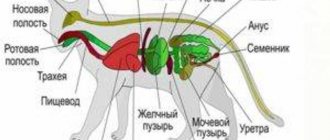
Internal organs
Of course, a dog's anatomy is not limited to the skeleton and sensory organs. Internal organs occupy a special place in the anatomy of a dog.
Expanded image of a dog's internal organs
Digestive system
It originates from the mouth, where food enters. Afterwards, the food moves through the esophagus and reaches the stomach, where the incoming food is digested. Gastric juice and enzymes grind food into a homogeneous mass called zymus.
Next, the small intestine comes into play, interacting with the pancreas, duodenum and liver. The walls of the small intestine transmit incoming substances into the blood.
Next in line is the large intestine, where the digested food goes. By the time it enters the large intestine, all useful vitamins, minerals and other substances have already been received. Feces are formed from the remains of waste food.
Circulatory system
The main circulatory organ in dogs, like in humans, is the heart. Through the arteries, blood enters each internal organ and circulates through the veins to the heart. The heart is located between the 3rd and 6th ribs in front of the diaphragm.
The heart consists of four chambers and is divided into the right part, where venous blood circulates, and the left part, where arterial blood flows. The parts of the heart are in turn divided into the atrium and the ventricle. The walls of the heart consist of an inner layer - the endocardium, an outer layer - the epicardium and the cardiac muscle of the myocardium.
Important! The size and rate of its contractions depend on the breed, sex and age of the pet.
Respiratory system
The respiratory organs provide the supply of oxygen. The respiratory system consists of two sections:
- upper. Consists of the nasal cavity, nasopharynx, trachea and larynx;
- lower. Consists of lungs and bronchi.
The physiology in this case is as follows: air enters through the nostrils, and their size depends on the breed of the dog. In the nasopharynx, the incoming air undergoes a warming process, and the nasal glands act in this process as a filter that rejects dirt and dust. The incoming air then continues to move through the larynx. After this comes the trachea. The air then reaches the lungs, which contain blood vessels.
For your information! On average, a dog takes 10-30 deep breaths per minute. Small breeds breathe more frequently than larger breeds. The frequency of inhalations may increase depending on the emotions that the animal experiences, and this indicator may look different.
Urinary system
The dog's urinary organs are designed to empty the body and maintain an acceptable level of water-salt balance. The kidneys produce hormones that control hematopoietin and renin. That is why disruption of the urinary system leads to a number of diseases and often death.
Excretory system
The activity of the excretory system depends on the kidneys. The kidneys work with the bladder through the ureters and end in the urethra. The main task of the excretory system is to remove urine from the body.
The kidneys are equipped with nephrons, which are surrounded by blood vessels. The older the animal, the more often problems with this internal organ may occur.
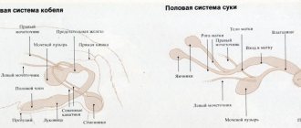
Reproductive system
The reproductive system is intertwined with the excretory system. In male dogs, the urinary tract is the vas deferens. In addition to the testes, male dogs have a prostate, which provides life to sperm.
The reproductive organ of females is the uterus. The dog's uterus is equipped with horns to which the ovaries, fallopian tubes and vagina are attached. Bitches go into heat twice a year, but it still depends on the type of breed.
Note! In northern breeds, estrus occurs once a year and lasts 28 days.
Once finished, the female can be bred.