What is a cat's heart?
An organ such as the heart is often compared to a pump. It pumps blood, after which it enters the vascular system to be cleansed and enriched with oxygen. The blood then supplies the cat’s entire body with nutrients and oxygen, removing waste and carbon dioxide from the body’s tissues.
© shutterstock
The physiology of all mammals, including cats, is such that this hollow organ consists of 4 parts: the upper two atria, left and right, and the lower ventricles, too, left and right.
The right side of the lung carries blood to the lungs, where it is oxygenated and returned to the heart, to the left side, which sends blood through the arteries to the organs. This is how you can briefly describe the circulatory system.
In order to carry out such necessary work, the pear-shaped heart is located in the chest, between the 4th and 6th ribs, closer to the left side of the chest wall. To more accurately determine the location of the cat’s heart, you can focus on the ulnar tubercle. When a cat stands, his heart is located right above this mound.
The anatomy of cats of different breeds may differ when it comes to the shape of the head or skeleton, the size of the tail or ears. But the location of the internal organs, including the heart, is the same.
But sometimes a cat can get sick. He is always tired, eats poorly, sleeps often, takes little care of himself, does not play, or some kind of pulmonary whistling comes from the sternum. These are all the first signs that the cat is suffering. And the cause of this may be heart disease. Does the position of the heart change due to illness? Perhaps it becomes larger and increases as a result of the disease. But then the heart will take up more space in the chest. How to help and what you need to know about cat heart disease?
Hello student
HEART POSITION
The longitudinal axis of the heart in a dog forms an open cranial angle of 40° with the sternum, in a cat it forms an angle of 25–30°, with the apex of the heart directed towards the diaphragm. The base of the heart in a dog is oriented craniodorsally and lies approximately at the level of the fourth rib. The right (“cranial”) ventricular edge runs along the sternum, from which it is a short distance away, the left (“caudal”) ventricular edge follows the cranial edge of the VII rib. Both edges meet at the apex of the heart, which faces slightly to the left and reaches approximately the VII costal cartilage. Thus, the heart is located 4/7 to the left and 3/7 to the right of the midline and occupies the space of the III-VI intercostal spaces in this plane, and in the frontal plane the space between the sternum and the middle of the chest cavity. The heart is close to the chest wall only with its left surface in the area of the cardiac notch of the left lung.
The topography of the lateral chest wall is important in such methods of clinical examination of the heart as palpation, percussion, auscultation, and radiography. The chest in its cranial part is covered by the scapula and part of the humerus, as well as the muscles associated with them. The line of the triceps muscle, linea musculi tricipitis, is formed by the caudal border of the triceps brachii muscle and stretches from the thoracic angle of the scapula to the ulnar tuberosity. This line is clearly visible through the skin. By pulling the thoracic limb forward, you can free the lateral chest wall in the area of the IV-VII ribs for examination.
Rice. 3. Position of the heart in a standing dog fixed in formalin (after Schummer, 1984)
A vertebra cervicalis VII; In vertebra thoracica I; With vertebra thoracica VI; D costa I; E costa VI; F scapula; Ghumerus; Hsternum; I radius; To ulna
a auricula cordis dextra, b conus arteriosus; with atrium sinistrum ef auricula cordis sinistra; d sulcus coronarius; e sulcus interventricularis paraconalis; f ventriculus dexter, g his margo ventricularis dexter; h ventriculus sinister, i his margo ventricularis sinister; k apex cordis; l diaphragm contour
1 truncus pulmonalis; 2 arcus aortae; 3 aorta thoracica with aa. intercostales dorsales; 4 v. cava cranialis; 5 v. cava caudalis; 6 a. pulmonalis sinistra, 6′ vv. pulmonales; 7 ligamentum arteriosum (Botalli); 8 a. subclavia sinistra; 9 truncus brachiocephalicus; 10 a.m. subclavia dextra; 11a. carotis communis dextra; 12 a.m. carotis communis sinistra; 13 a.m. vertebralis; 14 v. jugularis externa sinistra 15 a. et v. axillaris sinistra, 15′ a. et v. thoracica interna; the dashed line corresponds to the caudal edge of m. triceps brachii (linea mi. tricipitis or anconaea)
Knowledge of the topography of the heart allows the use of various methods of intravital heart diagnostics.
The apical impulse of the heart, which occurs as a result of contraction of the heart muscle and leads to a shaking of the lateral chest wall, in a dog is heard clearly on the left in the lower third in the IV-VI intercostal space, especially well in the V intercostal space, and slightly worse on the right in the IV-V intercostal space .
Using percussion, as a rule, it is possible to detect an area of absolute cardiac dullness in the part of the heart not covered by the lung on both sides in the IV-VI intercostal spaces. The dorsal border of this area is formed by the symphyses of the IV-V ribs. Auscultation of heart sounds or pathologically altered heart murmurs in a dog should be carried out in the 5th intercostal space for the left ventricle and in the 4th intercostal space for the right ventricle. For intracardiac injection, only the right ventricle is suitable and the needle should be inserted from the right side into the 5th intercostal space as close to the sternum as possible.
In a cat, the caudal edge of the triceps brachii muscle runs parallel to the IV intercostal space, the heart is located closest to the chest walls on the left side between the IV and VI ribs, on the right side - under the V rib. In these places, the apical impulse of the heart is clearly perceived and heart sounds are clearly audible. The 5th intercostal space is also suitable for intracardiac injection in cats. In a standing cat, this point is located directly above the elbow tubercle.
SIZE AND WEIGHT OF THE HEART IN CARNIVORES
Table 1. Heart mass in dogs of various breeds (according to Ballmer, 1937)
| Breed | Absolute mass | Average weight | Relative mass |
| Saint Bernard | 200—500 | 301, 0 | 0, 64% |
| Great Dane | 130—470 | 293, 1 | 0, 71% |
| Cop | 100—350 | 233, 8 | 0, 78% |
| Setter | 100—200 | 158, 6 | 0, 73% |
| German Shepherd | 100—300 | 199, 6 | 0, 75% |
| Airedale Terrier | 100—300 | 185, 0 | 0, 76% |
| Schnauzer | 40—150 | 95, 6 | 0, 71% |
| Spaniel | 30—120 | 92, 8 | 0, 76% |
| Spitz | 15—100 | 58, 4 | 0, 76% |
| Dachshund | 40—100 | 75, 2 | 0, 73% |
| Miniature Pinscher | 10—80 | 48, 0 | 0, 70% |
| Fox terrier | 24—120 | 67, 7 | 0, 73% |
Table 2. Mass and size of the cat’s heart (according to Sichert, 1935)
| Floor | Body weight, in g | Heart mass | Diameter | District hearts, in mm | ||
| absolute, in g | relative, in % | sagittal, in mm | transverse, in mm | |||
| male | 3360 | 18, 4 | 0, 55 | 33, 0 | 24, 3 | 90, 3 |
| female | 2480 | 12, 7 | 0, 51 | 25, 5 | 19, 3 | 77, 6 |
STRUCTURE OF THE HEART WALL
The heart wall consists of three layers: the epicardium, epicardium, myocardium, myocardium, and endocardium.
The epicardium corresponds to the visceral plate of the serous pericardium. It covers the surface of the heart with a smooth, shiny, transparent membrane. The outer layer of the epicardium consists of mesothelium, the cells of which, depending on the degree of stretching of the heart, have a flat or cubic shape. Below them is the lamina propria, lamina propria, in which bundles of collagen fibers are arranged to follow the changes in the shape of the heart. This is followed by a subepicardial layer of collagen and elastic fibers associated with the connective tissue skeleton of the cardiac muscle. Large vessels and nerves pass through this layer. In the coronary sulcus and in areas of the interventricular grooves close to the base of the heart, there are deposits of adipose tissue, the amount of which depends on the age and fatness of the animal.
The red muscle layer, the myocardium, consists of membrane-covered longitudinal muscle cells of a special structure with a nucleus located in the center. Individual cells are connected to each other through processes extending at sharp angles and form a kind of network. Lines of adhesion, or intercalary plates, disci intercalares, unite muscle cells into more extensive systems of fibers covered with sarcolemma, sarcolemma. Those, in turn, form bundles united into a three-dimensional mesh, the cells of which are filled with delicate connective tissue - endomysium, endomisium. This internal connective tissue of the myocardium in some places continues into the collagen fibers of the tendons of the papillary muscles, connecting the muscle fibers of the papillary muscles with the tendon strings of the leaflet valves. The number of blood capillaries in the myocardium approximately corresponds to the number of muscle fibers.
The endocardium is a membrane of connective tissue and elastic fibers and a layer of endothelium that lines the internal cavities of the heart and valves. The endocardium is movably connected to the base, which allows it to adapt to changes in the stretching of the walls during cardiac activity.
Heart diseases
Sometimes, there is no way to independently determine any deviations in the cat’s health in the early stages. Only the consequences let you know that something is wrong with your pet. Then you should consult a doctor. But it is advisable to undergo preventive checks regularly. The veterinarian is able to detect any deviations in the furry’s condition. How he does it? Two ways :
- Having examined for changes from the norm.
- Listen to the noises and sounds of the heart muscle.
For owners, their cat's weight gain can be a source of pride and a sign of their care. But the doctor knows how difficult it is for the heart to work if a cat develops various types of obesity. He will ask about the routine and inquire about feeding the cat.
By checking the pulse and how regular the heartbeat is, the intensity and characteristics of the heart murmur, the doctor will be able to draw a conclusion about the cat’s condition. And then she will determine whether she needs help. And, if necessary, then what kind.
Based on materials from www.merckmanuals.com
The cardiovascular system of cats includes the heart itself, as well as blood vessels - veins and arteries.
Heart failure in cats: prevention
Of course, after recovery or during the course of treatment, it is worth adhering to the mechanics of prevention. also follow these methods.
. You should not allow excess weight and sitting in one place. Cats should play, move, jump. After all, obese pets are primarily susceptible to heart and vascular diseases. It is also worth seeing a veterinarian to identify the disease and pathology at an early stage.
Owners of Maine Coons, Scottish Folds, Persians, British cats, and Sphynx cats should call a veterinarian. The disease does not depend on the breed, but these are the most susceptible.
Heart failure is common in
pets that have been neutered. After all, they become lazier and become obese. Therefore, such pets must be monitored even more carefully and intensively.
The main thing is to identify the disease at an early stage, only then can you take action and create a course of treatment that will bring benefit and recovery.
The structure of a cat's heart.
Heart
performs the functions of a pump - pumps blood.
The right side of the heart pumps blood to the cat's lungs, where the blood is oxygenated. The left side serves the rest of the organs, delivering blood and nutrients to them, as well as removing waste products (such as carbon dioxide). The cat's heart is a hollow muscular organ, which (like all mammals and birds) is divided into four chambers. The muscular middle layer of the heart is called the
myocardium.
The upper chambers of the left and right sides of the heart are called the atria
(left and right, respectively). Both lower chambers are called ventricles - also left and right.
The work of a cat's heart.
Each contraction of the heart occurs in two stages - diastole
and
systole
. The first stage is diastole, which can be tracked by the sound of the mitral and tricuspid valves closing. The second stage is systole, determined by the sound of the aortic and pulmonary valves closing. During diastole, the ventricles relax and fill with blood, and during systole, they contract and push out blood.
The frequency and strength of the heart's contractions, as well as the degree to which blood vessels are filled or constricted, are controlled by certain hormones and the autonomic nervous system (the part of the nervous system that controls unconscious activity).
Heartbeat.
A cat's heart beats because it receives very weak electrical impulses from the sinus (or sinoatrial) node. This node is the natural pacemaker of the heart. Periodic electrical impulses or discharges from the sinus node cause contraction of the muscle fibers of the heart. While the cat is at rest, the sinus node continues to function, delivering discharges - for a relaxed cat, the normal rate is about 200 discharges per minute.
Heart rate has an inverse relationship with blood pressure. When the pressure increases, the rhythm slows down, and when it decreases, the pulse frequency increases.
Sounds and heart murmurs of cats.
The heart makes sounds because the flow of circulating blood speeds up and slows down, causing vibrations in the heart. Heart sounds can be heard with a stethoscope. For cats, two heart sounds can be distinguished normally.
Heart murmurs
are vibrations that can be heard in the heart or large blood vessels. Typically, vibrations are caused by turbulence in the bloodstream or structures of the heart, such as parts of the valves. Noises are typically described by their timing (that is, whether they are heard constantly or only intermittently), their intensity (that is, whether they can be heard easily or with difficulty), and by the location of their source. Not all murmurs are signs of abnormalities; for example, they can almost always be heard in the hearts of kittens up to six months of age.
Arrhythmia in cats.
Arrhythmia
is a violation of the heart rate, regularity or correct form of heartbeat. Arrhythmia does not always indicate problems with the cat's heart. Many types of arrhythmia have no functional significance and therefore do not require special treatment. Some types of arrhythmia, however, can cause severe consequences such as loss of consciousness due to lack of blood flow to the brain or even lead to sudden death of the cat. Many diseases are associated with disturbances in normal heart rhythm.
Pulse of cats.
Pulse
is a rhythmic dilatation of the arteries that can be felt with your fingertips during a physical examination of your cat. In a cat, the pulse is usually checked in the hip area (at the femoral artery). In a healthy cat, you can feel the pulse in the neck in the jugular cavity. The pulse may be absent, strengthened or weakened - all of which may indicate a certain type of heart disease or defect.
Both wild and domestic cats are carnivores. Nature has endowed them with dexterity, acute hearing and smell, and the ability to move silently while tracking down prey. All representatives of the cat family are born hunters. This is evidenced by the structure of their body. Cats share some similarities with other mammals, but they also have unique traits.
What are the physiological features of domestic cats? Do they see colors? How many fingers does a cat have? What allows them to climb trees? How many teeth do kittens have? Which side is a cat's heart located on?
Symptoms
Symptoms of heart disease in cats are not always visible, but she cannot complain about her health. Therefore, the health of the pet is entirely in the hands of the owner. He must monitor his pet and, upon discovering the first symptoms of the disease, should seek advice from a veterinarian.
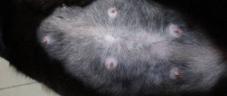
A cat's panting with a protruding tongue is a sign of heart disease.
- Fatigue is difficult to notice in a cat, since it leads a generally quiet lifestyle.
- Dyspnea. Breathing occurs through the abdomen, without the participation of the chest.
- An attack accompanied by loss of consciousness. The cat, at this time, can be mistaken for a dead animal. Usually the attack passes quickly, but it happens that pets die, as their body experiences an acute lack of oxygen.
- The animal wheezes and meows terribly.
- Heavy breathing indicates pulmonary edema.
- Complete or partial paralysis of the hind legs.
- Cardiopalmus.
- Cyanosis of the gums.
- Loss of appetite.
In cats, coughing is not a cardiac symptom.
First aid for a cat who faints
The onset of an attack requires quick and correct actions by the owner, since, sometimes, it can end in death.
- Lay the cat down, and it is necessary to give her a side position of her head.
- Pull out your tongue.
- Place a cool compress on your head.
- Place cotton wool soaked in ammonia to your nose.
- The paws must be fixed higher than the head, so there will be more blood flow to the head.
- Call your veterinarian.
How to distinguish a healthy cat from a sick one
Since cats generally lead a calm lifestyle, they are couch potatoes, not all owners can distinguish a healthy animal from a sick one. She can report any changes in well-being by changing her behavior, that is, if the cat used to behave independently of the owner, but now does not leave him, then this indicates that something is bothering her.
Some people think that purring in cats is a sign of health. This is wrong. Purring, abruptly replaced by aggression or growling, indicates that she is in pain.
A healthy animal has:
- Smooth wool.
- The nose is wet and cold.
- The mucous membranes of the eyes are pinkish in color.
- The animal is vigorous and active.
Sick animal:
- Lethargic, lies more than usual.
- He tries to get away from everyone to a secluded place.
- Can be very excited.
- The meow is pitiful.
- Movements are clumsy.
- The nose is warm with cracks.
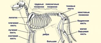
Skeletal structure of a cat
The cat skeleton contains approximately 240 bones. It has an axial and peripheral part. The structure of the axial section includes:
- Scull. Its front and brain parts are almost equal in size. The facial part is formed from 13 bones. The dentition of adult animals consists of 30 teeth. In the jaw of a one-month-old kitten there are 26 milk units, which change to permanent ones by 6 months.
- Spine. Movable vertebrae make the cat very flexible. The most massive bones are located in the cervical region. The thoracic part consists of 13 vertebrae, to 12 of which ribs are attached on both sides. The lumbar part includes 7 bones; muscles that support organs located in the abdominal cavity are attached to them. The sacrum consists of 3 fused vertebrae, the tail - of 12–28 movable ones.
- Rib cage. The breast bone unites 8 pairs of ribs in the front of the body. The animal's collarbones are rudiments, so they are not developed. This makes it easier for the cat to move around and gives it the ability to get into narrow crevices.
Internal structure of a domestic cat
Representatives of the cat family are mammals, so the structure of the internal organs of a cat is practically no different from similar structures of other creatures included in this class. The systems and organs of these animals work according to principles characteristic of most mammals and perform similar functions. However, there are features that are unique to cats. The internal structure of a cat can be seen in the photo, and a description of the most important systems is presented below.
Internal organs of a cat
The cardiovascular system
The cardiovascular system is formed by a network of blood vessels and the heart, which ensures the movement of blood cells and lymph. The main function of this system is to saturate tissues with nutrients and oxygen, as well as remove waste products.
The heart is a special muscle in a cat's body. It has 4 chambers: 2 atria and 2 ventricles. In an adult cat, the weight of the heart is about 15–30 g. The ventricles of the heart contract and force the blood to move through the vessels.
Large blood vessels - veins and arteries - carry blood away from the heart and return it back. Small vessels are capillaries that deliver blood to the organs. Thanks to them, tissues are saturated with oxygen and nutrients. Blood consists of blood cells (red blood cells, platelets and white blood cells) and plasma.
Digestive organs
The digestive system includes the oral cavity (tongue, teeth, salivary glands), pharynx, esophagus, stomach and pancreas, liver, gall bladder and intestines, which includes 4 sections: duodenum, small intestine, ileum and large intestine. The esophagus starts from the base of the mouth and connects to the stomach, the inner surface of which is formed by many folds. They increase the mechanical effect on food masses during digestion.
The food then passes through the pyloric sphincter and enters the duodenum. Here the food is mixed with pancreatic and liver enzymes. The digestion process continues.
In the small intestine, all nutrients are absorbed thanks to the many villi lining the inner surface of the intestine. The mass then turns into feces, passing through the ileum and colon, where excess moisture is sucked out of it.
What is anatomy?
Anatomy is a branch of science devoted to the study of the structure of the bodies of various creatures. Anatomy helps to establish the common features inherent in a particular species of animal. This science studies the external characteristics of species, the location of internal organs relative to each other, and clarifies their significance and functions.
Anatomy includes the following areas of science:
- osteology , which studies bone structures;
- myology , which studies the structure of muscle fibers, the location of muscles and features of work;
- syndesmology , which studies the elements connecting parts of the skeleton;
- angiology , which studies blood vessels, lymphatic and circulatory systems;
- neurology , aimed at studying the functions of nodes and parts of the nervous system;
- splanchnology , systematizing knowledge about the structure of the respiratory, digestive, excretory and reproductive organs;
- endocrinology , explaining the significance of the endocrine glands;
- aesthesiology , which studies the functioning of the senses.
These scientific disciplines make it possible to trace how the formation of the most important systems occurs, as well as to establish their interrelationships. By studying the anatomy of a cat, you can find out what distinguishes it from other mammals. Anatomical knowledge allows us to understand the purpose of certain structures of the body.
Cat's sense organs
Cats have well-developed senses. The following organs are responsible for the perception of the outside world in cats:
- Visual. Cat's eyes have a convex cornea, which makes it possible to obtain high-quality images. The pupils react to light by constriction. Cats see perfectly at a distance of 3–6 meters. In the dark, the eyes of these predators glow, which is explained by the presence of a tapetum - a special vascular layer located behind the retina. Color perception allows a cat to distinguish between 3 colors: blue, red and green.
- Auditory. Cat ears even perceive ultrasonic waves. The outer ear is a cartilaginous formation, it is very mobile due to its connection with 27 muscles. Cats have a fold of skin inside their ear.
- Olfactory. Cats have about 80 million olfactory receptors in their noses, which allow them to distinguish a huge number of odors.
- Tactile. The skin, mucous membranes, and vibrissae are responsible for the sense of touch. The sense of touch allows cats to determine the temperature of the environment, feel touch and experience pain.
- Flavoring. The receptors responsible for the perception of taste make cats excellent tasters. They sense and determine the quality of food well thanks to the Jacobson tubes located on the palate.