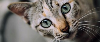
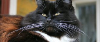
Eyes are the mirror of a cat's soul
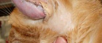
Eyes are not only a mirror of the soul, but also an indicator of health. And an experienced cat owner can easily determine from the cat’s gaze how his pet is feeling. If your cat has a dull look, or, even worse, his eyes are running or spinning, while the cat seems to be looking into emptiness, it’s time to sound the alarm. This phenomenon is called nystagmus.
We invite you to talk about all this in our publication today...
What causes the functioning of the vestibular apparatus to be disrupted, what are the symptoms?
The most common clinical symptoms of vestibular disease include: sudden falls of the animal on a completely level place, an unexplained body roll in one direction or another, a clear and constant curvature of the neck, as well as nystagmus - a rapid and involuntary oscillatory movement of the eyeballs. If the cause of the disease is serious, a “sagging” muzzle may be observed. This is explained by the fact that the facial nerves (and, accordingly, muscles) are closely connected with the hearing organs.
The causes of this pathology can be extremely diverse, ranging in severity from spontaneously resolving to fatal. Very often the disease develops against the background of advanced meningococcal infection; its cause can be any inflammatory disease. Also in veterinary practice, there are often cases of vestibular syndrome occurring against the background of the willful use by animal owners of certain “human” antibiotics that are toxic to cats.
Remember!
Giving your cat gentamicin, as well as any drugs from the tetracycline group, is strictly prohibited! Perhaps you will cure the infection, but after such “therapy” your pet may well remain disabled.
Why does the kitten walk poorly?
Cerebellar ataxia in cats is a congenital pathology. It manifests itself at an early age, when kittens begin to take their first independent steps.
Ataxia is a disorder of motor coordination. This deviation can have various origins. In this case, the cause of the pathology is underdevelopment of the cerebellum. This organ is responsible for the sensation of the body’s position in space and the coherence of movements.
In sick kittens, damage to the cerebellum occurs during fetal development due to various adverse effects on the mother’s body during pregnancy. Various factors can contribute to the birth of a sick kitten. Most often, cerebellar ataxia in babies occurs if a pregnant cat has had panleukopenia (distemper). Parvovirus causes which leads to organ hypoplasia.
Ataxia in kittens usually develops if the mother becomes ill with distemper in the later stages. Infection with panleukopenia early in pregnancy usually leads to the death of the embryos. If the infection occurs closer to birth, then both dead kittens and cubs with cerebellar hypoplasia may be born.
Other harmful effects on the mother’s body can also lead to congenital ataxia in a kitten:
- bacterial infectious diseases;
- food or poison poisoning;
- helminthic infestation;
- poor nutrition.
There is also a hereditary form of ataxia in cats. However, this pathology is rare.
Diagnostics and therapy
Diagnosis of vestibular syndrome itself is quite simple, since the diagnosis is made based on clinical signs. The difficulty lies in identifying the disease that caused it all to begin.
Your veterinarian will need a comprehensive medical history; the specialist will conduct a full examination of the pet, including neurological and otoscopic examination, which are needed to identify infectious diseases, identify inflammatory processes, and tumors. Please note that in complex or doubtful cases, MRI is strongly recommended. Unfortunately, not every hospital has such equipment. It is precisely because of the difficulty of identifying the primary pathology that many cases of vestibular syndrome are considered “idiopathic.”
Feline vestibular syndrome
is a disease of the inner ear that can negatively affect your cat's sense of balance. This disease mainly affects cats aged 12 years and older. In most cases, feline vestibular syndrome is idiopathic in nature, which means that the causes that caused it are difficult, and sometimes even impossible, to determine.
For vestibular syndrome
the animal may walk staggering, fall, or lose balance. His head sometimes tilts to one side, and his eyes, as they say, begin to “run” (nystagmus). If there are any manifestations of at least one of these symptoms, then this is a reason to worry about the health of your cat.
How to care for a Siamese cat
Due to the short hair and lack of undercoat, caring for the Siamese cat is reduced to a minimum. It is very easy to comb it, even the owner’s hand is suitable for this: moisten your palm and stroke the pet along the growth of the fur, towards the tail. All loose hairs will remain on the palm.
Like all felines, the Siamese cat should be regularly bathed, ears cleaned, and teeth brushed. It is better to start all these procedures while your pet is not yet grown up: firstly, he will get used to them and will accept them without resistance, and secondly, this breed is prone to dental diseases.
When cared for, Siamese cats can live up to twenty years. Their inherent diseases are the same as those of most felines: these are genetic problems, as well as common cat diseases. These include:
- liver amyloidosis, subsequently leading to liver failure;
- development of breast cancer;
- enlargement of the myocardium, or heart muscle (cardiomyopathy);
- dental diseases (gingivitis, tartar and others);
- strabismus (very rare, and in ancient times, like knots on the tail, was considered a sign of the breed);
- ordinary ailments (worms, lichen, fleas - can be cured by the owners).
Fortunately, serious diseases in Siamese cats are rare, and the absence of others depends almost entirely on the attentive and caring attitude of the owner towards his pet. Give your pet attention and love, and he will selflessly warm your heart.
And below is not a Siamese, but also a beautiful cat:
Vestibular disorders
The vestibular apparatus, as is known, is responsible for the correct orientation of the animal’s body and head in space in relation to ground level. Without going into details, it consists of many nerve fibers originating in the brain and ending in the inner ear. Disorders in the functioning of the vestibular system affect the brain's ability to accurately determine the position of the body. It is for this reason that a cat with vestibular disorder
loses sense of balance, tends to fall frequently, throws head back, and is unable to walk in a straight line.
Causes
There are a large number of reasons that can lead to the development of nystagmus in dogs. Most of them are associated with central nervous system disorders and related diseases. In some works devoted to this problem, one can find such a definition as “a disorder of the balancing system,” that is, a disruption of the functioning of the vestibular apparatus and the part of the brain responsible for maintaining proper balance of the head and body.
Also possible causes of the development of nystagmus in dogs can be, for example, hypothyroidism, traumatic injuries (for example, due to a car accident), various tumors, thiamine deficiency, viral infections (for example, carnivore distemper), and, as a result, inflammation, heart attacks, hemorrhage in the heart, exposure to poisons and toxins (for example, lead).
Types of vestibular disorders in cats
There are two main types of vestibular disorders in cats.
. They are divided into peripheral and central disorders. The central disorder occurs in the brain, and the peripheral disorder occurs in the inner ear. Usually, the central disorder has an organic cause and when it is eliminated, the disorder stops. The main symptoms of vestibular disorder in cats:
- falls;
- loss of coordination;
- tilting the head to one side;
- movement in a circle, rocking;
- stumbling on level surfaces;
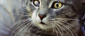
- constant and frequent movement of the eyes in different directions (nystagmus).
Description of the breed
The Siamese cat is medium in size and is characterized by a thin but muscular build. A female cat weighs three to four kilograms, and a female cat weighs four to five kilograms. The elegant, sleek Siamese cat has surprisingly long, narrow limbs, with the hind limbs slightly longer than the front. Its small paws are oval shaped. The tail is long, thin and ends in a pointed end.
The neck is similar to the rest of the body, long and narrow. The head is medium in size and wedge-shaped. The chin and ears form almost a triangle. The nose is long and straight, and the chin is barely defined. The pointed ears are large and slightly diagonal. The eyes are almond-shaped, slightly slanted and spaced far apart. Typical for a Siamese cat is a bright blue eye color. The coat is short and has little undercoat because the breed originated in warm Southeast Asia. The wool is very soft and shiny.
The Siamese cat is one of the most demanding breeds. She requires a lot of attention from her owner because she is very affectionate and prone to jealousy. She doesn't like other animals in the house, but she feels very comfortable in the company of other Siamese cats. Therefore, she cannot be left alone. Because of its temperament and pronounced love of play, the Siamese cat needs a lot of space in the apartment.
A common problem in keeping Siamese cats is their sensitivity to cold and wet conditions. You should never expose Siamese cats to drafts. Additionally, the Siamese cat tends to have poorer vision at night than other breeds. After washing, it must be dried thoroughly. But the shorthaired Siamese cat's coat is very easy to care for. It is enough to clean it with a brush. If you keep two Siamese cats, you have even less work to do because the cats love to groom and clean each other.
Diagnosis of a vestibular disorder in a cat
Only a physiological examination and study of the animal’s medical history can help in diagnosing disorders of the vestibular apparatus. Along with examining the cat's inner ear, it is also necessary to conduct a full range of neurological examinations. A blood test will assess the overall health of your pet. Perhaps it will help find out the cause of vestibular disorder
. It is recommended to conduct a complete blood test, including biochemical and sugar content. It would be nice to have the results of a urine test (general and Nechiporenko) to determine the normal functioning of the liver and kidneys, to rule out the toxic nature of the disorder. If the disease cannot be diagnosed even after these tests, then a spinal tap, MRI, or x-ray of the skull may be required. If the results of these tests are negative, then central vestibular disorder can be safely excluded from the list of possible causes of the disease, despite the similarity in the manifestation of symptoms. There are quite a few options for brain damage (tumors, cysts, infiltrates, etc.) from which you can start and establish the truth of these neurological disorders.
However, since we are talking about vestibular syndrome
, it is very important to determine what type of vestibular disorder your pet is suffering from. And all this is definitely a prerequisite for a pet treatment program.
Treatment of vestibular disorder in cats
In general, vestibular disorders do not require treatment or require only minor treatment. Symptomatic, as a rule. Usually, in case of nausea in an animal with this disease, drugs such as diphenhydramine, miklesin, no-spa, riabal, cerucal, etc. are recommended. They will help your pet cope with the consequences of “seasickness”. If the basis of the disease is idiopathic, then healing will take longer (several weeks).
The most important thing in case of vestibular disorder is to exclude dangerous situations when a cat can harm itself.
It is advisable to visit a veterinarian at the initial stage of the disease. He can prescribe medications that will help the animal endure the disease more comfortably, and will give you competent advice. Karataev Pavel Sergeevich, veterinarian, neurologist, VK “Zoolux”, Kiev. Abbreviations: PVS – peripheral vestibular syndrome. CVT – central vestibular syndrome.
Signs of pathology
The disease first appears in childhood, when the kitten begins to move actively. The baby walks with a strong stagger (“drunk gait”), often falls and spreads his paws wide when moving. This is the leading symptom of the pathology. In addition, the kitten's head trembles, especially when it tries to focus its attention on a toy or some other object.
Veterinarians distinguish several degrees of cerebellar ataxia in cats:
- Easy. The kitten has slight gait disturbances, and the baby periodically falls. But in general, the animal moves without any problems.
- Average. The pet's movement is very difficult, and frequent falls are observed. But the animal is still able to walk.
- Heavy. The animal cannot move at all.
However, the pets do not experience any other changes in their health status. This disease is not accompanied by pain. Cats with ataxia eat normally and do not feel ill.
A kitten with ataxia develops normally. Damage to the cerebellum does not affect the mental abilities of the pet in any way. The disease also does not affect life expectancy. A cat with ataxia can live to a ripe old age.
This disease does not progress. On the contrary, with age, the animal’s movements become more coordinated. If a cat’s gait worsens over time, then most likely this is due to other pathologies, and not to cerebellar ataxia.
Anatomy of the vestibular system
The vestibular system
is a component of the nervous system that is responsible for maintaining balance and posture of the body and head.
Functionally, the vestibular system consists of two parts
(Fig. 1) - peripheral (outside the brain stem) and central (located in the brain stem and cerebellum). Determining the location (peripheral or central vestibular syndrome) is a very important step in the diagnosis of patients with vestibular disorders.
The peripheral part of the vestibular apparatus (Fig. 2, 3) is located in the petrous part of the temporal bone (bone labyrinth). The bony labyrinth consists of the semicircular tubules, the vestibule and the cochlea (which takes part in the formation of hearing). Inside the bony labyrinth there is a membranous (membranous) labyrinth. The space between the walls of the bony and membranous labyrinth is filled with a fluid - perilymph, whose properties are similar to cerebrospinal fluid. Inside the membranous labyrinth there is another type of fluid called endolymph. The three semicircular tubules are located at right angles to each other. Each semicircular canal at one end has an extension - an ampulla, in which receptor cells are located (ciliated cells extending their villi into a jelly-like structure - the cupula). When the endolymph in the semicircular tubules moves, the cupula also moves and displaces the villi, and the dendritic endings of the sensory neurons of the 8th pair of cranial nerves (CN) are stimulated. The utriculus and sacculus also contain a receptor organ - the macula, which is covered with ciliated cells. The cilia extend into a jelly-like structure (otolithic membrane) and are embedded in crystals (otoliths). As the otolithic membrane moves relative to gravity, the villi are displaced and an action potential is generated. Nerve fibers arising from the nerve cells of the semicircular tubules and sacs are collected in nerve bundles and exit through the internal auditory canal along with the facial nerve into the cranial cavity (in the rostral part of the medulla oblongata). The combination of vestibular and auditory axons at this level form the 8th pair of cranial nerves. The axons then enter the brainstem at the level of the trapezius body and caudal cerebellar peduncles.
After entering the brainstem, the axons travel in different directions. Most axons form synapses with the vestibular nuclei (Fig. 4). A small number of axons bypass the vestibular nuclei and reach the cerebellum through the caudal cerebellar peduncles. Some axons form synapses in the fastigial nucleus of the cerebellum, others ascend to the cerebellar cortex, in the flocculonodular lobe. There are 4 vestibular nuclei on each side of the brain stem. They are located on the ventral wall of the 4th cerebral ventricle. Axons from these nuclei extend into the spinal cord (lateral vestibulospinal tract) and rostrally into the brainstem (medial longitudinal fasciculus). Fibers of the vestibulospinal tract influence the tone of the extensors of the limbs. The medial longitudinal fasciculus passes rostrally (axons end in the nuclei of the cranial nerves III, IV, VI and influence the position of the eyes) and caudally into the spinal cord (forms the medial vestibulospinal tract). Information from the vestibular system is also projected to other areas of the brain stem (vomiting center in the reticular formation) and the brain (including synapses in the thalamus).
Study of the vestibular system
The main function of the vestibular system is to maintain balance and body position in space.
The vestibular system also influences the extraocular muscles. The influence of the vestibular system on maintaining balance is achieved through its influence on the extensor muscles. Each side of the vestibular system enhances the function of the extensors ipsilaterally. In a normal state, the right and left sides act equally and therefore balance is maintained. If a problem occurs on one side of the vestibular apparatus, the function of the extensors on the opposite side increases, which leads to the main symptoms that can be observed with vestibular syndrome. With vestibular syndrome, ataxia is observed (possibly with a fall on the side), but there will be no paresis or paralysis. The vestibular system also influences the formation of the oculovestibular reflex, therefore the assessment of pathological eye movements is of great importance in the localization of pathology. The brain always focuses vision on some object. When the head moves from side to side, for example to the left, the extraocular muscles on the right will “pull” the eye back (to the right) to ensure focus on the object. When the muscle contraction limit is reached, the eye quickly moves in the direction of head movement (to the left) to focus on a new object. This reflex activity is called the oculocephalic reflex (physiological nystagmus), and the slow phase will be observed in the direction opposite to the movement of the head. Pathological nystagmus occurs independently, without head movements. Nystagmus can be without fast and slow phases (pendular - pendular nystagmus) or have a fast and slow phase (jerk - clonic nystagmus). Pendulum-like nystagmus is not a symptom of vestibular disorders; it is observed in some breeds (Siamese, Himalayan, etc.) as a congenital pathology of the visual pathways. With unilateral damage to the vestibular apparatus, an imbalance of nervous activity occurs, because the vestibular apparatus on the healthy side continues to constantly send impulses. This imbalance is interpreted by the brainstem as body movement, and nystagmus will occur with a rapid phase in the opposite direction of the lesion.
Nystagmus can be spontaneous and positional, the latter occurs in certain body positions (when raising the head up, lying on the back). Depending on the direction of movement, nystagmus can be horizontal, vertical and rotational. Another symptom that is often observed with vestibular syndrome is strabismus (pathological position of the eyes, usually ventral or lateral ipsilateral displacement). Strabismus can be spontaneous (always present) and positional (observed only in certain positions, for example when raising the head up) (Fig. 5). Nausea and vomiting are rarely observed in dogs and cats with vestibular syndrome. The vomiting center is located in the reticular formation in the medulla oblongata and has a direct connection with the vestibular nuclei.
Determining the location (peripheral or central vestibular syndrome) is a very important part in the diagnosis of vestibular disorders. A thorough history, physical and neurological examination provide significant assistance in this regard. The main task in determining localization is to identify signs that are characteristic only of central vestibular syndrome.
Head tilt is observed in both peripheral and central vestibular syndrome. With PVS, the tilt of the head always occurs in the direction of the lesion; with PVS, the tilt of the head can be in the direction of the lesion or the opposite (see below - paradoxical vestibular syndrome).
Tilt of the head can be combined with walking in a circle. Nystagmus – horizontal and rotational – is possible at any location (PVS or CVS). Vertical nystagmus indicates CVS. With PVS, the fast phase of nystagmus is directed in the direction opposite to the lesion; with CVS, the fast phase can be directed in any direction. Since animals with PVS are able to quickly compensate for lesions, nystagmus may be observed for only a few days. In CVS, nystagmus is often positional and can change direction (eg, rotational/vertical) with changes in head position. The number of eye movements per minute can also help with differentiation a little - with PVS the rate is usually greater than 66 movements per minute. With bilateral vestibular syndrome, there will be no nystagmus or head tilt. Strabismus in PVS is ventral or ventrolateral, and it is observed on the affected side. Often strabismus is positional. With CVS, strabismus can occur in any direction. The facial nerve (VII) passes through the internal auditory canal of the petrous bone and dorsal to the middle ear cavity. Due to its proximity to the peripheral part of the vestibular apparatus, some diseases can cause dysfunction of the facial nerve (paresis/paralysis). The sympathetic innervation of the eye also passes near the middle and inner ear, so Horner's syndrome may also be observed in peripheral vestibular syndrome (Fig. 6).
The presence of deficits in cranial nerves (except VII and VIII) indicates central vestibular syndrome. Changes in consciousness may also occur with CVS. Another sign of CVS is a deficit in proprioceptive reactions. Since the proprioceptive pathways have no connection with the peripheral vestibular system, decreased postural responses indicate CVS. Also, with CVS, there may sometimes be signs of cerebellar damage. Symptoms of forebrain damage (seizures, loss of vision, behavioral disturbances, etc.) in combination with vestibular syndrome indicate multifocal lesions. Paradoxical vestibular syndrome occurs when the central vestibular system is damaged. In this case, the head tilt will be observed in the direction opposite to the lesion. Normally, the cerebellum sends inhibitory impulses to the vestibular nuclei. If inhibition is lost, the vestibular nuclei on the same side will send more impulses to the extensors ipsilaterally, causing the body to move in the direction opposite to the lesion. In such cases, the localization of the lesion can be determined by proprioceptive deficit - a decrease in postural reactions will occur ipsilaterally. Paradoxical vestibular syndrome occurs when the flocnodular lobe of the cerebellum, the caudal cerebellar peduncles, and the rostral and medial vestibular nuclei in the brainstem are affected.
Diseases leading to peripheral vestibular syndrome (PVS)
Degenerative diseases/anomalies
Congenital malformations are more common in young animals and may be associated with deafness. With a bilateral problem, the patient may not have nystagmus or head tilt. In this case, symmetrical ataxia, wide stance of the limbs and side-to-side movements of the head will often be observed. Symptoms begin to appear when animals begin to walk. Many of the animals can compensate for vestibular disorders (to the point of being able to walk). In this case, deafness (if any) remains. Congenital anomalies of the vestibular system are more often observed in breeds such as the German Shepherd, English Cocker Spaniel, Doberman Pinscher, Siamese and Burmese cats.
Metabolic causes
Hypothyroidism is a common metabolic cause of vestibular syndrome in older animals. Involvement of the peripheral vestibular system may be the first sign of hypothyroidism. Typically, hypothyroidism has an acute onset and a non-progressive course. Nearly all animals exhibit head tilt and positional strabismus, and many patients also show other signs of facial nerve damage, including decreased threat response and palpebral reflex. Such patients may also experience systemic signs of hypothyroidism, such as hypercholesterolemia. Hypothyroidism can cause both peripheral and central vestibular syndrome; central may be associated with ischemic infarction. Treatment consists of replacement therapy; most patients return to normal with early treatment. Neoplasms can cause PVS by compressing or invading the vestibulocochlear nerve. Peripheral nerve neoplasms may initially appear as PVS, but over time the tumor may invade the brainstem. Ear neoplasms (eg, ceruminal gland adenocarcinoma) can invade the middle and inner ear and cause PVS.
The most common toxic causes leading to PVS are aminoglycosides and various ear cleaners. The onset of development of vestibular syndrome is usually acute. Treatment is to stop using the medications.
Trauma rarely results in PVS. In this case, vestibular syndrome will often be combined with signs of injury to other areas of the brain.
Diseases leading to central vestibular syndrome In principle, any disease of the brain can also involve the central part of the vestibular system. There are no diseases that involve only the central part of the vestibular system.
Neoplasms can be primary or secondary. Possible chronic course or acute onset. Neoplasms cause compression, ischemia or infiltration. The prognosis depends on the location, size, and type of tumor. Treatment is conservative (for example, corticosteroids to relieve peritumoral edema), chemotherapy, surgical removal, radiation therapy. Inflammatory diseases can be infectious (canine distemper, feline infectious peritonitis, toxoplasmosis, bacterial meningoencephalitis, etc.) and non-infectious in origin (considered autoimmune diseases). Treatment depends on the cause (for non-infectious meningoencephalitis, treatment involves the use of various immunosuppressive drugs). Forecasts are always very cautious. Metronidazole intoxication
– one of the possible causes of damage to the central vestibular system. The onset of the disease is acute, usually observed after long-term use of high doses of metronidazole. Symptoms of intoxication – generalized ataxia, nystagmus, vomiting; in more severe cases - depression, convulsions, opisthotonus. Treatment is discontinuation of metronidazole, after which patients return to normal, usually within 1–2 weeks. Using diazepam usually speeds up recovery. Thiamine deficiency can cause damage to the vestibular system. It can occur due to a lack of thiamine in the feed, the destruction of the vitamin during the cooking process, or the content of a large amount of thiaminase in the food (a substance that destroys thiamine; it is found a lot in fish). Thiamine deficiency causes bilateral foci of necrosis and hemorrhages in the brain stem. In cats, the vestibular nuclei are primarily affected, and there may also be evidence of cerebellar involvement. Treatment is the administration of thiamine; with early detection and treatment, the prognosis is usually favorable.
Projections to the brainstem
The vestibulospinal tract is the main projection into the spinal cord, descending from the vestibular nuclei in the medulla oblongata to all segments of the spinal cord as part of the ventral funiculus on the same side, the effect of which on motor neurons, mediated by segmental interneurons, is increased contraction of extensor muscles and inhibition contraction of the flexors on the same side with simultaneous inhibition of contraction of the extensor muscles on the opposite side (Fig. 1B).
Thus, the overall effect of activation of the vestibular system is to increase the tone of the muscles that counteract gravity on the same side and suppress the tone and extensor reflex on the opposite side. 2,3 These pathways help coordinate motor activity of the limbs, neck, and trunk in response to head movements.
Medial longitudinal fasciculus. The medial longitudinal fasciculus (MLF) (Fig. 1B) descends from the vestibular nuclei in the medulla oblongata to synapse on lower motor neurons in the motor nuclei of cranial nerves III, IV, and VI. 3,5 This pathway mediates coordinated eye movements as head position changes. In addition, the MPP is part of the pathway responsible for the physiological nystagmus induced when testing the vestibulo-ocular reflex.
READ Short-haired and smooth-haired cat breeds, their features
Reticular formation and vomiting center. Axons from the vestibular nuclei project to the vomiting center in the reticular formation. This pathway is involved in vomiting in motion sickness/vestibular disease. Vomiting is not typical for vestibular diseases in animals, unlike in humans. 1,3,4
Conscious perception of balance. It is clear that conscious perception of balance is important, based on verbal descriptions of spatial perception deficits in cortical dysfunction in people with vestibular disorders. 3,4 The afferent pathways that play a role in conscious perception of vestibular disorder are currently poorly understood but are thought to extend from the switching centers of the thalamus to the temporal cortex. 4
Vestibular axons from the vestibular nuclei and vestibular ganglion project to the vestibulocerebellar system (flulnonodular lobe and cerebellar tent nucleus) through the caudal cerebellar peduncle. 2,6 These axons support the coordination of eye, neck, trunk, and limb movements in relation to head movements and also when the head is stationary.
It is possible that spaniels and their mixes are predisposed to cerebellar infarctions. 45
If a heart attack is suspected, the animal should be evaluated for hypertension, hyperadrenocorticism, hypothyroidism, and heart or kidney disease. 30.44
Many animals with infarcts in this area improve with time and supportive treatment.
The risk of infarction and morbidity associated with neurological impairment is significantly higher in dogs with infarcts that have predisposing conditions. 44