- Animal cardiology
- Informational portal
- Gastroenterology of dogs
- Literature
Infusion therapy in cats and dogs
used for the treatment of shock, dehydration and correction of electrolyte and acid-base balance. Changes in electrolyte balance cannot be predicted accurately, so electrolytes must be measured. Vomiting of gastric contents often leads to the development of hypokalemia, hypochloremia and metabolic alkalosis. Loss of intestinal contents classically results in hypokalemia with or without acidosis, but hypokalemic metabolic alkalosis can occur. Vomiting in an animal leads to hypokalemia; however, animals with hyperadrenocorticism or anuria due to renal failure may have hyperkalemia. If it is not possible to immediately measure electrolytes, an infusion should be performed with saline plus 20 mEq of potassium chloride per liter, with the condition that the liquid will be administered 1 or 2 times depending on the condition. To exclude severe hyperkalemia, it is advisable to take an ECG.
The administration of bicarbonates is rarely required, because dilation of blood vessels and improved perfusion of the periphery facilitates the course of lactic acidosis. Bicarbonate is primarily prescribed to patients with severe acidosis (eg, pH
Types of Fluid Therapy in Cats and Dogs
Parenteral fluid administration is required if the animal is significantly hypovolemic
, or GI fluid absorption is questionable (eg, severe bowel disease, obstruction, vomiting, or ileus). Subcutaneous fluid administration is quite acceptable if the animal is not in shock, absorbs the fluid and tolerates subcutaneous administration normally. Subcutaneous depots have a capacity of 10 to 50 ml depending on the size of the animal. Before re-introducing into one depot, you need to make sure that there is no residual liquid there. Severely dehydrated animals do not absorb fluid from the subcutaneous space as quickly as required, so it is advisable to administer fluid intravenously initially. Animals with severe dehydration or shock require intravenous fluid administration, even if this requires venesection. Fluid can be administered intramedullary if intravenous infusion is necessary and a catheter cannot be placed. To do this, use large diameter hypodermic needles or bone marrow aspiration needles (preferably). They can be inserted into the femur (trochanteric fossa), tibia, iliac wing, or humerus. Fluids can be administered intramedullary at a maintenance infusion rate or faster.
Intraperitoneal fluid administration is also possible, but replenishment of the vascular bed occurs more slowly than intravenously or intramedullary.
Dogs in shock (with tachycardia, poor peripheral perfusion, cold extremities, prolonged VOC, weak femoral pulses, and/or tachypnea) may require isotonic intravenous crystalloid 88 mL/kg or more within the first hour. The "maximum" level may be exceeded if required to restore adequate peripheral perfusion; the patient must be under constant supervision. It is important to remember that dogs with systemic inflammatory response syndrome (SIRS) initially have brick-red mucous membranes, warm extremities, and a strong femoral artery pulse before classic signs of shock occur. Large dogs in severe shock, such as gastric volvulus, may require 2 catheters, 16 and 18 gauge, and the infusion bags must be pressurized to achieve an adequate infusion rate. Cats are easily overdone, so cats need to be monitored more closely when fluids are given rapidly. In general, for cats in shock, the volume should not exceed 55 ml/kg for the first hour.
To treat shock, ringer lactate or saline is used. However, you need to be sure that fluids used to treat shock do not contain a lot of potassium, due to its potential cardiotoxic effects.
Hypertonic saline (7%) can be used to treat severe hypovolemic or endotoxic shock. Relatively small volumes (ie 4-5 ml over 10 minutes) are approximately as effective as large volumes of isotonic crystalloid solutions. Hypertonic solutions remove fluid from the intracellular and interstitial space into the vascular bed and stimulate the vascular reflex. Hypertonic solutions should generally not be administered to animals with hypernatremia and dehydration, cardiogenic shock, or renal failure. Uncontrolled bleeding may also be a contraindication to their use. The doctor may give hypertonic saline in 2 mL/kg increments until a total of 10 mL/kg has been given or until the serum sodium concentration is 160 mg/eq/L or more. After administering the hypertonic solution, the physician may continue to administer other solutions, but in smaller volumes (eg, 10 to 20 mL/kg/hour) as long as shock is controlled. A mixture of 7% saline plus dextran 70 has a longer duration of action than a hypertonic solution alone.
This mixture can be administered in a volume of 3-5 ml/kg over 5 minutes. Dextran rarely causes allergic reactions or renal failure, but should be used with caution and is contraindicated in animals with coagulopathy.
Colloids are also useful in treating shock. Like hypertonic saline, colloids draw water from the interstitial space into the vascular bed; however, their effects last longer and do not increase the amount of sodium. Relatively small volumes can be administered rapidly (ie, 5 to 10 mL/kg, not to exceed 20 mL/kg per day) and subsequent fluid volumes should be reduced to prevent hypertension. Colloids should be used with caution in animals with a tendency to bleed.
If it is difficult to maintain maximum blood pressure above 80-90 mmHg, vasopressors may be necessary. IPA vasopressin was more effective for these purposes than administration of dopamine or dobutamine.
Infusion therapy for cats and dogs.
Approximately 44 to 66 ml of fluid per kilogram of body weight is required daily to support dogs weighing 10 to 50 kg, larger dogs requiring less. Dogs weighing less than 5 kg may require an infusion of 80 ml/kg per day. It is important to choose the correct solution to prevent electrolyte imbalances, especially hypokalemia. In general, potassium should be supplemented in animals with anorexia, vomiting, diarrhea, or receiving long-term fluid therapy. Possible development of iatrogenic hyperkalemia should be monitored (e.g., ECG or plasma potassium determination, and no more than 0.5 mEq/kg/hour should be administered. Oral potassium supplementation is often more effective than parenteral potassium supplementation, but only if the animal is not vomiting Cats receiving IV infusion initially show a decrease in potassium levels, even if it is contained in the solution at a concentration of 40 or more mEq/L.
Dehydrated animals that are not in shock are treated with fluid replacement. To do this, first of all, you need to assess the degree of dehydration. Prolonged straightening of the skin fold is observed in animals with 5-6% dehydration. However, in any cat or dog with weight loss, the skin fold may take longer to resolve, whereas in obese animals, it may resolve regardless of the severity of dehydration. Dry, sticky oral mucous membranes usually indicate 6-7% dehydration.
However, animals that are dehydrated but nauseous may have moist mucous membranes, and animals that are dyspneic or tachypneic (not dehydrated) may have dry mucous membranes. By multiplying the animal's weight by the percentage of dehydration, the amount of fluid needed to replenish the deficiency is obtained. This volume is administered over 2-8 hours, depending on the condition of the animal. The infusion rate should usually not exceed 88 ml/kg/hour. In general, it is better to slightly overestimate rather than underestimate fluid deficiency unless the animal has congestive heart failure, anuric or oliguric renal failure, severe hypoproteinemia, severe anemia, or pulmonary edema.
It is usually easier for a cat to be harmed by excess fluid than for a dog. Current fluid losses are assessed based on the presence of vomiting, diarrhea, and urination; however, losses are usually underestimated.
Weighing animals is one way to assess the effectiveness of infusion therapy. Progressive weight loss suggests inadequate fluid resuscitation. To evaluate results consistently, the same rating scale must be used. A change of 1 pound (0.45 kg) is approximately 500 ml of water.
The occurrence of wheezing in the lower parts of the lungs, a galloping rhythm, or pulmonary edema indicates that the animal is apparently overhydrated. The appearance of a heart murmur is not a sign of overhydration; In dogs with severe dehydration and valvular insufficiency, the murmur may not be detectable, but becomes audible when blood volume is replenished. Measuring central venous pressure is excellent for detecting excess fluid administration; however, this is rarely required except in animals with severe cardiac or renal failure receiving aggressive fluid resuscitation. CVP is usually less than 4 cmH2O and should not exceed 10-12 cmH2O, even during aggressive fluid resuscitation.
Oral rehydration is used to facilitate sodium absorption.
Additional administration of a monosaccharide (eg, dextrose) accelerates the absorption of sodium and subsequent water intake. This approach works if the animal can consume fluids (i.e., no vomiting) and the intestinal lining is functional (i.e., sufficient villi are present). Absorption occurs predominantly in mature epithelial cells at the tips of the villi. There are similar products on sale for people, as well as recipes for preparing similar solutions. Failure to adequately assess the animal's condition can lead to severe hypernatremia. Some dogs and cats with enteritis not caused by parvovirus infection can receive fluids orally.
The type of fluid therapy for animals with hyperproteinemia depends on the degree of hypoalbuminemia. Excess fluid administration may dilute serum albumin concentrations and cause ascites, edema, decreased peripheral perfusion, or a combination of these factors. In this regard, careful calculation of fluid requirements and ongoing losses is necessary. For animals with severe hypoalbuminemia (eg, serum albumin 1.5 g/dL or less), plasma transfusion (6 to 10 mL/kg) should be considered first to increase oncotic pressure. A common mistake is not getting enough plasma. Therefore, blood albumin concentrations should be measured every 8 to 12 hours to ensure that a sufficient volume has been administered.
Moreover, animals with severe enteropathy or protein-losing nephropathy rapidly lose infused protein, making repeated transfusions necessary to maintain plasma albumin concentrations.
Albumin replacement can be very expensive for large dogs
. Human albumin is used instead of canine plasma and is quite effective, although side effects have been reported. Hetastarch (5 to 20 ml/kg/day) and dextran 70 can be used instead of plasma and albumin. Hetastarch (presented as a 6% solution) is larger than albumin and may therefore remain in the vasculature longer than albumin, thereby helping to maintain plasma oncotic pressure in animals with protein-losing enteropathy. If hetastarch is used, the physician should reduce the rate and amount of fluid given to prevent hypertension. Sometimes the administration of hetastarch leads to massive edema and a significant worsening of ascites.
Intravenous infusion
Intravenous drugs are administered through a catheter attached to the patient's vein. Needles or butterfly systems are rarely used. Our patients usually refuse to sit still and hold the limb with the needle inserted into the vein motionless.
Solutions can be administered intravenously at the required speed; they directly enter the bloodstream, which is vital for patients in severe and extremely critical condition. If necessary, blood components and nutrients can be administered.
Intravenous infusion should be carried out under the supervision of a veterinarian. Usually it takes from an hour or more, if necessary it can continue around the clock (usually such animals are placed in a hospital). For patients who require extremely low or very precise rates of drug administration, syringe pumps (perfusors) are used. For everyone else, infusion systems are used, in which a solution through a tube enters the patient’s blood under the influence of gravity.
Infusion therapy is an indispensable tool in the treatment practice of a huge number of diseases, but requires thoughtful and careful use. The composition, volume, speed and type of infusion should be determined exclusively by a veterinarian.
Table - General recommendations for intravenous potassium administration
| Plasma potassium concentration (mEq/L) | Amount of potassium for additional administration |
| 3.7-5.0 | 10-20 |
| 3.0-3.7 | 20-30 |
| 2.5-3.0 | 30-40 |
| 2.0-2.5 | 40-60 |
| ≤2.0 | 60-70 |
* do not exceed a potassium dose higher than 0.5 mEq/kg/hour, except in animals with acute severe hyperkalemia, and only under constant ECG monitoring. Regular monitoring of plasma potassium concentration is required when administering a liquid with a potassium content of more than 30-40 mEq/L.
^Top
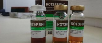
Device for infusion of infusion solutions and blood substitutes PR 21-07 and PR 23-07
System for infusion of infusion solutions and blood substitutes PR 21-07, sterile, single use. This device is intended for infusion of blood substitutes and infusion solutions from bottles with a 0.8 mm Luer injection needle and a metal needle for connection to the bottle. The PR 21-07 / PR 23-07 system includes: - Semi-rigid transparent dropper with a filter designed for filtering solutions during infusion. — The dropper provides the formation of 20 drops from (1.0±0.1) g of water. — The transparent material of the tubes makes it possible to detect air bubbles. — A metal needle with a diameter of 2.6 mm is used to pierce a bottle cap (model: 21-07) — A polymer needle combined with two channels for liquid and air with an air filter, used to connect to a polymer container or to pierce a bottle cap (model: 23 -07) — The injection unit allows for additional injections of medications and is used to connect an injection needle with a Luer cone and a silicone coating with a diameter of 0.8 mm or 1.2 mm. — The device has a roller clamp that allows you to smoothly adjust the infusion speed - from jet to drip. — The air duct is designed to allow air to enter the bottle during the pouring process; it has a needle with a diameter of 2.0 mm. — Working length of the main part of the device: 1400 mm — The connection of the device parts can withstand an excess pressure of 40 kPa. Sterilization by radiation method. The product is certified. Shelf life Shelf life from the date of sterilization is 3 years. Manufacturer: Russia (PR 21-07) Price: TEMPORARILY NOT SUPPLIED!
Instructions system for infusion of infusion solutions and blood substitutes PR 21-07 method of administration and dose
Good to know
- Other tests for diagnosing gastrointestinal diseases in dogs and cats
- Atrophic myositis in cats and dogs. Myositis of masticatory muscles
- Benign muscular hypertrophy of the pylorus. Pyloric stenosis in cats and dogs
- Special diet for pathologies of the gastrointestinal tract for dogs and cats
- Immunoproliferative enteropathy in Basenji dogs
- Foreign objects in the stomach in cats and dogs
- Intussusception in cats and dogs
- Bacterial diseases of dogs and cats - causative agents of enterocolitis
- Hypertrophy of the mucous membrane of the antrum of the stomach in dogs and cats (etiology, symptoms, diagnosis, treatment)
- Histoplasmosis of dogs, cats and humans (diagnosis and treatment)
- Granulomatous enteritis and gastritis in dogs and cats (symptoms, diagnosis, treatment, prognosis)
- Idiopathic megacolon in animals
- Idiopathic decreased gastric motility in dogs and cats
- Specific methods for analyzing feces in dogs and cats
- Disease of dogs and cats caused by bacteria of the genus Helicobacter
- Diseases of dogs and cats associated with malnutrition
- Volvulus of the mesentery and intestines in cats and dogs
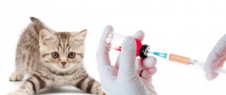
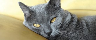
Putting an IV on a cat
The main instrument of the IV is a needle, which is inserted into the vein, and the entire system is connected to it. The pharmacy will provide you with:
- Butterfly needles are essentially the same needle, but with a smaller diameter. Designed for injection into small veins, which is important for the treatment of cats. At the base of the needle there is a “butterfly” made of flexible plastic - allows you to securely fix the system, “wings9raquo; folded to the paw and wrapped with adhesive tape.
- Intravenous peripheral catheters (brownulls) are a mini-system made of plastic. A flexible, thin latex tube is inserted into the vein, and the catheter itself is fixed to the skin - a device into which a needle attached to a syringe with medicine is inserted. Of course, administering medications through a catheter is easier and more convenient, however, the system can only be installed by a doctor.
Note! With proper care, the catheter can remain in the vein for up to 6 days, infusion with a simple needle is performed only once!
Before placing an IV on a cat at home, you need to take into account that the container with the medicine should be located 40–50 cm above the animal, think in advance where to attach the bottle. From what you have at hand, a cellophane T-shirt bag is suitable, make a hole in the bottom, thread the neck of the bottle, tie the handles above the bottom, and hang it by the resulting loops. The structure of all systems is similar; the infusion set contains:
- Aspiration needle or bag - the needle is inserted into the bottle of medicine, the bag is filled with liquid for infusion.
- Intake chamber - equipped with a filter, designed to separate oxygen from the liquid.
- Dispenser – equipped with a plastic runner wheel that regulates the speed of liquid falling. Before infusion, you need to know exactly how to administer the drug - drip or jet.
- The cannula is most often located on a rubber bag in front of the needle, which is inserted into the skin. If necessary, additional drugs are injected into it using a regular syringe.
Note! In addition to the sampling needle, you need to insert another needle into the bottle (included in the kit) for “suction”. air, otherwise the liquid will not fall into the tube. In droppers with bags, a hole for air intake is most often provided.
Let’s make a reservation right away that during emergency care you may need an intravenous injection, but believe me, giving an injection is easier than putting an IV on a cat at home - we are considering the most difficult option:
- Remember to be sterile and use alcohol.
- We attach the bottle in any convenient way. We unseal the system and insert the sampling needle into the rubber stopper of the bottle. We insert a second needle into the cork to allow air to flow. The temperature of the drug should not be low, at least room temperature.
- We move the dispenser to a place convenient for you and pinch the conductor with the adjusting wheel. If you skip this step, air will get into the tube!
- We squeeze the plastic container (collection chamber) several times, the medicine will be filled into it.
- We open the system slightly (lower the wheel) until the drug begins to drip from the cannula or needle and again press the tube with the dispenser.
- We put the cat in a comfortable position, and if necessary, shave the hair on the front paw - the area between the elbow and wrist. The needle can also be inserted into the vein of the hind leg; it all depends on the convenience and obedience of the animal.
- We pull the paw with a tourniquet closer to the elbow. If necessary, squeeze and unclench your fingers, bend and straighten your wrist - you should see how the veins become convex and clearly visible under the skin.
- We treat the skin with alcohol and the most interesting thing is that carefully, progressively, slowly, we insert the infusion needle parallel to the paw. Be careful and calculate your strength; it is more difficult to pierce a vein than a muscle, especially if the animal is dehydrated (the skin is more elastic).
- We fix the needle with adhesive tape in several places. We hold, calm, and entertain the pet until the end of the procedure. Remember that an IV for cats is a considerable stress and discomfort; do not leave your pet alone, monitor your pet’s breathing and general well-being.
We invite you to read: Ringworm in cats - photos, signs and treatment
Please note! Constantly check the area of skin where the needle is inserted. If you notice a slight mild swelling, the medicine goes under the skin and the needle needs to be reinstalled. Even a calmly lying cat, with deliberate contractions of the paw muscles, can “remove the vein” from the needle.
If you need to place an IV in the withers, for example, you need a subcutaneous injection of saline solution for dehydration or Ringer's solution to relieve toxicity - the scheme is the same, but strictly ensure that the needle is inserted parallel to the cat's spine!