The pain shock experienced by the animal, as well as the consequences of the injury, can cause death.
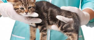
The preliminary diagnosis is confirmed in a veterinary center by puncture of the chest cavity with a syringe.
An alternative is an X-ray examination. An x-ray will detect existing accumulations of air in the pleural cavity; they will be indicated on the images by light spots.
The injured animal is recommended to rest and rest throughout the entire period of treatment. As a result of the course of therapy, the air in the pleural area resolves and the animal returns to normal. Otherwise, surgical correction may be prescribed.
For a week after stabilization of the animal’s health, the animal should be limited in its movements and not disturbed.
Is it possible to prevent closed pneumothorax in cats and dogs?
Etiology of pneumothorax in animals
Air can enter the pleural cavity through a wound in the chest wall.
Secondary pneumothorax occurs when the pulmonary pleura is ruptured, it happens in the case of a rib fracture, a sudden increase in intrapulmonary pressure during coughing or heavy work, through the rupture of pulmonary abscesses, gangrenous or tuberculous foci.
In dogs, pneumothorax often occurs due to trauma (bites by large dogs, car injuries, strong blows to the chest area, gunshot wounds, puncture wounds to the thoracic area).
Pneumothorax may be a consequence of purulent-putrefactive pleurisy.
Pathogenesis
There are open, closed and valve pneumothorax.
With open pneumothorax in animals, when inhaling, air enters the pleural cavity through a wound in the chest wall, and when exhaling, it comes out. Gradually, the pressure in the pleural cavity equalizes with atmospheric pressure, which leads to collapse of the lungs. On the damaged side, the mediastinum shifts to the healthy side, and intrathoracic pressure increases.
Pneumothorcus in horses is an extremely dangerous pathology!
Closed pneumothorax in dogs and other animal species is characterized by a similar development, but after the hole is closed, the air resolves and all symptoms disappear. Closed pneumothorax also occurs in cases where gas in the pleural cavity is formed from putrefactive exudate.
Valvular pneumothorax occurs in cats, dogs and other animal species. This type of air accumulation in the pleural cavity is characterized by the fact that air enters the pleural cavity with each breath, but its exit is difficult, so the air pressure in the lungs can exceed atmospheric pressure.
We invite you to familiarize yourself with Horse breeding: development in Russia, directions, products, breeds
Pneumothorax is accompanied by compression of the lungs, difficult gas exchange in the lungs, can lead to death from asphyxia, and is complicated by pleurisy and pneumonia.
Overview of Pneumothorax in Cats
Pneumothorax is the abnormal presence of air in the chest cavity, which limits the normal inflation of the lungs during inspiration. Air is usually restricted to spaces in the lungs.
Pneumothorax can be divided into the following categories:
Common Causes of Pneumothorax
Pneumothorax may result from any of the following:
Cats with pneumothorax experience difficulty breathing, and in severe cases, if left untreated, pneumothorax can be fatal.
What to watch
Diagnosis of pneumothorax in cats
Treatment of pneumothorax in cats
Treatment of pneumothorax may be required as an emergency procedure and may include any of the following:
Home care and prevention
Cats showing signs of difficulty breathing should be seen by a veterinarian immediately. Limit exercise initially after leaving the hospital.
Keeping cats indoors can reduce the risk of pneumothorax caused by traumatic injuries from car accidents or attacks by other animals.
There is no way to prevent spontaneous pneumothorax from occurring.
Traumatic pneumothorax
Traumatic pneumothorax is the most common pneumothorax in dogs, occurring in approximately half of all cases of thoracic injury (Kramek and Caywood, 1987). Traumatic pneumothorax is defined as open if its cause is a penetrating wound to the chest wall due to a bite or cut wound, gunshot or burn.
Closed traumatic pneumothorax is caused by blunt force trauma that results in rupture of the airway or pulmonary parenchyma. Simple closed pneumothorax is characterized by a nonprogressive loss of intrapleural negative pressure. Conversely, tension pneumothorax occurs when the ruptured airway, lung parenchyma, or chest wall acts as a one-way valve.
Treatment Treatment of traumatic pneumothorax depends on many factors, including the volume, source and direction of air flow within the pleural cavity (Orton, 1993), as well as the clinical condition of the patient, the severity of associated injuries and the availability of first aid. The most important goal of first aid is to stabilize the patient's condition, regardless of the cause of the pneumothorax, remove free air from the pleural cavity and restore the normal intrathoracic pressure gradient.
There are a variety of techniques for this, including thoracentesis, thoracostomy tubes with continuous or intermittent aspiration, or a vibrating Heimlich valve (Bulter, 1975). Additional measures will include oxygen supply, infusion therapy and antibiotics, depending on the general condition of the animal.
Open pneumothorax During examination and transportation of the animal to the clinic, the thoracic wound must be immediately covered with a clean swab, bandage or glove. The wound should be examined quickly, after removing hair and uneven edges. Then apply a pressure bandage with antiseptic ointment.
In case of dyspnea, thoracentesis should be performed immediately to remove air from the pleural cavity. After stabilizing the animal's condition, careful treatment and reconstruction of the wound is performed. If pneumothorax progresses after the first thoracentesis, a thoracostomy tube is placed with continuous or intermittent suction.
Simple pneumothorax Simple pneumothorax, with the exception of severe cases, is easily treated with conservative treatment. Bilateral thoracentesis should be performed if there is a clinical indication of tachypnea (respiratory rate greater than 80 breaths per minute), dyspnea, or hypoventilation (cyanosis, abnormal arterial blood gases, or decreased oxygen saturation).
Tension pneumothorax Tension pneumothorax causes severe pathophysiological consequences and can lead to rapid death. Dogs with tension pneumothorax develop severe and progressive dyspnea, weakness, shallow breathing, abdominal breathing, and a barrel-shaped chest.
Chest x-rays show large amounts of free air, flattening, and caudal displacement of the diaphragm. Urgent care is required, consisting of thoracentesis and then the installation of thoracostomy tubes with intermittent or continuous suction. A one-way valve can be used as an alternative to continuous suction, but its effectiveness is reduced by the presence of blood or effusion and is not suitable for dogs weighing more than 15 kg (Orton, 1985).
A diagnostic thoracotomy to clarify the diagnosis is performed only if aggressive drug treatment is not able to stabilize the animal's condition. During surgery, it is often very difficult to find the area of damage that caused the pneumothorax, and general anesthesia is a rather risky procedure. To visualize both sides of the chest, it is best to perform a midline sternotomy (Bjorling, 1994).
Perforation of the esophagus Perforation of the esophagus, leading to pneumothorax, is usually caused by trauma to a sharp foreign body that has entered the lumen of the esophagus; The cause may also be a bullet wound, a bite wound, or iatrogenic endoscope trauma. Pneumothorax due to esophageal injury is often accompanied by pneumomediastinum and inflammatory pleural effusions.
The diagnosis of perforation is confirmed by endoscopy or contrast X-rays using an organic (iodinated) contrast agent. Medical treatment of esophageal perforation has already been discussed (Johnson and Sherding, 1994). Pneumothorax and pleural effusions are treated initially with chest tube drainage.
Types and varieties of pneumothorax in cats. Their reasons
There are closed (internal) and open (external) pneumothorax. There are also several more varieties, and they all have different etiologies.
The following types of pneumothorax are distinguished:
- valve (in some sources it is designated as tense);
- spontaneous.
traumatic;
The causes of traumatic pneumothorax are automobile injuries, gunshot or stab wounds of the chest wall, injuries during intubation and thoracentesis, etc.
Diagnosis
The diagnosis is made based on history and clinical symptoms, using instrumental diagnostic methods, for example, radiography (Fig. 1).
Rice. 1. Severe pneumothorax resulting from an accident. The edges of the lung are very distant from the chest wall and diaphragm due to the accumulation of a large amount of air in the pleural area. The increase in the density of the lung parenchyma is due to compression atelectasis and, presumably, pulmonary contusion. The radiograph indicates the presence of valvular pneumothorax.
Pneumothorax in a cat (X-ray).
Spontaneous pneumothorax
Most of these falls happen in the spring or early summer when owners open their windows. Birds and insects flying by awaken the hunting instinct, and the cat can rush after prey and forget about everything. The outcome of such a hunt can be disastrous.
The nature of damage when cats fall from a height may depend on the height of the fall, the surface on which the animal fell, or the position of the body in space during the fall.
I recommend: Iritis and iridocyclitis in cats
Very often, when falling from a height, cats experience polytrauma, that is, simultaneous damage to several organs or parts of the body, and at least one of these injuries or a combination of them threatens the life of the animal.
Consequences of cats falling from a height
The most common occurrences when cats fall from a height are:
Shock
– a pathological process that develops in response to exposure to extreme irritants and is accompanied by a progressive disruption of the vital functions of the nervous system, circulatory system, respiration, metabolism and other body functions. In a state of shock, the animal's blood pressure decreases, tachycardia, anxiety, breathing problems and other signs are observed.
Pneumothorax
- this is the accumulation of air in the pleural cavity; it can occur in the bottom case due to a rupture of the lung or bronchus, as well as due to an open wound of the chest. Often pneumothorax may be accompanied by bleeding into the chest cavity or pleurisy. Pneumothorax can be diagnosed using an x-ray. The main clinical signs of pneumothorax: rapid breathing, cyanosis of the mucous membranes, rapid heartbeat. In this condition, the animal may die from suffocation due to collapse of the lungs. This requires immediate contact with a veterinary clinic for help. In this case, the veterinary clinic performs thoracentesis - a puncture of the chest and removal of air from it, as well as placing the animal in an oxygen box (oxygen therapy).
Fractures of the spine, pelvis, limbs, jaw
If a fracture is suspected, an X-ray examination is performed in a veterinary clinic to confirm the diagnosis, establish the type of fracture and subsequent treatment. For limb fractures in cats, splints can be used, but this type of fixation is often not effective due to specific characteristics. That is why in veterinary medicine, surgical treatment of a fracture (osteosynthesis) is most often carried out using an external fixator, plates, pins, and wires. For spinal fractures, the possibility of treatment is determined after an X-ray examination. The degree of vertebral displacement, fractures and the degree of spinal cord injury (ruptures or compression) are diagnosed.
Cleft palate
When diagnosing this pathology, a visual examination is sufficient.
In some cases, surgical treatment is carried out, in others – therapeutic (local treatments, diet).
Rupture and bleeding of internal organs
When falling from a height, parenchymal organs (liver, kidneys, spleen) and bladder are most often affected. Minor disturbances, as well as severe post-traumatic bleeding, may occur here. In this case, the animal will experience anemia - visible mucous membranes become white, body temperature and pressure decrease, and tachycardia develops. Bladder rupture is a serious problem, as the animal can die from peritonitis. To make a diagnosis, X-ray and ultrasound examinations are performed in a veterinary clinic. A bladder rupture can only be treated with emergency surgery.
Traumatic brain injuries
This is mechanical damage to the skull, brain and its membranes. With this pathology, animals usually experience disorientation in space, impaired coordination of movement, and in rare cases, nausea and vomiting.
Treatment of pneumothorax in cats
Pneumothorax is the accumulation of air or gas in the pleural cavity as a result of a penetrating wound to the chest, rupture of the pulmonary pleura due to rib fractures, bruises of the chest or severe coughing, opening of abscesses in the chest cavity, rupture of the bronchi.
With a closed pneumothorax, the air trapped in the chest cavity usually resolves within a few days, which leads to the animal's recovery without treatment.
Add a comment Cancel reply
Veterinary services
Permits of the veterinary clinic:
Diagnosis of pneumothorax
The priority diagnostic methods for suspected pneumothorax are thoracoscopy and radiography. A cat's examination is carried out only in a veterinary clinic; at home, a veterinarian can only suspect a pathology based on symptoms. And you definitely can’t make a diagnosis over the phone or on forums or on the Internet.
An X-ray image visualizes the condition of the lungs, the presence/absence of collapse of one or two lobes. Signs of damage to the pulmonary parenchyma, the presence/absence of foreign bodies, rib fractures and other injuries that could cause pneumothorax are noted.
Using thoracoscopy, you can examine in detail the surface of the pleura, identify injuries, ruptures of the esophagus, trachea or bronchi. Be sure to conduct a blood gas analysis to identify acid-base imbalance (acidosis and hypercapnia).
Pneumothorax in a cat
Why is it developing?
Classification
Veterinarians divide pneumothorax in cats into the following types:
Return to contents
How to recognize?
Symptoms of the pathological condition appear exclusively in a situation when the accumulation of air becomes more than 40% of the volume of the chest cavity. If this happens, cats experience the following symptoms:
- dyspnea;
- shallow and rapid breathing;
- anxiety;
- change in the shade of the mucous membranes to blue;
- open mouth;
- increase in heart rate.
Shortness of breath appears in a pet with significant damage to the chest.
Diagnostic measures
To remove excess air from the respiratory organs, the animal undergoes thoracentesis.
The final stage of diagnostic measures is to determine the level of oxygenation. For these purposes, blood is taken from the animal from an artery, mainly from the femoral one.
How is the treatment carried out?
| Video (click to play). |
Symptoms of air accumulation in the pleural cavity in animals
Depending on the severity and cause of pneumothorax, animals undergo therapeutic puncture of the chest cavity, thoracotomy with suturing of the lung wound and chest wall wound, drainage of the pleural cavity, lobectomy (for neoplasms), resection (for pulmonary bullae). In case of cardiovascular collapse, droppers are prescribed, and in case of respiratory failure, the animal is placed in an oxygen box for oxygen therapy. At the same time, the underlying disease is treated.
Complete cure for pneumothorax is observed in most patients if the owners of their pets consult a veterinarian in time.
As a result of an animal falling from a height, bites of other animals, bullet and knife wounds, broken ribs, puncture wounds and other penetrating injuries, as well as as a consequence of the development of various types of lung diseases (tuberculosis, echinococcosis, etc.), a closed pneumothorax may occur, which characterized by rupture of the lungs and unnatural accumulation of air in the chest cavity. When the air becomes contaminated, purulent complications may occur.
How to recognize closed pneumothorax in cats and dogs that have undergone any trauma?
The pain shock experienced by the animal, as well as the consequences of the injury, can cause death.
What to do in such cases? To ensure a speedy recovery and prevent the death of the animal, you should immediately seek advice and help from a veterinary clinic. If an open wound is found on a cat or dog, it must be closed before transportation with a sterile bandage or bandage to prevent infection. In order not to once again disturb a wounded animal that may have broken bones, it is possible to call a veterinarian at home.
The preliminary diagnosis is confirmed in a veterinary center by puncture of the chest cavity with a syringe.
As an alternative, examination using X-ray examination. An x-ray will detect existing accumulations of air in the pleural cavity; they will be indicated on the images by light spots.
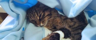
Treatment of closed pneumothorax under the supervision of a veterinarian lasts on average from seven to fourteen days. To relieve pain shock, the veterinary doctor prescribes a sedative or anesthesia; to normalize the functioning of the lungs and heart, conservative treatment is prescribed. If you have a severe cough, medications that reduce its manifestation are indicated.
The injured animal is recommended to rest and rest throughout the entire period of treatment. As a result of the course of therapy, the air in the pleural area resolves and the animal returns to normal. Otherwise, surgical correction may be prescribed.
For a week after stabilization of the animal’s health, the animal should be limited in its movements and not disturbed.
Is it possible to prevent closed pneumothorax in cats and dogs?
Exactly as much as it is possible to create safe living conditions for them. Constant control by the owner of the house and on the street, walking on a leash, as well as keeping an eye on the cats while airing the room will help minimize the possibility of injuries, bites, falling out of the window and other troubles.
Pneumothorax
Pneumothorax is a pathological process accompanied by the accumulation of air between the visceral and parietal layers of the pleura. Found in all animal species. It can be complete, partial, unilateral and bilateral, closed, open, valvular, encysted and recurrent.
Etiology. Pneumothorax occurs when a penetrating wound to the chest by a shrapnel, bullet, piece of wood, horn or other objects occurs during chest surgery. Sometimes it appears during the opening of abscesses, rupture of the esophagus or damage to the lung parenchyma in animals suffering from tuberculosis, interstitial emphysema, or pulmonary echinococcosis.
Often, pneumothorax occurs as a result of rupture of the lung due to pneumosclerosis and penetration of foreign bodies from the proventriculus. Pneumothorax can also be caused by rib fractures, a sudden increase in intra-alveolar pressure with severe coughing, pushing, strenuous work, prolonged mooing and falling.
Pathogenesis. With a closed pneumothorax, the air trapped in the pleural cavity is absorbed and the lung returns to normal. If infected air enters, especially in animals with reduced resistance, a complication develops in the form of purulent or putrefactive pleurisy. If there is a gaping hole in the chest wall or a rupture of a large bronchus, an open pneumothorax occurs when air freely flows into and out of the pleural cavity. A large amount of microflora enters the pleural cavity, and preconditions arise for the complication of pleurisy.
In cases where conditions are created for air to enter the pleural cavity at the time of inhalation or movement, and the exit of air from the cavity is difficult or impossible, valve pneumothorax develops, accompanied by a constant increase in pressure in the chest cavity, resulting in increased compression of the lungs and the diaphragm. is pushed back. With unilateral valve pneumothorax, except as noted, the organs of the chest cavity are displaced in the opposite direction.
With all types of pneumothorax, the lung tissue is compressed, blood circulation and gas exchange are hampered, oxygen starvation occurs, and the heart and respiratory center are stimulated.
Symptoms. Sick animals are very depressed, sometimes sweat, the pulse is rapid, the mucous membranes are cyanotic or pale. The most characteristic sign of pneumothorax is the sudden onset of severe shortness of breath, which progresses quite quickly without an increase in body temperature and nasal discharge.
In parallel with increased shortness of breath, cyanosis of the mucous membranes and conjunctiva increases. With valvular pneumothorax, a bulge of the chest is found, shortness of breath quickly increases, the degree of which depends on the force of compression of the lungs. If pneumothorax is associated with damage to the lung tissue, a cough occurs, during which a reddish mass is thrown out.
When percussing the affected side, an atympanic sound is detected, and with valve pneumothorax, a tympanic sound with a metallic tint, similar to a box sound, is detected. This sound is amplified outside the normal pulmonary field, that is, the pulmonary field seems to expand. The respiratory movements of the chest of the affected half are weakly expressed, while the healthy half is enhanced.
Breathing sounds on the affected side are sharply weakened or cannot be heard at all, while on the healthy side increased vesicular or mixed breathing can be heard. When pneumothorax is complicated by pleurisy, pain, increased body temperature are noted, percussion sound is dull in a horizontal line, with a metallic tint. Fluoroscopy shows clearing at the location of the gases. When bleeding into the chest cavity, signs of posthemorrhagic anemia are detected.
We suggest you read: The turtle's eyes do not open - causes, diseases, treatment - symptoms, treatment, medications, causes
Flow. With closed pneumothorax, recovery is possible in 7-14 days, with valvular pneumothorax – death after a few hours or days. Pneumothorax, which occurs when a lung abscess is opened or an echinococcal bladder ruptures, is fatal.
Pathological and anatomical changes. A hole, sometimes blood, is found in the chest or lung tissue; an encysted bladder and a purulent mass are found in the pleural cavity. In all types of pneumothorax, the lungs are collapsed on both or one side. Collapse of the lungs is more pronounced with valvular pneumothorax and less pronounced with other types.
Diagnosis. Pneumothorax is diagnosed based on anamnestic data, clinical signs and fluoroscopy. Of the clinical signs, the sudden appearance of severe and rapidly progressing shortness of breath without an increase in body temperature, a change in the shape of the chest, the absence of respiratory sounds and the presence of an atympanic sound on the affected half of the chest are of diagnostic importance. Fluoroscopy is used to establish posterior displacement of the diaphragm and atelectasis of the lung.
It is necessary to exclude cavity, swelling and hyperemia of the lung, entry into the chest cavity of the stomach or intestines due to rupture of the diaphragm. In the latter case, the atympanic sound is detected only in the peri-diaphragmatic part of the chest, the strength of the percussion sound changes, and peristaltic noises are established by auscultation at the site of the atympanic sound.
caffeine, strophanthus, glucose - intravenously, camphor oil - subcutaneously. In case of open pneumothorax, the wound is sutured and air is pumped out from the chest cavity using the Samkovich, Delafoy apparatus or using a test trocar and needle connected through a rubber tube with an Agali tap to a Janet syringe. Air is pumped out for valve and closed pneumothorax. After removing the air, the pleura is irrigated with a penicillin solution.
Prevention. Avoid penetrating wounds of the chest, development of processes in the lung tissue that contribute to spontaneous rupture of the lung parenchyma, as well as rupture during periods of physical stress, straining and coughing. For severe coughs, it is useful to prescribe substances that reduce the excitability of the cough center.
Pneumothorax is the accumulation of air inside the pleural cavity. Air usually enters the cavity due to a rupture of the chest wall, bronchial tree, pulmonary parenchyma, or esophagus. Clinical symptoms of pneumothorax may include tachypnea, dyspnea, abdominal breathing, or cyanosis. Auscultation reveals muffled lung sounds and increased resonance to percussion.
X-rays show air inside the pleural cavity, protrusion of the edges of the lungs through the hole in the chest wall and diaphragm, and displacement of the heart silhouette from the sternum on lateral views. The pathophysiological manifestation of pneumothorax is a decrease in pulmonary expansion with subsequent impairment of ventilation and perfusion, hypoxia and loss of intrapleural pressure gradient, which impairs thoracic absorption and venous return.
Treatment of animals with pneumothorax
Treatment of dogs, cats, cattle, horses and other animals with pneumothorax is aimed primarily at closing the hole (suturing, applying a log bandage, etc.) in the pleural cavity so that the open pneumothorax is converted into a closed one.
Air and gases are removed from the pleural cavity with a Janet syringe through a large-diameter needle, to which a rubber tube with a clamp is attached. When treating pneumothorax in a dog, it is possible to install a chest drainage to permanently remove air from the pleural cavity.
After pumping out the air, antibiotic solutions or 0.2% ethacridine lactate are injected into the pleural cavity without removing the needle.
After surgery, sick animals are prescribed rest, cardiac and tonic medications, and antibiotics to prevent pleurisy.
Treatment for pneumothorax in a cat is pain relief, rest (placement in a cage), removal of air from the pleural cavity, pain relief, antimicrobial drugs, fluid support for the body. Treatment of possible complications.
In severe cases of the disease, oxygen therapy is used.
Prevention
It is important to remember that if a cat jumps out of a window once, it will do so a second and third time, regardless of the severity of the injuries. It is very important to install protective nets on the windows to prevent your pet from escaping from the house.
If you find a lost cat in front of your house, you need to take it to a veterinary clinic as quickly as possible. Many injuries are extremely painful. It is best to wrap your pet in a blanket or thick towel and place it in a box. This type of transportation is optimal for the victim.
Video about high-altitude injuries in cats - interview with a veterinarian:
Can pneumothorax be treated at home?
No and no! In addition to emergency care (often surgical), an animal with pneumothorax requires constant monitoring, as complications can quickly develop.
Frankie Frankie, as the doctors named the cat, had the worst type of pneumothorax – valvular pneumothorax. The air slowly accumulated in the chest, increasing the mortal threat with every minute. A surgeon was urgently called back from the holiday break. After Frankie was put under anesthesia, a drain was inserted between his seventh and eighth ribs (this is mandatory!) and some of the air was pumped out.
The surgeon then attended to his prolapsed bowel and bladder.