What are they?
Neoplasms are primary and secondary. Primary, as the name suggests, arises directly in the brain. Sometimes they grow inside the brain itself, sometimes in the depths of the meninges. In turn, primary tumors are divided into malignant and benign. Secondary neoplasms are a consequence of cancerous pathologies inside other internal organs. Abnormal cells enter the brain along with metastases. It should be remembered that all tumors that arise in the optic or auditory nerves (and similar tissues) are always primary. As we have already said, neoplasms can be benign (without metastases, with clear boundaries) and malignant. The latter quickly spread throughout the body through metastases and do not have clear localization or boundaries.
Diagnosis of neoplasms
First of all, a veterinary neurologist studies the medical history and analyzes what the owner tells him. Evaluates how much the pet’s behavior has changed and over what time.
| Forebrain | Cerebellum | Vestibular apparatus | Brain stem |
| When the cerebral cortex is damaged, the cat moves in a circle with an inclination in the direction in which the tumor is formed. The head is lowered, appetite is changed, thirst, depression, convulsions, loss of orientation. If the hypothalamus is affected, vision loss occurs. | Hypermetry, tremors that increase with sudden movements, the cat stands unnaturally, paws are widely spaced. | Strabismus, nystagmus, circling, poor appetite and vomiting, ataxia, head tilted. | Paralysis, ataxia, weakness of muscles, paws on one side, changes in the functioning of the heart and lungs, coma. |
Next, the cat is completely examined, everything is done to visualize the neoplasm and make a diagnosis. Neoplasms must be differentiated from other disorders, infections, and metabolic disorders. Do:
- biochemistry and blood testing;
- radiography;
- Ultrasound of internal organs;
- CSF analysis, CT, MRI.
A biopsy is a reliable method that can confirm the presence of tumors in the brain and their malignancy. It is done under the control of CT and MRI.
Main manifestations
It should be remembered that in the initial stages this disease practically does not manifest itself in any way. A cat may show signs of mild illness, but even the best doctor is unlikely to link these symptoms with the formidable processes occurring in the animal’s brain.
When the first specific manifestations appear, the process has already gone very far:
- A cat can wander around the apartment for a long time. The movements are “numb”, the gaze is empty. The animal often bumps into furniture.
- From time to time the cat stops and begins to circle aimlessly in one place. Sometimes he rests his forehead against the owner’s chair or leg. It is especially scary if the animal not only walks, but meows hoarsely and anguishedly.
- When the process goes very far, the cat experiences convulsions and seizures almost every day. Sometimes there are paralysis and paraplegia (more typical for spinal cord tumors).
- Blindness may occur from time to time. Sometimes it goes away, but more often it remains.
- Other disorders of nervous activity: impaired swallowing, attacks of aggressiveness, sudden affection.
Important! It should be borne in mind that manifestations of aggression or any other sudden change in behavior, strange taste preferences and other “quirks” can be caused by something worse... for example, rabies! So, under no circumstances try to make a diagnosis yourself; call a veterinarian immediately!
Clinical signs
Clinical signs of intracranial tumors are very varied and usually include disturbances in mental status, disorientation, and loss of habitual activities. Most specific clinical signs depend on the location of the tumor. In the initial stages, clinical signs may be short-lived and intermittent, but as the tumor grows they become more pronounced and permanent.
Epileptiform seizures may be one of the first signs of a tumor of the cerebral cortex. Multiple cranial nerve deficits are characteristic of ventral brainstem tumors. Dysmetria, convulsive readiness and ataxia can be associated with cerebellar tumors. Visual impairment and blindness – hypothalamus or optic nerve meningioma.
Primary brain tumors are not usually accompanied by paraneoplastic syndromes. An exception may be a pituitary adenoma, leading to hyperadrenocorticism. Also, clinical signs are mainly due to increasing intracranial pressure, mass effect and cerebral edema. An extremely dangerous complication with a fatal outcome is wedging of the brain through the foramen magnum.
Table No. 2. Possible clinical signs of intracranial tumors depending on location.
Diagnostics
1. Routine hematological and biochemical examination to exclude extracranial causes (uremia, etc.), concomitant diseases.2. X-ray of the skull to detect osteolysis or hyperostosis of the skull bones, which is typical for meningioma in cats, a primary neoplasm of the nasal cavity and paranasal sinuses, or a neoplasm of the skull bones.3.
Survey radiography of the chest and abdominal cavity to identify primary neoplasms and concomitant diseases.4. Ultrasound of the chest and abdominal cavity to detect the primary tumor and diagnose associated diseases.5. Analysis of cerebrospinal fluid for the purpose of diagnosing inflammatory diseases of the brain, which is very important for differential diagnosis.
Precipitation methods are preferred. In some cases, this makes it possible to detect atypical cells. This method can also be used to diagnose neuroleukemia. Spinal puncture in such animals should be performed with extreme caution. With increased intracranial pressure, there is a danger of brain wedging in the event of a sharp drop in pressure.
Possible measures to prevent such a complication are slow drainage of cerebrospinal fluid, pre-infusion of mannitol and hyperventilation.6. Electroencephalography. This diagnostic method for tumor lesions of the brain is based on the fact that, as a rule, tumor tissue is electrically neutral.
better visualization of intracranial soft tissues, the ability to differentiate more subtle changes in tissues (edema, vascular changes, hemorrhages and necrosis).8. Biopsy. Intravital biopsy is quite complex and not feasible in all cases. For a biopsy, it is necessary to know the exact location of the tumor, so a biopsy is performed only after an MRI. The possibility of performing a biopsy depends on the location of the tumor and the general status of the animal. Often a biopsy is performed only postmortem.
Table No. 3. Presumptive diagnosis of brain tumors based on MRI.
Supportive and symptomatic treatment includes anticonvulsant therapy (phenobarbital 2-4 mg/kg orally every 12 hours) and corticosteroid therapy (methylprednisolone 10-15 mg/kg).
What to do?
If you notice any of the signs described above in your pet, contact your veterinarian immediately! Even cancer today is not a death sentence, including in pets. In some cases, you can even do without major operations, since modern drugs give good results during chemotherapy. If we are talking about a neoplasm of benign etiology, then it (with a high degree of probability) can be removed surgically, after which the animal will live a long and happy life! In short, brain tumors in cats can be cured today, so in no case should you delay going to the veterinary clinic.
At the moment, many cases of brain tumors in animals have been recorded (Simon R. Platt., Natasha J. Olby., 2004). However, despite this, information about this disease is insufficient and inaccurate. In Russian-language literature such reports are rare. Often, in the early stages of the disease, the tumor is asymptomatic, which complicates its diagnosis, despite the availability of diagnostic methods such as computed tomography (CT) and magnetic resonance imaging (MRI). In this regard, the problems of diagnosis and treatment of brain neoplasia are relevant.
Brain neoplasia is more common in dogs than in cats. Its incidence in dogs is 14.5 per 100,000 animals. (Withrow SJ, MacEwen EG 2007) Brain tumors can occur in all breeds, at any age and regardless of gender. Despite this, some features of the manifestation of this disease have been identified. It is known that brachycephals are most predisposed to glial tumors of the pituitary gland, and dolichocephals to meningiomas. Breeds such as Boxer, Golden Retriever, Doberman Pinscher, and Scottish Terrier have a greater risk of developing neoplasia. The tumor most often occurs in dogs over 5 years of age.
The incidence of neoplasia in cats is 3.5 per 100,000 animals. (Withrow SJ, MacEwen EG 2007) No breed predisposition has been identified.
Brain tumors originate either from the tissues of the brain itself or from its membranes. The tumor can also penetrate into the brain from adjacent tissues (for example, from the bones of the skull) or be metastases from other organs. The causes of brain tumors in dogs and cats are currently unknown, although dietary, environmental, genetic, chemical, viral, traumatic, and immunological factors may be considered.
The most common primary tumors in dogs and cats are gliomas and meningiomas. Ependymoma, choroid plexus papilloma, medulloblastoma, plasma cell tumor, lymphoma, and neuroblastoma are also common in cats. The most common secondary tumors in dogs are locally invasive nasal adenocarcinoma; metastases from the mammary glands, prostate gland, or pulmonary adenocarcinoma, metastases from angiosarcoma; and germination of pituitary adenoma or cancer. In cats - macroadenoma and macrocarcinoma of the pituitary gland. (Withrow SJ, MacEwen EG 2007) The data is presented in the summary table below.
Brain tumors in dogs and cats
Neurological signs occur when the brain is compressed by a tumor. In this regard, they are not specific to the tumor itself and other diseases that can cause the same symptoms cannot be excluded. Many dogs and cats with brain tumors may not show serious clinical signs for a long time, but subtle symptoms may be noticed, such as changes in behavior, reluctance to sit in the owner's arms, a decrease in the frequency of purring, and a decrease in activity. This is because the tumor can grow slowly and surrounding tissues adapt to the increased pressure without causing serious signs of brain dysfunction. Neurological symptoms mainly depend on the location, size and growth rate of the tumor. (Withrow SJ, MacEwen EG, 2007)
Clinical signs depending on the location of the tumor.
Weakness and sensory disturbances mean damage to the frontoparietal cortex of the cerebral hemispheres. Visual deficit refers to damage to the visual pathways of the optic nerve in the occipital lobe of the telencephalon. Loss of smell is associated with damage to the olfactory brain.
The veterinarian's first step should be a complete physical and neurological examination to determine the diagnosis and approximate location of the tumor. Once again, I would like to draw your attention to the fact that these clinical signs are not specific to neoplasia. The same symptoms are caused by congenital disorders, infections, immunological and metabolic disorders, injuries, vascular disorders and other diseases. Therefore, it is important to rule out these diseases before making a final diagnosis. To do this, use diagnostic methods such as:
Hematological and biochemical studies. This is done to rule out non-brain diseases and assess anesthetic risk.
Radiography. Plain radiography of the thoracic and abdominal areas can exclude a primary tumor in other parts of the body and metastasis to the lungs. X-ray of the skull provides scant information, but it can detect a tumor of the skull or nasal cavity, lysis or hyperostosis of the skull.
An abdominal ultrasound is also performed to exclude extracranial causes of signs of cerebral dysfunction.
CSF analysis is recommended as an aid in diagnosing a brain tumor. Changes in the cerebrospinal fluid are not detected in all types of tumors. Used to exclude inflammatory causes of brain dysfunction. Cannot be performed with increased intracranial pressure.
CT and MRI are the main diagnostic methods. Allows you to determine the presence, size, location of the tumor. Performed under general anesthesia. MRI is considered preferable to CT. CT scan can detect bone changes (skull fractures). MRI provides clearer visualization of soft tissue and detection of brain edema, cysts, hemorrhage and necrosis. Images of the brainstem or cerebellum obtained with MRI are superior in quality to images obtained with CT.
Biopsy. The veterinarian usually relies on CT or MRI to diagnose intracranial lesions. However, although they can provide information about the nature of the lesion (size, location and relationship to other structures), these diagnostic methods cannot provide evidence of the underlying pathology (Simon R. Platt., Natasha J. Olby., 2004). Some lesions caused by infection (fungal granuloma) may look like a tumor (take up a large area) but may not be one. Therefore, a biopsy is the only method for definitive diagnosis of a brain tumor. It allows you to identify the type of tumor and its malignancy. Tumors well suited for intracranial biopsy: meningioma, astrocytoma, oligodendroglioma, ependymoma, metastatic adenocarcinoma, some lymphoid tumors. Tumors located in deep areas of the brain, such as cerebellar medulloblastoma, tumors of the brain stem, thalamus and hypothalamus, are poorly suited for biopsy (Simon R. Platt., Natasha J. Olby., 2004). The most well-known biopsy methods are ultrasound-guided biopsy and CT-guided biopsy.
Currently, there are a large number of treatment methods - palliative treatment of symptoms, surgical removal, radiation therapy, chemotherapy, boron-neutron capture therapy (one of the founders of this method in veterinary medicine was Prof. V.N. Mitin), immunotherapy, gene therapy, etc. .
Chemotherapy. Plays a minor role (Withrow SJ, MacEwen EG, 2007) in the treatment of tumors. Factors limiting the use of this method:
Radiation therapy is the most common treatment method. The goal is to destroy the tumor while minimizing harm to surrounding, healthy tissue. Radiation can be used alone or in combination with other methods. When selecting the radiation dose, you need to pay attention to the type of tumor, its location and the tolerance of surrounding, healthy tissue. Radiation therapy is also prescribed to reduce relapses after surgery.
Surgical removal. The goal is to cure the disease by completely removing the tumor (this is quite rarely possible), or to reduce symptoms by reducing pressure on brain tissue. This treatment method is not always possible. In order to carry out surgical treatment, you need to know the exact location, size and degree of invasion of the tumor. Also, the possibility of surgery depends on the general condition of the patient and the accessibility of the tumor.
Palliative treatment of symptoms is aimed at reducing intracranial pressure, swelling and slowing tumor growth. For this therapy, glucocorticosteroids are used. Also includes antiepileptic therapy, in which phenobarbital is prescribed.
The blood-brain barrier does not allow many chemicals to pass through.
Heterogeneity of tumor cells. As a result, only certain tumor cells are sensitive to the drug.
The tumor may only be sensitive to high doses, which can be toxic to normal brain tissue and other organs.
5. Immunotherapy. A method that activates cellular immunity against brain tumors by culturing and stimulating lymphocytes has been tested in dogs. Five dogs with brain glioma showed tumor shrinkage and clinical improvement. Immunotherapy is currently being investigated. (Withrow SJ, MacEwen EG, 2007)
6. Gene therapy is a treatment method in which DNA or RNA is transferred to target cells to change their genotype for therapeutic purposes. Although gene therapy was conceived as a treatment for genetic diseases, its potential for use in tumor treatment was quickly recognized. This method is currently being researched. (Withrow SJ, MacEwen EG 2007)
The prognosis for brain tumors depends on many factors. These are the type of tumor and its location, the degree of secondary action of the tumors and the neurological status of the patient. Animals with brain herniations have a very poor prognosis, while animals with easily accessible tumors and minor neurological deficits have a very favorable prognosis. (Richard A.S. White, 2003)
Unfortunately, most tumors cannot be completely cured, so the main goal of treatment is to ensure a good quality of life for the animal for as long as possible.
There are few data on life expectancy with palliative care alone. The results of one study show that the average survival time with this treatment is 56 days. (Withrow SJ, MacEwen EG, 2007) The prognosis can be significantly improved by surgery, radiation, chemotherapy or immunotherapy, used alone or in combination.
General principles that are used when considering a forecast:
The larger the tumor size, the worse the result.
The stronger the manifestation of clinical signs, the worse the prognosis.
Supratentorial tumors (tumors of the forebrain) have a better prognosis than infratentorial tumors (tumors of the brain stem and cerebellum). (Withrow SJ, MacEwen EG, 2007)
The later the disease is diagnosed, the worse the result.
Case from practice
In December 2010, to the Department of Veterinary Surgery at the Moscow State Academy of Veterinary Medicine and Biotechnology. K.I. Scriabin received a cat named Peach, 7 years old, male, British Shorthair, neutered. (Fig. 1.) The owners complained of poor support ability of the pelvic limbs.
During the diagnosis, proprioceptive ataxia of the pelvic limbs was revealed, manifested by impaired postural reactions, increased panniculitis reflex and pain in the sacral region. All cranial reflexes were normal. The animal underwent a survey radiography of the lumbar and sacral region - no changes were found. As a result, to diagnose the causes of ataxia, the cat underwent a CT scan of the skull (Fig. 2) and myelography, contrast was administered through the lumbar approach. A myelographic examination did not reveal any pathological changes in the spinal cord. A CT scan revealed a primary intracranial brain tumor. The owners were recommended to undergo surgical treatment of the animal.
Stages of surgery
Premedication used atropine 0.04 mg/kg IM, cefazolin 5-10 mg/kg IM, dicinone 10 mg/kg IM, methylprednisolone drip IV 20 mg/kg, then mannitol 1 g/kg. During the surgical intervention, neuroleptanalgesia was performed with the drugs xylazine 2 mg/kg and zoletil. An infusion of Ringer's solution 20 ml/h, plasma expanders (Voluven) and saline solution were also administered. Before surgery, a urinary catheter was installed in the animal and tracheal intubation was performed.
Based on CT data, a rostrotentorial right-sided approach was chosen at the tumor location. An arcuate skin incision was made from the frontal bones to the interparietal bone. (Fig. 3.) The temporal muscle was separated with a sharp preparation, and the bones of the skull were subsequently skeletonized. The area of bone that will be removed was identified and marked. For this purpose, we used a high-speed pneumatic drill with a 3 mm carbide bur and drilled grooves along the marked lines (Fig. 4). Then they bit it out with Kerrison's nippers. In this way, a quadrilateral was removed from the bone tissue. The branches of the middle meningeal artery were coagulated with a bipolar coagulator. Initially, the incision of the dura mater was carried out by lifting the membrane with Adson tweezers and a pointed scalpel; later, a dental scraper was brought under the dura mater and an incision was made with a No. 11 scalpel to the size of the craniotomy. Hemorrhages of the arachnoid and soft membranes, parenchyma were controlled using bipolar cauterization and a homeostatic sponge. The location of the tumor was determined, it was separated from the adjacent tissues and enucleated. (Fig. 5, 6.) The vessels of the “brain bed” were coagulated with a bipolar coagulator. Complete excision of the tumor was determined according to intraoperative ultrasound data. To prevent hematoma formation, drainage was installed. Each layer of tissue was sutured with a non-absorbable monofilament suture (Prolene 4-0). (Fig. 7.) The dura mater was sutured separately, on top of which a hemostatic sponge was placed. Immediately after the operation, Kvamatel 2 mg was administered intravenously (hereinafter referred to as IV), Gordox 2000 units. IV, furosemide at a dose of 4 mg IV.
Histological examination of pathological material.
The tumor was sent for histological examination. Diagnosis: meningioma. Histological picture: the tumor consists of voluminous spindle-shaped cells forming intertwining, twisting or palisading bundles. The cells are penetrated by a moderate amount of dense, sometimes edematous, fibrous stroma, unevenly expressed in different areas. Multiple small foci of hemorrhage were noted in the tumor tissue. Tumor cells have a large, oval nucleus with thin strands of chromatin, 1-2 prominent nucleoli and mild eosinophilic cytoplasm. The degree of anisocytosis is moderate, mitoses are rare.
After the operation, a positive recovery trend was observed. A day after the operation, the animal's ataxia disappeared. During the rehabilitation period after surgery, no manifestations of neurological syndromes were noted. A clinical and neurological examination of the animal was carried out - a week after the operation, after 2, after a month, after 2 months, no neurological changes were found. The animal has now fully recovered.
Richard A.S. White. Oncological diseases of small domestic animals.// Aquarium, Moscow, 2003, - 350 pp.
Kuleshova O.A., Yagnikov S.A., Kemelman E.L., Leonova T.A., Mitrokhina N.V., Trubnikova E.A. Clinical case of surgical treatment of intracranial meningioma in a cat. // Russian Veterinary Journal. Small domestic and wild animals, 2010; 3:29-34.
BSAVA Manual of Canine and Feline Neurology. Third edition.,Editors: Simon R. Platt., Natasha J. Olby., 2004, 432 pp.
Withrow SJ, MacEwen EG Small animal clinical oncology.// Saunders Elsevier, US, 2007, - 846 p.
Jaggy A. Small Animal Neurology - Hannover. Schlutersche,2010 – 580p
Glioma of the frontal lobe of the brain involving the olfactory bulb
Brain tumors in dogs and cats represent an important cause of morbidity and mortality in companion animals, primarily dogs.
Given practical limitations in veterinary medicine, it is likely that the incidence of brain tumors in dogs is underestimated. Although data from individual studies vary, meningiomas account for approximately 50% of primary tumors in dogs, gliomas represent 40–70%, and choroid plexus tumors constitute the third most common group.
Secondary tumors account for approximately 50% of all brain tumors in dogs, with the most common types being hemangiosarcoma, pituitary tumors, lymphoma, metastatic carcinoma, nasal extension tumors, and histiocytic sarcoma. Most primary and secondary tumors occur in older dogs, most cases over 5 years of age. The ages for dogs with meningiomas, gliomas and choroid plexus tumors are 10-11 years, 8 years and 5-6 years, respectively. Primary tumors (especially gliomas) can sometimes be seen in younger dogs.
Brain tumors are more common in larger breed dogs, and meningiomas are more common in Golden Retrievers, Boxers, and Miniature Schnauzers. Astrocytomas and oligodendrogliomas are significantly more common in brachiocephalic breeds (boxer, Boston terrier, and bulldog), and choroid plexus tumors are more common in golden retrievers.
Most brain tumors in cats are primary, with meningiomas predominating. Lymphoma and pituitary tumors are the most common secondary tumors, with other primary and secondary tumors such as gliomas being observed relatively rarely compared to dogs.
Clinical signs
Neurological signs occur when the brain is compressed by a tumor. In this regard, they are not specific to the tumor itself and other diseases that can cause the same symptoms cannot be excluded. In the initial stage, the clinical picture is asymptomatic. Then, as the tumor grows, a pattern of compression of individual areas of the brain develops, which depends on the location of the tumor growth site.
• walking in circles
• violation of the act of swallowing, etc.
When localized in the anterior lobe of the brain:
— Cortex of the greater hemispheres: Circular (maneuver) movements (usually in the direction of damage), changes in behavior, depression, lowering of the head, decreased or increased appetite and thirst, disorientation, convulsions, changes in postural reactions contralaterally (on the opposite side).
— Hypothalamus: Change in mental status, damage to the optic nerve (II pair).
When localized in the vestibular apparatus: Head tilt, ataxia, manege movement, loss of appetite and vomiting, strabismus, nystagmus.
When localized in the cerebellum: Head tilt; hypermetry (dysmetria); ataxia; tremor, which intensifies with some movements and decreases when the animal rests; not a natural pose (with limbs widely spaced).
When localized in the brain stem: Paralysis (hemi-, tetraparesis), weakness on one side, ataxia, disruption of the respiratory and cardiovascular systems, deficiency of cranial nerves (V,VI,VII,IX,X), coma.
When localized in the frontal and parietal zones: Weakness and sensory disturbances.
Visual deficit refers to damage to the visual pathways of the optic nerve in the occipital lobe of the telencephalon. Loss of smell is associated with damage to the olfactory brain.
Diagnostics
Initially, a history is taken and a complete physical and neurological examination is carried out, from which the diagnosis and approximate location of the tumor can be determined.
Clinical signs are not specific for neoplasia. The same symptoms are caused by congenital disorders, infections, immunological and metabolic disorders, injuries, vascular disorders and other diseases. Therefore, it is important to rule out these diseases before making a final diagnosis.
For differential diagnosis the following is carried out:
Hematological and biochemical studies. This is done to rule out non-brain diseases and assess anesthetic risk.
Radiography. Plain radiography of the thoracic and abdominal areas can exclude a primary tumor in other parts of the body and metastasis to the lungs. X-ray of the skull provides scant information, but it can detect a tumor of the skull or nasal cavity, lysis or hyperostosis of the skull.
An abdominal ultrasound is also performed to exclude extracranial causes of signs of cerebral dysfunction.
Cerebrospinal fluid analysis is recommended as an aid in diagnosing a brain tumor. Changes in the cerebrospinal fluid are not detected in all types of tumors. Used to exclude inflammatory causes of brain dysfunction. Cannot be performed with increased intracranial pressure.
CT and MRI are the main diagnostic methods. Allows you to determine the presence, size, location of the tumor. Performed under general anesthesia. MRI is considered preferable to CT. CT scan can detect bone changes (skull fractures). MRI provides clearer visualization of soft tissue and detection of brain edema, cysts, hemorrhage and necrosis. Images of the brainstem or cerebellum obtained with MRI are superior in quality to images obtained with CT.
Biopsy. The veterinarian usually relies on CT or MRI to diagnose intracranial lesions. However, although they can provide information about the nature of the lesion (size, location and relationship to other structures), these diagnostic methods cannot provide evidence of the underlying pathology. Some lesions caused by infection (fungal granuloma) may look like a tumor (take up a large area) but may not be one. Therefore, a biopsy is the only method for definitive diagnosis of a brain tumor. It allows you to identify the type of tumor and its malignancy.
Tumors well suited for intracranial biopsy: meningioma, astrocytoma, oligodendroglioma, ependymoma, metastatic adenocarcinoma, some lymphoid tumors.
Tumors located in deep areas of the brain, such as cerebellar medulloblastoma, brain stem, thalamic and hypothalamic tumors, are not suitable for biopsy. The most well-known biopsy methods are ultrasound-guided biopsy and CT-guided biopsy.
Treatment
Currently, there are a large number of treatment methods - palliative treatment of symptoms, surgical removal, radiation therapy, chemotherapy, boron-neutron capture therapy.
Palliative treatment: Corticosteroids to treat peritumoral edema, together with anticonvulsants to control seizures, form the mainstay of palliative treatment for canine brain tumors. In general, the prognosis for survival with palliative care is 1-10 weeks.
Radiation therapy: The goal is to destroy the tumor while minimizing harm to surrounding, healthy tissue. Radiation can be used alone or in combination with other methods. When selecting the radiation dose, you need to pay attention to the type of tumor, its location and the tolerance of surrounding, healthy tissue. Radiation therapy is also prescribed to reduce relapses after surgery.
In general, the survival prognosis for radiotherapy alone for all tumors, intra-axial tumors and extra-axial canine brain tumors is approximately 33-39 weeks, approximately 40 weeks and approximately 40-49 weeks, respectively. Although in most cases, the combination of surgical treatment with radiation in the treatment of canine brain tumors provides improved outcomes compared to radiation therapy alone.
Surgical removal: The goal is to cure the disease by completely removing the tumor (this is quite rarely possible), or to reduce symptoms by reducing pressure on the brain tissue. This treatment method is not always possible. In order to carry out surgical treatment, you need to know the exact location, size and degree of invasion of the tumor. Also, the possibility of surgery depends on the general condition of the patient and the accessibility of the tumor.
Chemotherapy: The main barrier to the use of chemotherapy in the treatment of CNS neoplasias is the blood-brain barrier. This endothelial and astrocytic barrier eliminates the use of the vast majority of chemotherapeutic agents. Exceptions are the alkylating agents: lomustine (CCNU), carmustine (BCNU), and temozolomide. Asparaginase and 5-hydroxyurea also successfully penetrate the BBB.
Other approaches have been used to overcome the BBB, including intrathecal injection (into the cerebrospinal fluid system) of drugs such as methotrexate and injection of drugs into the tumor. This can be done by local injection, but the delivery of the drug to the tumor is often insufficient. Therefore, convection-assisted delivery, which was developed for use in dogs by Dr. Dickinson's group at the University of Davis, is becoming popular.
Other approaches include binding the drug to be administered to nanoparticles or liposomes. All of these protocols are still at the development stage, but will become clinically relevant in the near future.
Unfortunately, most tumors cannot be completely cured, so the main goal of treatment is to ensure a good quality of life for the animal for as long as possible.
Veterinarian-intern Zbarakh Elizaveta
Cancer is a killer of cats and dogs. This is the number one disease for animals and is often fatal. Many symptoms of cancer in cats are subtle and could be caused by another condition, but if you notice any of the following potential warning signs of cancer, it is highly recommended that you talk to your veterinarian.
Types and symptoms
This disease is divided into several subtypes, each of which differs in some nuances in the course of the pathological process. There are cytotoxic, interstitial, vasogenic and filtration edema. In veterinary practice, they most often deal with the vasogenic and cytotoxic varieties. In the first case, edema begins due to increased vascular permeability, and in the second, the reason is the “swelling” of the neurons and astrocytes themselves. It happens with improperly prescribed drug treatment, parasitic and infectious diseases, as well as poisoning.
Be that as it may, you need to remember one thing - this condition is extremely serious and dangerous, regardless of the reason that provoked the development of edema. If you see any of the symptoms described below, immediately (!) go to the vet. The consequences of non-intervention will be too serious. So, here are the most critical signs:
- Nervous attacks. The cat cannot sit, it sways from side to side, it hits its head against the wall, etc.
- Loss of consciousness.
- Unnatural body position, uncoordinated, unnatural movements.
- If the cause is hypo- or hyperthermia, then bloody discharge may be observed from the ears, nose or mouth.
- Redness of the eyes.
- Cyanosis of visible mucous membranes and skin (that is, their blueness).
- Heavy or rapid breathing (dyspnea or tachypnea, respectively).
- Bridycardia or tachycardia (faster or slower heart rate).
The cat is constantly hiding
Cats love a good hiding place, but if you notice that your pet is spending more time under the bed or in hard-to-reach places, there may be something wrong with him. “Owners often tell me that they notice when their cat is sick. If animals are social but spend more time looking for new hiding places or stop coming out during feeding time, then you should be wary,” says Jake Seidel, founder of . The veterinarian runs an animal hospital in upstate New York and is also a member of the American Veterinary Medical Association.
“A cat hiding too often is a common sign that something is wrong with it, but it may not necessarily indicate cancer. Therefore, a visit to the veterinarian is a good idea, says the specialist. “Know that your pet is trying to secretly tell you something.”
Weight loss
Weight loss is a very clear symptom of cancer in a cat. Dr. Seidel says this sign is evidence of a gastrointestinal tumor.
“When cats are offered food during feeding hours but turn away from the bowl or refuse food, this should not go unnoticed,” he says. “Cancer can also cause a cat to lose weight but still retain her appetite. If you notice your cat is losing weight, it's time to make an appointment with your veterinarian."
Oral lesions
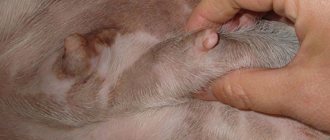
Ulcers, growths, bleeding gums or changes in their color can be a sign of cancer in a cat, especially in an adult. These signs in cats often go undetected for too long. “We usually detect visible oral tumors too late because people don't look at their pet's mouth,” Seidel says. “Many oral growths can be really devastating because people don’t notice them until they are large.” Your veterinarian also suggests brushing your pet's teeth on a regular basis.
"It's good to observe when your pet yawns or eats," advises Timothy Rocha, DVM, an oncology specialist in New York City.
The cat has diarrhea
"In some cases, stomach upset is not a sign of cancer in the animal," says Dr. Rocha. "But if it persists or gets worse, take your cat to the vet." According to the specialist, excessive use of a box with sand by a cat, difficulty urinating, and the presence of blood in the urine or feces are potential signs of cancer.
“Don’t ignore vomiting. A simple urge and an upset stomach are also signs of a tumor,” says Dr. Seidel.
Symptoms
Animals with cerebral edema may go into a coma, depending on the severity of the disease. Pets with this disease are often blind, powerless, unable to walk, and may experience seizures.
When your cat has cerebral edema and you, not knowing what to do, are looking for advice on this topic on the Internet on forums, we recommend not to self-medicate and experiment on your beloved cat. The fact is that there are many reasons for cerebral edema in an animal, and the consequences of your experiment may disappoint you and your family.
Presence of seizures
“Seizures can be a sign of a brain tumor in cats, which is most common in older cats,” says Dr. Seidel. “If you begin to notice sudden and uncontrollable bursts of activity, such as chopping and chewing food, leg twitching, or foaming at the mouth, you should visit your veterinarian immediately.”
Cats may also suffer from atypical seizures, which are classic seizures and manifest as bouts of strange behavior such as sudden rage or hysteria, excessive licking, chewing or scratching, and biting their owner.