Dangerous conditions include necrosis in cats. There are several types of tissue necrosis, which are accompanied by changes in skin color, the appearance of non-healing ulcers, and an unpleasant odor. The necrotic process is irreversible, so at the first symptoms, the owner should take the cat to the veterinarian, who will prescribe treatment and give preventive recommendations.
According to veterinarian statistics, 99% of animals with gangrene require amputation of the affected limb.
Why does necrosis occur?
The pathology is characterized by irreversible tissue death and cessation of cell activity due to impaired blood circulation, nerve conduction, and the activity of anaerobic or pyogenic bacteria. Most often, areas of the body where the blood vessels are small are affected - ears, paws, muzzle, tail, tongue. Often internal organs are affected - intestines, stomach, kidneys, liver. Depending on the location, the causes of necrosis are as follows:
- Internal organs: entry of foreign objects into the gastrointestinal tract;
- helminthiases;
- cirrhosis;
- volvulus.
- frostbite;
- glossitis (inflammation of the tongue);
Allergies or exposure to toxic chemicals can lead to tissue death.
In addition, the following factors cause tissue death in any area:
- autoimmune failure;
- allergy;
- prolonged inhalation of pesticides by animals;
- heart attack;
- electrical injury;
- disturbance of nerve conduction.
Causes
- Sometimes there is a violation of sterility during the provision of medical care in a veterinary clinic or at home.
- Fights between animals, during which damage is caused to each other (a pinpoint tear of the skin occurs and infection occurs on the fangs or claws). By the way, a cat bite is one of the dirtiest injuries; it does not heal for a long time and festers.
- Punctures or cracks on the paw pads while being outside (from broken glass, chips, thin wire) and climbing trees, buildings (from thorns), and at the same time introducing a foreign body into the wound (splinter, sand);
- Breaking off a claw due to injury (possibly purulent inflammation of the corolla, after which it will fall off on its own, or the help of a surgeon will be required).
What types of necrosis are there?
Veterinarians distinguish the following types of pathology shown in the table:
| Type of necrosis | Characteristic |
| Dry | Affects mainly internal organs with obstructed blood flow, lack of oxygen, fluid |
| Wet | Most often occurs on the skin |
| Areas of the body and organs rich in fluid are affected | |
| At the site of pathology, rotting and swelling develops | |
| Gangrenous | Characterized by putrefactive decay and drying of tissues due to pathogenic microbes |
| Myonecrosis primarily affects muscle and adipose tissue | |
| Straight | Develops due to injury, exposure to microbes or toxic chemicals |
| Indirect | Formed due to disruption of the cardiovascular, nervous or endocrine systems. |
| Sequestration | It is formed in internal organs, does not undergo decomposition for a long time and is found among living tissues |
| Bedsores | Occurs at the site of greatest tissue compression |
Diagnostics
It so happened “historically” that cats are very restless creatures. They love to walk and sometimes find themselves in quite dangerous situations. For example, they encounter dogs, find themselves bitten by a dog or even by their own relatives. The result is wounds. The bitten variety is a “gift” in itself, but a weeping wound on a cat is even worse.
A little theory
The wound in this case is a violation of the integrity of the skin layer, affecting the underlying tissue. It can be superficial (essentially a serious scratch) or deep, affecting even the muscle layer and large vessels. In pets, such pathologies are most often the result of traumatic exposure.
They occur during fights, attacks by other animals, after collisions with cyclists or cars, and also include surgical injuries that have to be inflicted on the animal during surgery. In short, there are many reasons. But in this case, it is much more important to us what happens to the wound in the future.
Its healing can occur in two types, first described by the luminary of Russian medicine, N. I. Pirogov. He distinguished between healing by primary and secondary intention. With primary intention, wounds heal only after surgery: their edges are smooth, regenerative processes start immediately.
- Granulation. To protect itself from the penetration of microflora and debris into the wound cavity, the body begins to “darn” the wound with granulation. Fibroblasts and endothelial cells participate in the process, and new blood vessels grow from the latter.
- Increasing the epidermal layer. Epithelial cells along the edges of the wound channel gradually begin to “slide” onto the upper surface of the granulation, forming a new layer of skin epidermis.
- Myofibroblasts also play an important role in the healing process. It is from these cells that muscle fibers subsequently develop. They play the role of a kind of “ties”, literally tightening the edges of the former wound channel.
In principle, wound healing by secondary intention proceeds similarly, but in this case pyogenic microflora interferes with the process: complete granulation of the wound canal will not occur until all dead tissue and microbes are removed from the wound along with pus and other secretions. And it is with this process that difficulties may arise...
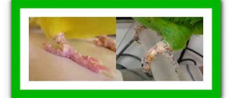
This is a wound whose surface is not contracted by granulations and does not heal. It literally oozes ichor or necrotic exudate. The color and smell of the exudate resembles rotting meat slop. It is necessary to distinguish a weeping wound from an ulcer: in the first case, a skin defect occurs as a result of mechanical action, in the second it is only a consequence of some severe metabolic disorders or hormonal metabolism in the body (a good example is ulcers due to diabetes or uremia).
However, sometimes the boundary between an ulcer and a weeping wound is not so obvious: in particular, with dermatitis, both of these pathologies can develop, simultaneously and in parallel. We list the main causes of this type of injury:
- Thermal burns (occurring under the influence of an open flame, steam). This also includes electrical shock to the skin.
- Burns caused by prolonged exposure to sunlight.
- Local circulatory disorders, which can be caused by diseases of the cardiovascular system, blood clots, parasites, etc.
- Dermatitis, eczema, other inflammatory skin diseases, including those of fungal origin.
- Long-term mechanical irritation (from an incorrectly fitted flea collar, for example).
- Often a simple abrasion “evolves” into a weeping wound if it was subsequently contaminated with pathogenic microflora.
| Video (click to play). |
In addition, weeping wounds are the “calling card” of long-haired breeds. Thick and long fur creates suitable conditions for the development of a weeping wound (especially when the cat constantly licks the affected area). The danger here is that the owners may not notice a small wound on the back until their pet simply starts to stink.
In the mildest cases, ichor leaks onto the surface of the wound. Its appearance indicates a normal healing process. In this case, the liquid is plasma. It leaks out of the vessels because the body itself increases the degree of their permeability. Contains many healing factors including oxygen, nutrients, cytokines, growth factors, chemotactic factors, WBCs (white blood cells), enzymes that help resist bacteria entering the wound. The wound looks good, there are no signs of severe inflammation or necrosis, there is no smell, the general condition of the animal is normal.
Cloudy exudate is formed by decomposed necrotic tissue. There may also be pus mixed in; in this case, the wound smells very unpleasant. A persistent, cloudy and smelly effusion is a good “hint” to the need for surgical debridement to remove dead and decaying tissue, foreign bodies, etc. In addition, a “fragrant” violation of the integrity of the skin indicates the danger of developing a septic process.
We suggest you read: Worms in cats and kittens - symptoms, photos and treatment of worms in kittens
Symptoms: how to recognize the disease?
An increase in blood pressure and body temperature in an animal is a sign of necrosis.
Necrosis of internal organs in a cat develops mainly against the background of other diseases, so signs of the underlying disease are observed. The presence of tissue death is indicated by the following symptoms:
- stool disorder;
- increased blood pressure;
- heart rhythm disturbance;
- refusal to eat;
- periodic increase in temperature;
- difficulty breathing or shortness of breath;
- lethargy, weakness.
On the external parts of the body the symptoms are pronounced:
- local increase or decrease in temperature;
- bluishness or redness, and eventually blackening of the skin;
- sweetish-putrid smell;
- decreased or absent sensitivity;
- hair loss in the affected areas;;
- persistent swelling, ulcers;
- hyperthermia;
- weakness, deterioration of health;
- pain, swelling and limited mobility if the bone is affected.
Treatment: what to do with necrosis?
Cell death can be stopped using Chlorhexidine, which is applied to the border of the lesion.
Tissue necrosis in cats can be cured by surgery. To prevent the spread of cell death due to bacterial infection, antibiotics are effective, which the veterinarian prescribes individually. To dry out necrosis, disinfect and identify clear boundaries of the lesion, dead tissue is smeared with brilliant green or Chlorhexidine. To prevent the cat from licking the medicine and getting poisoned, the limb should be bandaged and a special collar should be put on the neck.
Necrotic areas on internal organs are excised or a complete ectomy is performed. External lesions are treated by necrectomy, in which areas of necrosis in the cat are removed with a small inclusion of healthy tissue. In case of wet type of pathology or gangrene, amputation of the limb is performed. After the stump has healed and scarred, a prosthesis is installed.
Symptoms and treatment of gangrenous stomatitis in cats
- The heat exchange of the skin is disrupted, it becomes cold to the touch;
- the sensitivity of the skin is impaired, a feeling of numbness appears in the affected area;
- weakness and fatigue appear;
- movements and their coordination are impaired; if it concerns the lower extremities, lameness appears; if the upper limbs, then everything falls out of hand;
- pain and burning appear in the affected areas.
Dry and wet gangrene initially have in common
, the only difference is in the timing of their development. Dry gangrene begins gradually, slowly, sometimes over months and years, and the development of wet gangrene occurs over hours or several days. Further treatment depends on the type of gangrene - dry or wet.
Gangrene is always an indication for hospitalization in the surgical department of a hospital. Treatment of gangrene must be started urgently.
It is quite difficult to cure this condition. Treatment is always comprehensive, aimed at preserving the patient’s life, the cause of the development of gangrene, restoring blood circulation and preventing the spread of the process.
The amount of treatment directly depends on the type of gangrene.
Anesthesia, novocaine blockades.
, Neuroxon,
), Vazaprostan,
, Perftoran and other infusion solutions.
Drugs that destroy blood clots: Streptokinase, Actilyse, Retavaza, Levostor,
And so on.
oxygen.
Surgery:
- intravascular (endovascular) operations;
- bypass surgery and stenting of blocked vessels;
- amputation of dead tissue - affected limbs are routinely removed above the demarcation line from healthy tissue.
Antibacterial therapy.
Surgical treatment – removal of all “dead tissue”, amputation if necessary.
Detoxification therapy: intravenous infusion of various solutions.
Treatment of concomitant diseases: insulin therapy for diabetes mellitus, drugs that improve blood circulation,
and so on.
Surgical treatment - removal of affected tissue or amputation, local surgical treatment of the wound, access to fresh air for the wound (it is not recommended to bandage the wound).
– placing the injured limb in a pressure chamber under high oxygen pressure. Oxygen is detrimental to clostridia, the causative agent of gas gangrene.
Antibiotics.
Antigangrenous serum is a drug containing
to the main types of clostridia.
- Antibiotics intravenously and intramuscularly.
- Introduction of antibiotics and antiseptics into the bronchi using a bronchoscope.
- Detoxification therapy – intravenous drip administration of solutions.
- Drugs that dilate the bronchi: inhalations of Salbutamol, Ventolin, Berodual, injections of Eufillin.
- Drugs that enhance immunity.
- Surgical treatment: removal of part or amputation of the entire lung when a lung abscess (ulcer) forms, freeing the pleural cavity from pus. Surgical treatment is resorted to only if there is no effect from drug therapy.
- urgent surgery to remove the affected area of the intestine;
- antibiotics.
- surgical removal of the affected organ;
- antibiotics.
Indications for antibiotic therapy are any wet gangrene.
Considering that with tissue necrosis there is usually not just one type of bacteria, but a whole spectrum, antibiotics must act on all possible microorganisms, so not one antibiotic, but two or even more are often prescribed. The drugs are administered in the form of intravenous or intramuscular injections, and maximum doses are used. Recently, the method of administering antibiotics inside the lymphatic plexuses and vessels has proven itself to be successful.
We invite you to read: Abyssinian cat: 7 character traits
The most commonly used antibacterial medications to treat gangrene are:
- Penicillins - but there is a high risk of infection resistant to this group of drugs. Benzylpenicillin is not used for intestinal gangrene.
- III, IV and V generation cephalosporins: Ceftriaxone, Cefotaxime, Ceftazidime, Cefepime, Ceftaroline and others.
- Lincosamides: Clindamycin (Dalacin) - drugs used when the infection is resistant to penicillins.
- Aminoglycosides: Amikacin, Gentamicin.
- Tetracyclines: Tetracycline, Doxycycline.
- Levomycetin.
- Metronidazole.
Once the results of bacterial culture and antibiotic sensitivity testing are obtained, therapy can be adjusted.
used for any type of gangrene, as patients are bothered by unbearable pain. But, unfortunately, even narcotic drugs are not able to alleviate the patient’s suffering, as surgeons joke: “The best pain reliever is amputation.”
Types of pain relief for gangrene:1. Narcotic drugs (Morphine, Tramadol, Omnopon) have a good short-term effect, but their use can develop drug addiction, especially with long-term use.2. Non-narcotic painkillers (Analgin, Ibuprofen, Dexalgin) - have a very weak and short-term effect.3.
Novocaine blockades - the affected areas are injected with novocaine. This method not only reduces the intensity of pain, but also dilates blood vessels, improving their patency. 4. Epidural anesthesia - the introduction of anesthetics into the spinal canal. Suitable for gangrene of the lower extremities and scrotum.5.Physiotherapy - neurostimulation of the spinal nerves.
Ointments for gangrene
In traditional
ointments are rarely used to treat gangrene, as they may not only not help, but also cause harm.
Among those used, ointments containing antibiotics or antiseptics can be distinguished. This is Vishnevsky ointment, Levomekol, Iruksol, Solcoseryl. But these ointments cannot be used alone; they can help in combination with other treatment methods.
aimed at restoring the patency of a blood vessel:
- Thrombolysis is the removal of a blood clot blocking a vessel.
- Stenting is the installation of a special device - a stent - into the lumen of a narrowed vessel, which becomes a kind of frame for it, through which blood circulates unhindered.
- Bypass surgery is the creation of an artificial vessel through which blood can circulate to bypass the blocked vessel.
- Vascular prosthetics is the replacement of a non-functioning vessel with an artificial prosthesis or a transplanted vessel.
– excision and removal of “dead” tissue, can only be used for shallow necrosis of the skin and soft tissues. This operation allows you to save the limb, but increases the risk of complications.
3. Limb amputation - removal of the limb above the affected areas followed by the formation of a stump. Amputation is necessary in case of rapidly progressing gangrene, lack of effect from other treatment methods, and is carried out for health reasons. After complete formation of the stump, limb prosthetics is possible.
Half of patients with gangrene undergo amputation of the affected organ. Amputation is not a whim of a surgeon, but an event aimed at saving a life; this is the last thing a doctor resorts to when nothing else helps. It can be avoided with timely consultation with a doctor, a good response to drug treatment, and the elimination of factors that worsen blood circulation in the affected organ.
Gangrene is not treated at home, as this disease threatens the patient’s life. Every hour counts, the more time of inactivity, the higher the level of amputation. Time to experiment with herbs and other remedies
Traditional medicine will come to the rescue in combination with other traditional methods of treatment, but these should be means that increase the body's defenses, containing useful substances, vitamins and microelements.
In the article I will talk about stomatitis in cats, the reasons for its development and its varieties, including gangrenous. I will list the symptoms of the disease. I will tell you how to diagnose and treatment methods, how to care for a sick animal, and what kind of prevention needs to be carried out.
Stomatitis is an inflammation in the oral cavity that affects not only the gums, but also the cheeks, tongue or palate.
Here are the main reasons for the development:
- Injuries to the mucous membrane from sticks, hard toys, bones, etc.
- Chemical burns from detergents or other substances.
- Poor oral hygiene. Lack of dental care leads to the deposition of tartar on the enamel, which provokes the development of inflammation.
- Cold or very hot food.
- Some infections. Often the disease develops against the background of panleukopenia, parvovirus enteritis, and calcivirosis.
- Hormonal imbalance and diabetes. Metabolic disorders and problems with hormone levels can lead to infection.
- Candida fungus or others. In this case, whitish deposits appear in the cat’s mouth, which also contribute to the development.
This disease can also accompany some autoimmune diseases. In this case, healthy mucosal cells are rejected, which leads to the development of severe inflammation.
There are four types:
- Catarrhal. This is the initial form of inflammation. This type is usually a consequence of injury. The cat is drooling profusely and the temperature may rise slightly. The mucous membrane turns red and swells, and a whitish coating appears on the gums.
- Ulcerative. With this form, small and painful ulcers appear in the cat’s mouth. As the disease progresses, they increase in size. The mouth smells very unpleasant.
- Gangrenous. This type is a complication of ulcerative. Pathogenic microorganisms penetrate the ulcers, resulting in tissue necrosis. The cat is drooling profusely, has no appetite, and her cheeks are very swollen. The smell from the mouth is very pungent, putrid. The pet's lymph nodes are enlarged and there is general malaise.
We invite you to read: Sterilization of a pregnant cat: pros and cons
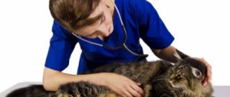
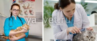
It is very important to show your cat to a doctor if these symptoms occur.
First of all, the veterinarian examines the cat’s oral cavity for inflammation, ulcers, plaque, and necrosis. Before treatment, it is necessary to exclude other, more serious diseases - leukemia, immunodeficiency, calcivirosis, panleukopenia, etc. To do this, blood is taken from the pet for testing.
Prevention
To avoid external necrosis in a cat, veterinarians recommend not leaving your pet in the cold, protecting it from burns, and putting away household chemicals or dangerous objects that the pet can swallow (rubber bands, ribbons, New Year’s rain, polyethylene). Exposed wires should not be left unattended if the house is undergoing renovations. Even minor wounds and scratches should be treated promptly, and deeper cuts or bites should be treated under the supervision of a veterinarian. The owner should carefully care for the pet after abdominal surgery and monitor the condition of the sutures. All systemic diseases in cats must be treated promptly. It is also recommended that the cat be vaccinated and dewormed in a timely manner.