Despite the small size of cats, ultrasound diagnostics is widely used in veterinary medicine to diagnose certain pathological conditions.
An ultrasound examination is a painless and quick method of examination that does not require anesthesia or additional injections of drugs.
For stress-sensitive animals such as cats, the ability to avoid additional procedures and unpleasant manipulations during research is very important. As a rule, cats tolerate an ultrasound examination quite calmly if they lie on a special soft table and are held by their beloved owner.
Indications for ultrasound of a cat
Routine ultrasound examinations in cats most often include examinations of the urinary system, liver and gallbladder. They are often prescribed based on the results of blood tests.
For example, an increase in the level of urea and creatinine in the blood indicates damage to the kidney parenchyma, the level of bilirubin and liver transaminases - pathological processes in liver tissue, inflammation or blockage of the gallbladder and bile ducts. In cats with symptoms of hyperthyroidism and elevated levels of the hormone T4, an ultrasound examination of the thyroid gland is performed.
Ivanova Nadezhda Viktorovna Veterinarian. Specialization: therapy, reproduction
In an emergency situation, for example, a fall from a height, a car injury, or a state of shock, a veterinarian can conduct rapid screening examinations of the cat’s abdominal and thoracic cavities. In this case, an assessment and detailed description of each organ is not carried out. A quick study is aimed at searching for free fluid (blood, urine, ascites fluid) in cavities, which can appear due to internal bleeding, organ ruptures, and pathologies of the cardiovascular system. This method, along with radiography, allows the doctor to assess the need for emergency surgery and further treatment tactics at the first visit.
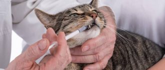
Another situation, typical mainly for cats, in which an ultrasound examination should be carried out immediately is acute urinary retention. If the owners notice that their cat has not been fully in the litter box for 24 hours, while he repeatedly tries to urinate, shows signs of anxiety, and screams, the condition of the wall of the bladder and urethra needs to be assessed using an ultrasound machine to eliminate the possibility of blockage with a urolith (stone). , as well as kidney pathologies.
Treatment of pyometra
There are two main treatment strategies for pyometra: surgical and conservative.
Surgical treatment is the most common and safest method: it is guaranteed to stop the development of the disease and remove the source of infection from the cat’s body.
The operation to remove the ovaries and uterus (ovariohysterectomy), which is performed for purulent inflammation of the uterus, begins after stabilizing the patient with the help of an intravenous infusion, carried out to relieve intoxication and restore fluid balance in the body.
During surgery, monitoring is aimed at maintaining normal blood circulation, gastrointestinal function, maintaining breathing and the required depth of anesthesia. The removal of the uterus is carried out very carefully to prevent its contents from entering the abdominal cavity. The bladder can be emptied if necessary by puncturing its wall (cystocentesis).
How to prepare a cat for an ultrasound
According to our personal statistics, most often cats come for an ultrasound of the kidneys and bladder. If this is a planned study, it is necessary to try to ensure that the animal does not urinate for several hours before the procedure. A full bladder is a good guide for a doctor; moreover, for cats, due to their species-specific propensity for diseases of the urinary system, a detailed examination of the bladder wall and its contents is very important.
If an examination of the cat’s liver, gallbladder, pancreas and other organs of the digestive system is planned at the appointment, it is very important to reduce the amount of gases and feces in the intestinal lumen. A full intestine makes it quite difficult to visualize other internal organs. To prevent this, we recommend that owners give the animal Espumisan (a drug from a regular pharmacy that reduces gas formation) a few days before a visit to the clinic. If you notice a lack of bowel movements in the last 24 hours before the test, a few hours before the ultrasound you can give the animal a microenema (Microlax) or give drugs that stimulate bowel movements (Lactulose, Vaseline oil).
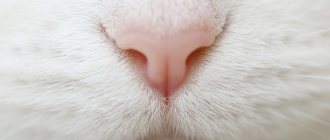
If the cat is not a hairless breed, for better visualization of the internal organs, you will have to shave off a section of fur on the belly (when examining the heart, the hair is shaved under the armpit on one side).
Unlike dogs, cats, as a rule, are not very willing to agree to lie on their back for several minutes of the procedure, but our ultrasound room is equipped with a special table with a soft surface, this allows us to reduce the animal’s discomfort during fixation to a minimum.
Ultrasound diagnostics of animals. Interview with a diagnostician
October 1, 2013
Lidiya Sergeevna Kamenskaya, a veterinarian for visual diagnostics at the Biocontrol clinic, spoke in her interview about how an ultrasound is performed on a dog and a cat, why an ultrasound examination is needed, and how to prepare an animal for ultrasound diagnostics.
— What does the ultrasound diagnostic method provide?
— Ultrasound diagnostics is a non-invasive research method that allows you to assess the condition of organs and the changes occurring in them. Ultrasound is a diagnostic method complementary to the initial examination of the animal (auscultation, palpation). If it is difficult to differentiate the diagnosis, if severe acute or chronic pathological processes in the animal’s body are suspected, the doctor will give a referral for ultrasound diagnostics to confirm the diagnosis. This method is used to monitor acute inflammatory processes, changes in the chronic course of diseases, oncological processes and in other similar situations.
— Is it possible to examine, for example, the eyeball using ultrasound?
- Certainly! In many cases where ophthalmoscopy is not possible, ultrasound is performed. The presence of neoplasms and blood clots inside the eyeball is detected, the presence and degree of retinal detachment, lens loss, and the presence of a neoplasm of the ciliary body or retrobulbar space are determined. Very often this study is done to confirm the diagnosis.
— Why are other research methods sometimes required after ultrasound diagnostics?
It is worth remembering that this method is non-invasive and in some cases an accurate diagnosis cannot be made based only on ultrasound results. On the monitor, the doctor sees the structural changes that have occurred in the organ, can describe their nature, and identify precise changes in the size and structure of organs and tissues. But the morphological composition of formations or the type of cells when the structure of organs changes can only be clarified by invasive research methods - taking a sample of cells or tissues by puncture of an organ or neoplasm for further cytological or histological examination.
— Do they do ultrasound diagnostics for cancer?
- Undoubtedly. First of all, to understand how widespread the neoplasm is, what structure it has, its size, boundaries, degree of invasion into nearby organs and tissues, and other data that is extremely important for the attending physician. In the presence of intra- and extraorgan formations, the ultrasound method is the initial study necessary for oncologists in order to understand the tumor of which organs and systems they are dealing with and what the treatment tactics will be in the future.
— Can all oncological diseases be seen and differentiated by ultrasound?
- It depends on the size of the tumor. Anything larger than 5 millimeters can be seen. Of course, subject to certain training and experience of the diagnostician himself, the availability of high-quality modern equipment and high-frequency sensors. These may be metastases, enlarged lymph nodes, extraorgan tumors. What is on the surface of the skin (cutaneous and subcutaneous neoplasms, breast tumors, thyroid tumors) can be viewed and assessed using ultrasound. In this case, the visual diagnostician will be able to answer the oncologist the following questions: what is the structure of the tumor in terms of tissue homogeneity or heterogeneity, cysticity, areas of necrosis and decay, the presence of blood or pus, areas of calcification and fibrous changes. Dopplerography (this is one of the methods of ultrasound examination of vessels and blood flows in them) can also assess the degree of vascularization (weaving of the vascular network) of the tumor. Ultrasound of an animal gives veterinary oncologists the opportunity to create an algorithm for further actions to study and clarify the diagnosis: aspiration of tumor cells for cytological examination or taking a tissue biopsy for histological examination.
— What diseases need to be monitored using the ultrasound method?
- Many. These include acute inflammatory processes and chronic diseases. Also age-related diffuse changes in internal organs. In addition, ultrasound is done as part of the medical examination of animals, which is recommended to be carried out on a regular basis, once a year, especially after cats and dogs reach the age of six. Control over oncological diseases is beyond discussion - any process of this kind should be monitored once every 3-4 months, and possibly more often, since everything depends on the type of tumor, its location, the aggressiveness of its spread, the response of the tumor itself to therapy and the response to whole body therapy. There are diseases that do not require frequent diagnosis, but some owners conduct control ultrasound examinations of the animal for their own peace of mind. The reason for ultrasound diagnostics is the physiological conditions of animals: estrus, pregnancy, postpartum changes.
— Quite often, pet owners, in order to consult with the doctors of the Biocontrol clinic on the forum, post ultrasound images there. How informative are they? Is it possible to conclude anything from them or should a repeat ultrasound be performed?
— For a doctor, static images on an ultrasound image carry almost no information. We obtain maximum information during research, during the research process. Here, the experience of the doctor himself plays a huge role: the correct use of sensors, device settings, projections, sections, viewing angles and other points. Of course, a lot depends on the quality of the thermal printer on which the scanogram is printed. However, if we are talking about data sent by patients to clarify the diagnosis or consultation, then the most informative will be not the image, but the protocol drawn up by the doctor after the ultrasound diagnosis. This is what needs to be posted on the forum.
— How can owners prepare an animal for an ultrasound examination?
— Ultrasound is a method that has certain physiological limitations. Thus, everything that is under the gas (in the stomach, intestines) cannot be seen - the gas reflects ultrasound, gives a persistent acoustic shadow effect and the image becomes uninformative. In order for the study to be successful, you must undergo a fasting diet (12 hours). The use of carminative and adsorbent drugs is mandatory: espumizan, activated carbon, enterosgel. In this case, the dosage of activated carbon is 1 tablet per 5 kg of animal weight at intervals 24 hours before the study, then 6 hours and another 2-3 hours before the study. Depending on the system you want to take a closer look at, there are additional conditions. For example, if an ultrasound of the bladder is required, then it should be moderately full, but not overstretched. If the remaining organs of the abdominal cavity are examined, an empty bowel and an empty stomach are needed.
— Is it necessary to cut the animal’s hair in the area where the sensor will be located during the study? What to do with show animals for which it is not advisable to have areas of their fur clipped?
“I would like to note that trimming the area to be examined is always safe. We perform it with a special electric machine that does not cut the skin and painlessly removes hair. For show animals, it is possible to make an exception and try to moisten the fur generously with alcohol, water or gel. However, this does not guarantee correct and fully informative research. When the owner asks not to cut his animal, the doctor always warns him about this. After all, the health of the animal is more important than the upcoming exhibition; accordingly, the diagnostic procedure must be carried out correctly! Typically, all owners agree to prepare the area for exploration. The fact is that in order to properly conduct an ultrasound examination, it is important for us to prevent the presence of a layer of air between the point of contact of the sensor and the surface of the animal’s body. But hair cutting was not introduced in vain, since sometimes wool gives us “shadows” and artifacts, misleading, or does not allow us to view certain areas or organs at all. In addition, do not forget - the doctor’s experience, a high-frequency sensor and proper preparation for ultrasound examination make it possible to give the animal the correct diagnosis as fully as possible.
— Do domestic animals have breed predispositions to diseases that can be differentiated by ultrasound?
- Of course have. Kidney problems most often occur in Abyssinian and Persian cats and Shar Pei dogs. Every second animal suffers from diseases or pathological changes, and these are congenital changes. Persian cats often end up in Biocontrol with polycystic disease - this is a genetic, congenital disease that primarily affects the kidneys, possibly the liver, and other organs. Rhodesian Redbacks, Zinenhunds, and Boxers often suffer from lymphoma at the age of 3-5 years, to varying degrees of generalization, up to metastases in the spine.
But it is incorrect to talk only about breed predispositions. After all, the state of health is determined not only by genetics, but also by the diet and lifestyle of the animal. Therefore, ultrasound diagnostics, like other diagnostic methods, is a powerful weapon in the hands of a veterinarian and a caring owner in the prevention and treatment of diseases of our pets.
Tatyana (02/24/2016 at 14:40): Hello. Help!!! What can you say?!! Our doctors are already in heat or endometritis for the third time, but this doesn’t make me any better (((I can’t find standard indicators to compare!(Thank you all!!!???
Reply | to write a message
- Biocontrol (02/24/2016 at 14:58): Tatyana, it’s better for you to communicate directly with our doctors on the forum:
Reply | to write a message
Elvira (07/29/2016 at 12:09): Hello. Please tell me, can a cat have no uterus?
Reply | to write a message
- Biocontrol (07/29/2016 at 14:56): Yes, theoretically this could happen.
Reply | to write a message
Svetlana (05.25.2020 at 13:05): Hello! Tell me, how many days after worming does the dog have a bloated belly?
Reply | to write a message
- Biocontrol (05.25.2020 at 15:40): Hello, Svetlana! It is better for you to consult our therapists directly about this. This can be done here:
Reply | to write a message
Click to cancel reply.
Ultrasound during cat pregnancy
Cat owners often schedule an ultrasound to confirm pregnancy. If the diagnosis is carried out in the early stages (20-25 days after mating), the most accurate calculation of the amniotic sacs is possible. At a later date, the doctor can assess the viability of the fetuses, demonstrate to the owners the heartbeat of the kittens, and monitor the possibility of kittens with severe developmental pathologies.
Ivanova Nadezhda Viktorovna Veterinarian. Specialization: therapy, reproduction
If a cat gives birth in a clinic, doctors use ultrasound to monitor the fetal heart rate, constantly assessing its viability, this allows them to make a timely decision about the need for a cesarean section.
Using ultrasound, it is also possible to plan mating of cats; examination of the uterus and ovaries gives a detailed picture of the growth and development of follicles, the condition of the endometrium of the uterus and the presence of inflammatory phenomena.
Special requirements for preparation for the study
Ultrasound examination of the abdominal organs can give accurate and informative results only if the person being examined begins to fulfill in advance all the requirements prescribed by the doctor to prepare the patient. Otherwise, during diagnostics, noise may appear in the image, distorting the ultrasound picture.
The doctor may verbally list all the preparatory requirements to the examinee, and in some medical institutions these lists are given to patients in printed form for convenience.
The procedure can only be performed on an empty stomach. If it is scheduled for the first half of the day, the patient needs to refuse breakfast. If the ultrasound is performed after 15:00, you can afford a light breakfast no later than 9:00 am. It is not recommended to consume any liquid an hour to an hour and a half before the start. Also, an hour or two before the ultrasound, you should not chew chewing gum or smoke - these actions provoke spasms of smooth muscles.
People who are prone to constipation should take a laxative the evening before the procedure as part of their preparation.
All medications taken at this time must be reported to the diagnostician in advance. During the ultrasound, the patient takes with him the results of all previously performed tests and examinations, if any.
Those who have a tendency to increased formation of gases in the digestive tract are prescribed a special slag-free diet with a reduced content of coarse dietary fiber. They begin to adhere to it 2-3 days before the examination date.
In addition to dietary restrictions, taking sorbents, for example, activated carbon after each meal, helps reduce the level of gas formation.
A slag-free diet, as a component of preparation for the procedure, involves avoiding the following foods:
- milk and dairy products;
- fresh bread, especially black bread;
- fresh fruits and vegetables;
- fatty and fried foods;
- smoked, spicy and pickled products;
- baking and muffins;
- alcoholic and carbonated drinks, strong tea and coffee;
- legumes
The diet before the study consists of lean meat, poultry and fish, cereals, yeast-free stale white bread, baked or boiled vegetables, vegetable broths, non-concentrated clear juices, compotes. Be sure to drink at least one and a half liters of clean water per day.
All these rules are irrelevant in cases where urgent diagnosis is necessary, when it is not possible to wait 2-3 days, for example, in case of acute appendicitis.
Ultrasound of a cat's heart
An ultrasound examination of the cardiovascular system may be prescribed to a cat after identifying some characteristic symptoms associated with cardiac pathologies (shortness of breath, discoloration of mucous membranes, increased blood pressure). A cardiac examination is also mandatory before surgical interventions performed under anesthesia for such cat breeds as British, Scottish, Ragdoll, Persian, Maine Coon, due to a genetic predisposition to cardiac pathologies.
Who can get pyometra?
Cats are less likely to develop pyometra than dogs.
This may be due to less progesterone being released. Unfortunately, due to the lower prevalence of the disease in cats, fewer factors are known to contribute to or prevent the development of pyometra in cats. However, it is known that older animals are most susceptible to the disease; the risk of the disease increases significantly after 7 years, the average age of onset of pyometra is 5.6 years. Despite this, pyometra can also occur in very young animals - even a ten-month-old cat can get sick.
Only castration can completely prevent the development of purulent inflammation of the uterus. A cat’s history of childbirth, according to research, also does not in any way protect it from the disease.
It is also reliably known that previous hormonal therapy associated with the intake of progesterone contributes to the development of pyometra, therefore the use of drugs to suppress sexual estrus in animals is extremely undesirable.
I did an ultrasound - does that mean treatment can be prescribed?
Many owners believe that such a study replaces other tests and, based on ultrasound, a diagnosis can be made and treatment can be prescribed. This is a very big misconception.
Ultrasound diagnostics allows us to see structural changes in organs, so to speak, to assess the scale of the problem and suggest or exclude a diagnosis.
But to make a final diagnosis, other laboratory and instrumental diagnostic methods are also required.
Therefore, if a doctor prescribes a further examination, you should not think that he is not sure about something; the doctor needs additional data to create a more complete picture of the disease. After all, the result of treatment directly depends on the correctness of the diagnosis.
Features of preparing and conducting diagnostics for children
Preparing a child, just like an adult, includes dietary restrictions. For 2-3 days, it is necessary to exclude from the menu foods that provoke increased gas formation, as well as candies and sweets. If a small patient has a tendency to flatulence, he is prescribed enzyme and absorbent drugs.
For children of any age, ultrasound is performed on an empty stomach. Mothers of infants need to calculate feedings so that the last one before the procedure is carried out no later than 3 hours before. It is not advisable to give your child fruit or vegetable purees before the test, as they take a long time to digest.
The process of performing an ultrasound examination of the abdominal organs in children is no different from a similar procedure in adults. Diagnosis is carried out with a special ultrasound probe using conductive gel.