(c) Veterinary center for the treatment and rehabilitation of animals “Zoostatus”.
Varshavskoe highway, 125 building 1.
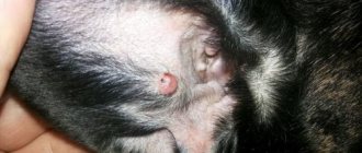
Atheroma is a cyst that occurs as a result of blockage of the sebaceous gland duct.
Normally, the sebaceous glands in dogs and cats, located throughout most of the body, produce an oily secretion that lubricates and moisturizes the surface of the skin. The ducts of the sebaceous glands are located in the hair follicle, so it is believed that the development of sebaceous cysts is associated with obstruction of the follicle, which leads to the accumulation of secretions and the formation of atheroma. Follicle blockage can occur due to various reasons, such as improper hair growth, poor hygiene, or skin infections.
Treatment of atheroma in dogs and cats
Atheroma, which is small in size and does not interfere with the animal, does not require any treatment. Sebaceous cysts that cause discomfort are removed surgically.
Atheroma is a benign skin disease that should not worry the owner much. However, if it grows, you should show the animal to a veterinarian and perform a puncture of the formation to exclude malignant skin tumors.
Read reviews about our veterinary center. Call the number and schedule a consultation right now or request a call back. (c) Veterinary center for the treatment and rehabilitation of animals “Zoostatus”. Varshavskoe highway, 125 building 1.
A number of diseases in dogs do not cause concern to owners at first glance because they seem quite harmless, but the consequences can be irreparable. Such diseases include pathological formations. In most cases, cysts in dogs are not dangerous, but some types can deprive the pet of health and even lead to death.
Signs of atheroma in cats
Benign sebaceous gland tumors in cats are usually hard, raised, cauliflower-like in appearance, and warts range in size from a few millimeters to several centimeters.
The lesions may be yellowish or pigmented, lack hair, have a greasy appearance, or be in the form of an ulcer. Nodules with hyperplasia of the sebaceous glands can be multiple.
Malignant tumors, sebaceous adenocarcinomas, tend to appear as solitary, hairless, erythematous intradermal nodules that invade the subcutaneous tissue and are up to four centimeters in diameter. Tumors of the sebaceous glands in cats are mainly found on the head, less often on the upper legs and back.
Types of cysts and causes of their occurrence
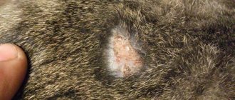
Photo of a cyst in a dog
A cyst is a cavity in which fluid—a secretion—forms. The secretion consists of natural decay products, as well as microorganisms and even parasite eggs. If a cyst with such filling bursts, nearby organs and tissues will be seriously damaged and the body will become infected.
In some cases, the cyst can degenerate into a malignant neoplasm, so veterinarian diagnosis and regular monitoring are necessary.
The type of cyst is determined by many signs - the location of formation, the structural features of the cavity, the tendency to grow and develop, and others.
Epidermal or cutaneous cysts are quite common. Its distinctive features include the following:
- Location: upper layers of the epidermis.
- A cavity in the shape of a circle or ball.
- The softness of the formation, this can be checked by palpation.
- The cyst does not cause pain to the dog.
- The size of the formation rarely exceeds 50mm.
The causes of cysts formed on the surface of the skin are very diverse. They often appear against the background of a clogged pore, which does not allow skin secretions to come out. As a result, a cavity is formed, which gradually fills.
When is skin cytology prescribed?
With the help of a cytological examination of a dog's skin, it is possible to detect neoplastic (atypical), infiltrating (inflammatory) and bacterial cells, parasitic organisms, and fungi.
Attention! The word “Cito!” written on the referral form for any analysis. has nothing to do with cytology, it means that the study must be performed urgently.
Indications for cytological analysis of skin in dogs are:
- papular or pustular rashes;
- skin itching of unknown etiology;
- alopecia (hair loss or uneven growth);
- formation of crusts or scales on the skin;
- nodular or tumor neoplasms localized in the dermis;
- changes in skin pigmentation.
Helpful information! Many of the skin diseases give a similar clinical picture; cytological studies of the skin make it possible to differentiate symptoms and diagnose dermatitis, bacterial pyodermatitis, parasitic skin diseases, benign and malignant neoplasms.
Symptoms of the disease
1. Cyst located on the surface of the skin
A cyst located on the surface of the skin is determined visually. Formation on the gums can be detected at the initial stages. The tissues where the cyst appears swell and have a bright red tint. Over time, the tumor may spread throughout the gum.
During this period, quite often the dog feels unwell, gets tired quickly, may refuse to eat, and in some cases the body temperature rises. When examining the mouth, a white or yellow neoplasm filled with pus will be revealed. If you do not help your pet, he will suffer from pain, asymmetry of the mouth, and gumboil.
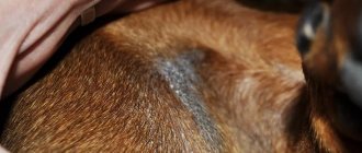
2. Sweat gland cyst
A sweat gland cyst is blue, blue or grayish in color, its size rarely exceeds 1 cm. Often a sweat gland cyst occurs in the ear cavity; in most cases it is not dangerous, and the pet does not experience discomfort.
3. Follicular cyst
A follicular cyst occurs at the hair root and is filled with dead cells. The characteristic color is gray, the size varies from 1 mm to several cm.
4. Dermoid cyst
Dermoid cyst is a congenital defect. It is a closed soft cavity of a round shape, rising above the hairline. In some cases, there is a risk of infectious damage to the nervous system, so veterinarians recommend timely treatment.
5. Cyst in the sublingual area
A cyst may also appear in the sublingual area. It looks like a round purulent sac of white and pink color. When a cyst forms in this place, the dog often refuses to eat because it feels uncomfortable in the process of eating food. If the size of the formation reaches a critical size, the dog may even stop drinking and begin to waste away from exhaustion.
6. Interdigital cyst
In most cases, an interdigital cyst does not cause pain in the pet; it looks like a round swelling. The formation is soft, the temperature in the area of the cyst is not elevated, and there are both single and multiple swellings. Short-haired dogs are especially susceptible to this disease. The disease is caused by ingrown hair into the skin.
Skin cysts have the same symptoms:
- Slowly increase in size.
- They do not cause pain, the dog behaves calmly while probing the cavity, does not whine, and does not try to push the owner away from him.
- When palpating the cyst, its smooth structure, heterogeneity, granularity are revealed - a dangerous “bell”.
- The pet gets tired quickly, sleeps more than usual, and does not want to walk for a long time.
- In some cases, body temperature rises.
- Loss of appetite.
Inflammation of the sebaceous glands: typical manifestations
The age of the dog at which the disease manifests itself is from one year to 5-6 years. According to observations, there is no gender predisposition; both males and females are affected.
Clinical signs are extensive, and the severity of sebadenitis or the extent of the affected area is not the same among representatives of the same breed, sex or age. But one of the obvious signs can be identified: the presence of skin scales (dandruff), which seem to be glued to the hairs; they are called “follicular casts”.
First, the pathological process involves the head, ears, muzzle, tail, and passes along the ridge. The lesions are symmetrical, forming equally on both sides of the animal's body, but almost never involve the paws and stomach. The Poodle, Samoyed and Akita Inu develop leaf-like keratinized discharges from the hair follicles that tightly grip the hair, forming a durable layer of dead tissue. If you examine the plucked hair, it is noted that the secretion “takes root” tightly at the root of the hair.
Locations of damage:
“Follicular casts”
- poodle: muzzle with transition to the chest;
- Akita Inu: many symmetrical lesions are formed, first on the head, ears, which go down to the neck, back and tail. There is practically no itching, but in the absence of timely treatment, the inflammation becomes a generalized form with the addition of secondary pathogenic microflora. In this case, the itching may intensify, and an unpleasant odor emanates from the pet;
- in the Belgian Shepherd: skin pathology is combined with an inflammatory process in the outer ear, in which dry deposits of sticky scales are formed.
In dogs with short hair (Vizslas), the inflammation resolves with the formation of nodules, coupled with confluent or arcuate alopecia. Outwardly, it looks like a skin eaten by a moth, on which there are skin flakes scattered in a chaotic manner. In this case, swelling of the muzzle almost always occurs. Such individual clinical signs lead experts to believe that inflammation of the sebaceous glands is an independent disease.
Diagnosis and treatment of cysts in dogs
With this problem, you can contact a general veterinarian or an oncologist. The doctor will conduct the following diagnostic tests:
- Examination of the dog, during which the veterinarian feels the external formations. If any, he will conduct a general assessment of the animal’s health.
- General analysis of urine and blood, checking its coagulability and hemoglobin level.
- Ultrasound research to study the nature of formation.
- X-ray.
- CT scan.
After the diagnosis is made, the issue of treatment is decided. In some cases, when there is no obvious danger to the health and life of the pet, no active action is taken. The veterinarian suggests that the owner observe the development of the cyst; if it does not grow or cause discomfort to the dog, then treatment will not be required. In the event that the formation progresses or there is a threat to the dog’s health, the doctor will take active action.
Drug treatment of cysts rarely gives a clear positive effect, but in some cases the dog is prescribed subcutaneous administration of drugs aimed at resolving the formation. This method is used only in the case of small formations; medications do not work on large cysts.
The most effective way out of the situation is surgery . The operation is performed both under local anesthesia, for example, when it is necessary to remove a cyst under the tongue, and under general anesthesia, in the case of formations on internal organs. After surgery, the doctor gives recommendations that will help the dog recover in a short period.
The owner must treat the seams himself, for this they use a solution of manganese or iodine, furatsilin. It is important to feed your pet properly. In case of oral surgery, the dog is not allowed to eat dry food for several hours.
Lump under the skin of a dog
If your dog has a lump under the skin at the site of a fresh surgical suture, then there is no cause for concern. Slight swelling, puffiness, redness of the tissues, dry compaction in the area of skin excision - this is a variant of the norm, a protective reaction of the animal’s body.
As the incision site heals, the lump decreases and disappears completely, swelling and swelling subside, and the tissue returns to its normal color. If the post-operative lump oozes blood, ichor or pus, then the dog should be shown to a doctor immediately.
A lump-like lump at or near the incision site may also be an overgrowth of granulation tissue. Granulation tissues are formed by young cells of the connective tissue in places of injury and damage; Over time, these tissues fill the surgical incision site and form a scar. Their growth is also considered a variant of the norm. But in this case, the dog must be taken to a veterinary clinic for cell collection to exclude atypical growth that could develop into cancer.
A small bump on the surface of the skin may turn out to be atheroma - inflammation of the sebaceous gland. This is a relatively rare disease in dogs, but does occur. Treatment consists of surgical removal of the inflamed gland.
Cause: atheroma
Atheroma is also suspected when a dog has a lump under the eye. But a lump or neoplasm in the periocular area can also be a sign of a basal dental abscess, especially in older dogs or representatives of breeds with genetically “bad” teeth: Russian Toys, Chinese Cresteds, Mexican Hairless. In this case, the lump will not go away even with local therapy and will begin to appear again after removal until the diseased tooth is cured or pulled out.
Atheroma, hematoma, abscess, histiocytoma (vascular connective tissue tumor) are suspected when a dog has a lump on the ear. A hematoma is usually the result of a blow or bruise to the head. Therefore, the seal can also be localized on the head, in the muzzle area, on the neck - depending on the dog’s build. The hematoma usually goes away on its own. An abscess often occurs as a result of a bite, scratch, or other damage to ear tissue and is treated with antibiotics, sometimes with surgery.
Histiocytoma requires surgery, as it can develop into a cancerous tumor. Due to blunt bruise trauma, inflammation of the lymph node or salivary gland, a lump also appears on the dog’s face, in the mouth or nose. A lump on a dog's leg is often the result of a bruised injury. On the hind leg, a lump often forms after a poorly performed intramuscular injection. In older dogs, bursitis, an inflammation of the elbow and knee joints, is also a cause.
Disease prevention
Since the causes of most types of cysts are unknown, veterinarians give general recommendations for preventing this disease. First of all, the owner must provide the dog with proper and balanced nutrition. Doctors recommend specially formulated foods that contain the required amount of vitamins and minerals for every day.
It is important to maintain dog hygiene, brush your pet regularly, cleaning the fur from possible accumulations of dirt, and also trim the fur. The dog should be washed with special products that will wash the coat and skin. Long-haired breeds are washed 2 times a month; dogs whose coat has medium pile need only be bathed once every 2 months. Washing too frequently dries out your pet's skin and disrupts its water balance.
It is necessary to regularly examine the dog’s skin; if neoplasms appear, a visit to the veterinarian cannot be postponed.
Causes of atheroma
The exact cause of the cyst formation has not been established. It is assumed that the following may contribute to the formation of a cystic body:
- microorganisms penetrating from outside;
- abnormal hair growth.
There is no genetic predisposition to atheroma in certain breeds of dogs. But the pathology most often manifests itself in animals from 4 to 8 years old.
Some experts talk about traumatic origin. This is due to the location, since the outer, open areas of the dog’s body are more often affected.
Symptoms of the disease
The dog's body is completely covered with hair. However, atheroma can be localized both in densely covered areas and in those where there is not much hair at all. More often, atheroma can be found on the head, limbs, and neck of the animal.
Initially, upon discovering a small pea, the dog owner does not pay attention to it. It is generally accepted that if the atheroma is small and does not cause concern to the animal, then there is no point in focusing on the pathology. However, when placed in “inappropriate” places, the animal can scratch with its paws, which will lead to premature autopsy.
When palpating the seal, the following is determined:
- clear contours;
- mobility;
- painlessness.
The process can remain in this state for years, with a slight increase. Size varies from pea to walnut.
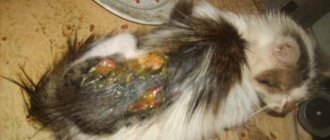
Under certain circumstances, atheroma begins to increase in size and “ripe.” In this case, the following may occur:
After ripening for years, atheroma can break out on its own. However, the leakage of a “curdled” purulent mass does not mean recovery. Without assistance, the process develops into a long-term non-healing ulcer.