To make an oncological diagnosis, all possible methods of examining the patient are often used (examination by an oncologist, ultrasound, X-ray, CT/MRI). However, cytological and histological tests play a special role. Microscopy of cells and tissues obtained during a biopsy allows you to confirm or refute not only the presence of a tumor process, but also to detect signs of inflammatory and specific non-tumor changes, which will allow you to develop a plan for further diagnosis and treatment of the patient.
Cytology in animals today is one of the main methods for making an accurate diagnosis.
When is cytology used?
Cytology is most often used to diagnose the nature of tumors and lumps on the surface of the body. However, cytology is also used to evaluate:
- Internal organs such as the liver, lungs, lymph nodes and kidneys
- body fluids such as urine and synovial fluid
- Abnormal fluids (effusion) that may accumulate in the animal's body, especially in the chest and abdomen
- Various surfaces of the body, both external and internal, such as the mouth, eyes, respiratory tract or vagina.
Treatment of histiocytoma in dogs
Treatment of this disease is possible only after an accurate diagnosis has been made. The most acceptable methods of treatment: excision (excision) or cryosurgery. If the tumor is inoperable due to its size or shape, the use of hormonal drugs is necessary. They are used systemically in an immunosuppressive dose or locally in the form of blockades. The essence of this technique is to introduce a large concentration of hormonal agents into the area of the tumor, which block the growth of the tumor and promote its regression. The prognosis is favorable. If systemic histiocytosis develops, a course of chemotherapy is prescribed and the prognosis worsens. The main drugs for chemotherapy are antitumor antibiotics of the anthracycline series and alkylating agents in recommended regimens.
What information will help the veterinarian in treatment?
Once the veterinarian knows the nature of the disease, he will take steps to find the specific cause (bacteria, parasites, fungi, allergies, etc.) in order to prescribe the appropriate treatment.
Cytology can predict how a tumor will behave in the future. This can help determine whether surgery is warranted and whether it is urgent.
If surgery is scheduled, a general understanding of the tumor type will influence the next steps needed to prepare the pet for surgery. It will also be easier for the surgeon to plan how to remove the tumor if its nature is known in advance. Finally, information about the tumor will allow the veterinarian to predict what will happen after surgery, and he will be able to answer the following questions:
- Are relapses possible?
- Will it spread to other parts of the body?
- When will the treatment end?
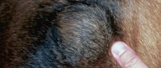
Symptoms and signs of cutaneous histiocytoma
Dogs of smooth-haired breeds are more prone to skin histiocytoma, regardless of gender. Education rarely makes itself known and leads to inconvenience for the animal. Cutaneous histiocytosis from Langerhans cells is characterized by an abundance of such skin formations (sometimes the mucocutaneous frame of the mouth), which can be of different sizes: from minor nodules to large tumors. Mostly Shar-Peis suffer from it.
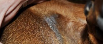
Cutaneous histiocytosis from IDC also makes itself felt by multiple approximately 4-centimeter formations in the head, neck, torso, and paws (sometimes lymph nodes are also affected). Moreover, the disease is typical for dogs of any breed.
A distinctive feature of systemic histiocytosis is lymphohistiocytic destruction of the walls of blood vessels, which makes itself felt in the form of dermatitis and panniculitis (inflammation of subcutaneous fatty tissue). This process results in anorexia, weight loss, eye lesions such as swelling of the apple and conjunctiva, and conjunctivitis. Mostly representatives of large breeds are affected.
Histiocytic sarcoma in dogs manifests itself as single and multiple tumors, which can be in one place or spread to other organs. The disease is typical mainly for Bernese Mountain Dogs, Rottweilers, Golden and Straight-Coated Retrievers. Hemophagocytic histiocytic sarcoma is a proliferation of macrophages in the spleen in dogs. It is characterized by regenerative destruction of red blood cells by anemia, thrombocytopenia, and bilirubinemia.
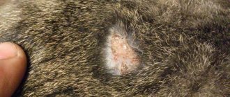
How is histiocytoma diagnosed in dogs?
The generally accepted understanding of this disease is that such types of neoplasms are exclusively benign. There have never been any deaths from histiocytomas in practice.
Skin ailments of this type are easily identified using clinical signs and diagnostic biopsy, the results of which must undergo histological examination. A detailed description of the onset of the disease, as well as the presence of signs provided by the pet owner, will help to correctly identify the disease. As well as conducting biochemical tests of blood, urine, ultrasound, x-rays, etc.
How is a cell sample taken?
There are several ways to collect cells from tissues, depending on where the problem is and what type of tissue it is. The most common method is to use a fine needle or biopsy needle. This is a simple method where a tissue sample is taken using a sterile, narrow needle attached to a syringe.
Other methods are typically used to collect cells from the surface of the body. Scaly or bare skin can be collected by scraping and scooping out some of the top layers of skin cells. The ulcerated skin can be collected using a clean glass and swab. Discharge from the nostrils, eyes, or vagina can be collected with a cotton swab.
No.AN404SKIN, Cytological examination of a smear-imprint from the skin
DESCRIPTION
Cytological examination of a skin smear is a fast and diagnostically valuable research method. This type of study is often used for exudative lesions and seborrhea. There are direct and indirect methods for obtaining a sample. With the direct method of obtaining fingerprint smears, a glass slide is pressed tightly directly onto the affected area (ulcers, erosions, crusts, pustules, plaques, papules, nodules, fistula tracts). Before this, the surface is lightly processed to first identify representative cells. Direct impression smears can also be made from the surface of removed lesions for rapid identification in cases of suspected neoplastic or inflammatory process. The indirect method of collecting the sample uses a swab, curette, or scalpel blade. The affected area is lightly scraped with a tool soaked in mineral oil, a glass slide is applied to it and gently “stretched” without causing damage to the cells. Indirect fingerprint smears are more often used for oozing injuries and in hard-to-reach areas of the skin (for example, ears and paws). Fine needle aspiration is a fast, inexpensive, and minimally invasive way to examine nodular lesions, skin tumors, and enlarged lymph nodes. During aspiration, a small number of cells are released into the sample, which may be non-indicative and will not provide any information about tissue architecture and invasiveness. Therefore, cytological examination of the skin is a screening method and does not replace biopsy and histological examination.
INTERPRETATION
The study results contain information for physicians only. The diagnosis is made based on a comprehensive assessment of various indicators and additional information.
The drug is examined for the presence of leukocytes, erythrocytes, nuclear epithelium, phagocytosis, the presence of microflora, Malassezia and, possibly, the presence of other material. The notes indicate the presence of other types of cells (acantholytic, mast cells, plasma cells, fibroblasts), and specify the predominance of leukocyte populations. When interpreting the results of a cytological examination of skin impression smears, the composition and ratio of cellular composition, as well as microflora, are assessed. The finding of a large number of different cell types (neutrophils, eosinophils, lymphoid cells, monocytes/macrophages, epithelial cells and fibroblasts) suggests the presence of acute inflammation. The detection of a significant number of monocytes, as well as lymphocytes and plasma cells, indicates a chronic course of the inflammatory process. The presence of a small number of lymphocytes and plasma cells is common in any form of inflammatory process in the skin, but the detection of a large number of them may indicate neoplasia or lymphocytic-plasmacytic pododermatitis. Reactive fibroblasts and epithelial cells can also appear during the development of neoplastic processes. Loose bacteria indicate contamination of the sample, but phagocytosis of bacteria indicates the pathogenicity of the microorganism and is a sign of an infectious process. Degenerative neutrophils in the smear (karyorrhexis and large, fragmented nuclei without cytoplasm) and the presence of microflora suggest pyoderma, whereas the presence of nondegenerative neutrophils with pyknotic nuclei (dark-stained with clustered segments) suggests sterile inflammation. The detection of acantholytic cells may indicate the development of an autoimmune disease. Although acantholytic cells can also occur in bacterial and fungal infections. The detection of a large number of bacteria and epithelial cells may indicate the growth of resistant microflora and a decrease in the protective properties of the body’s immune system. The presence of eosinophils in the smear confirms an allergic reaction, furunculosis, eosinophilic granuloma complex in cats. Eosinophils and mast cells in the smear are found in mastocytoma. The presence of a significant number of Malassezia dermatitis indicates the presence of Malassezia dermatitis. Pyogranulomatous inflammation may be associated with less common bacterial and fungal infections. If cells with signs of atypia are detected, a cytological examination by a qualified cytologist is recommended.
Units:
not detected or detected (the result is indicated in crosses or in numbers in the fields of view).
what stage follows after cytology?
The next stage after cytology is histology. Histological examination is the examination of whole tissue samples that are collected surgically. Histology focuses on tissue structure, development, and function.
In most cases, histology provides a definitive diagnosis and is generally considered the gold standard for diagnosis. Histology is often required to determine whether a tumor is benign or malignant and to confirm cytology findings. If your pet has undergone surgery to remove a tumor, it is advisable to request that the tissue be examined using histology.
How is material collected for research?
Collecting material for research is almost painless. More often, a tissue sample is taken for biopsy using a special needle (sometimes it can be a regular syringe needle) or a trephine instrument.
Cytological studies are carried out immediately after the material is taken, which can be a smear or puncture of the tumor contents or any biological fluid. If necessary, after cytological diagnosis, the doctor may refer the patient for histological examination.
If this process was at the initial stage or in its infancy, then measures can be taken in time and the animal’s health will be safe.
A cytological examination of animals done on time can save or prolong the life of your pet; he will live and delight you for many more years.
Experienced pathomorphologists of the Laboratory of the Veterinary Center of Dr. Bazylevsky A.A. will be able to quickly and accurately analyze cells and tissues to diagnose your pet. You can get more detailed advice by calling veterinary clinics.
What deviations from the norm may mean
To decipher a blood test in dogs, the difference between the norm and the resulting deviation is used. Self-diagnosis is complicated by an integrated approach to interpretation. It will not be possible to determine the disease by just one of the deviations.
Possible results depend on which direction the value deviates. It can cross the upper or lower boundary.
In clinical analysis
Small deviations within one tenth or even whole values are acceptable. This can be explained by an error in collecting material, improper preparation of the dog, or other reasons.
Serious pathology is indicated when the indicator differs too much. Possible diagnoses can be found below.
| Name | Below normal | Above normal |
| Red blood cells |
|
|
| Hemoglobin |
|
|
| ESR |
|
|
| Hematocrit |
|
|
| Reticulocytes |
|
|
| Platelets |
|
|
| Myelocytes | — |
|
| Plasmocytes | — |
|
| Leukocytes |
|
|
| Eosinophils | — |
|
| Lymphocytes |
|
|
| Neutrophils |
|
|
| Basophils | — |
|
| Monocytes |
|
|
The MCH and color score are used to determine the severity of the suspected disease. They are not considered outside of hemoglobin and red blood cells.
In biochemical analysis
During diagnosis, the veterinarian compares the hemograms of both studies. Possible deviations in the LHC are presented below.
| Name | Below normal | Above normal |
| Glucose |
|
|
| Urea |
|
|
| Creatinine |
|
|
| Total bilirubin | — |
|
| Direct bilirubin | — |
|
| Cholesterol |
|
|
| General lipids | — |
|
| Acid phosphatase | — |
|
| Alkaline phosphatase |
|
|
| Gamma-glutamyl transpeptidase | — |
|
| Creatine kinase | — |
|
| pH |
|
|
| Total protein |
|
|
| Serum albumin |
|
|
| ALT | — |
|
| AST |
|
|
| Triglycerides |
|
|
| Lipase |
|
|
| Alpha amylase |
|
|
| Lactate dehydrogenase | — |
|
| Calcium |
|
|
| Sodium |
|
|
| Chlorine |
|
|
| Potassium |
|
|
| Phosphorus |
|
|
| Magnesium |
|
|
To obtain reliable indicators, the owner needs to prepare the animal in advance. Restrictions are imposed on physical activity, nutrition and medication.
What is skin scraping used for?
Skin scraping is used in dogs and cats for any skin condition, especially hair loss and itching. There are no real contraindications to performing this test.
Mites burrow into the skin or hair follicles, some hiding deeper than others. Sometimes your veterinarian will need to do several scrapings in different places to find mites. Some mites tend to affect certain areas, so these areas may need to be given special attention when performing the test. A negative skin scraping does not guarantee that your pet is tick-free. Demodex mites are relatively easy to find under a microscope, while Sarcoptic mange mites can be very difficult to find even under a microscope.
What else can you find with skin scraping?
Skin scrapings can reveal the presence of abnormal cells in the superficial layers of the skin. It can identify certain fungi, bacteria, cancer cells and parasites. By identifying the root cause of a skin condition, effective and appropriate treatment can be initiated.
Is skin scraping painful for dogs?
Any possible pain is mainly due to the deep scraping of the skin. And so the level of pain varies from one dog or cat to another. Rather, the feeling from the procedure can be called discomfort rather than pain. And the area from which the scraping is performed usually heals very quickly after the procedure.
Is anesthesia used for skin scraping?
Skin scraping does not require anesthesia or sedatives. Most dogs and cats tolerate this procedure quite easily.