Disease prognosis
If the owner does not treat the pet, then the outcome of the animal is a foregone conclusion. Rarely does it survive more than two to three months. If a pet's mouth is affected by a neoplasm and the dog is treated with radiation and chemotherapy, then it is possible to extend its life to two years.
With surgical intervention not associated with other treatment methods, the pet’s lifespan will be no more than a year. The most favorable prognosis for melanoma is when the dog has the initial stage of the disease. Then there is some chance of a full recovery. At the moment there is no vaccine that would completely cure a pet without consequences. Leading pharmaceutical companies are working in this direction, but the drugs being created are in a state of testing and their effectiveness has not been fully proven.
Problem Definition
To begin with, it is wise to become familiar with your dog's skin, and therefore what is normal and where any moles are, by grooming and examining them regularly, ensuring that you are splitting hairs directly into the skin and not just brushing the surface layers! This will help you find existing moles and so know where to look when checking them, as well as ensure that you pick up new moles that form at an early stage.
It's important to note that moles are generally not a problem for dogs, and malignancies are not very common for them - also that melanoma is just one form of malignancy that moles can develop, and others exist. When you are examining your dog's moles or notice a new one, keep an eye out for the following indications that the mole may be changing and therefore may have become cancerous:
When you are examining your dog's moles or notice a new one, keep an eye out for the following indications that the mole may be changing and therefore may have become cancerous:
- Changes in the texture of the pier; for example, if it was previously smooth, but has become rough.
- Hair growth from a mole that was previously hairless.
- The mole, which was previously flat, rose slightly.
- Darkening or other changes in the color of the mole.
- The mole is getting wider and wider.
- The boundaries of a mole vary from clearly defined to more ambiguous in terms of where the mole stops and the rest of the skin begins.
- Any bleeding or crying from the mole.
It's important to remember that these types of changes don't always mean melanoma, but they do mean your dog should be checked by your veterinarian
Diagnostics
Diagnosis of basal cell carcinoma on a leg, arm, face or other place involves a number of diagnostic measures. This:
- detailed examination by a doctor and medical history;
- dermatoscopy. It is usually not enough for an accurate diagnosis;
- blood and urine tests;
- histology of the diseased area;
- cytology;
- CT, ultrasound, radiography. These studies are carried out if the tumor has already grown into cartilage or bone tissue.
Be careful! Since there are many forms of this disease, and some of its manifestations are similar to wounds, ordinary moles and nevi, only a doctor can make a diagnosis after a thorough examination! If you notice something suspicious and unusual on your skin, be sure to consult a specialist.
What is melanoma?
Melanoma is a type of skin cancer, and when Rodin becomes cancerous, it will almost certainly be the cancer responsible for the change. As mentioned, most moles are not cancerous and few moles will develop a malignant tumor at any stage of a dog's life, but since melanoma or skin cancer is one of the most common cancers affecting both dogs and humans, be vigilant about Potential signs of a problem are important.
Looking for free pet advice for your dog?. Click here to join the UKs favorite pet community - PetForums.co.uk
Main types of melanomas
Melanomas are divided into several types.
- Benign skin melanoma. It accounts for 75% of all cases of melanoma on dog skin and is localized in areas covered with hair. It has a dome shape with clear outlines. The formation is mobile, color: black with brown. The size of melanoma reaches 2 centimeters. Usually this is a single spot, less often multiple formations. In most cases, the prognosis for the dog is favorable.
- Malignant melanoma of the skin. Occurs in places where the skin meets the mucous membrane. Often localized in the scrotum area or near the pet's claws. This formation is characterized by rapid growth, the formation is immobile. Often not pigmented, its size exceeds 2 centimeters. This type of melanoma metastasizes to the lymph nodes and lungs.
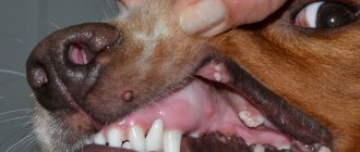
- Melanoma localized in the oral cavity. Often malignant. Visually, it appears as ulcerated or necrotic nodules or flat areas. Often, as it develops, it covers the entire area of the oral mucosa, lips, and gums. Often pigmentless, but may have dark pigmentation. Often, if the gums are affected, the neoplasm grows into the bone tissue of the jaw. If melanoma is localized on the tongue, then metastases develop, covering the lungs or nearby lymph nodes.
Scientists have not fully studied the prerequisites for the appearance of a neoplasm. While in humans exposure to light is a determining factor, in domestic animals UV radiation is not considered a disease-causing factor.
Skin Cancer Symptoms
Symptoms of basal cell carcinoma are extremely variable and depend on the form of the disease. The tumor manifests itself with the following manifestations:
- Shiny pink nodules with visible blood vessels.
- Papules or nodules covered with ulcers.
- Plaques. Dense, scar-like.
- Psoriasis-like plaques.
Stages of basal cell skin tumor:
- Stage 1 - the tumor is no more than 2 cm in greatest dimension.
- Stage 2 - the size of the tumor exceeds 2 cm, but there is no germination (invasion) into the underlying tissue.
- Stage 3 - Skin cancer has grown into underlying structures, such as muscles or bones. Or a less invasive tumor, but with regional metastases.
- Stage 4 - the tumor gives distant metastases.
Prevention
In most cases, papillomatosis in dogs is mild, and pets recover without medical intervention. An animal that has once had papillomatosis acquires immunity. She does not develop new warts, but the virus remains in the body forever, and the dog can infect animals with reduced immunity. The likelihood of catching the virus while walking is very high, so it makes sense to vaccinate your dog against papillomatosis.
In addition, you need to take care of your pet’s immunity. You can increase the body's resistance to viruses if you follow all the rules for keeping a dog and create a balanced diet for it.
Benign skin tumors in dogs are a disease that in most cases does not require treatment. However, any skin problems are a reason to consult a veterinarian.
What does melanoma look like in dogs? This is a malignant tumor based on melanocytes (cells growing in the deep layers of the skin, which protect them from exposure to light and give it pigment). Localization of the tumor: mouth, limbs, eyes. Males are more predisposed to the disease than females. The most common breeds affected by melanoma are setters, retrievers, and cocker spaniels. Neoplasms of this type often penetrate deep into tissues and metastases develop.
Symptoms:
- a crusty or bleeding skin sore that doesn't respond to antibiotics or creams;
- ulcers that do not heal within several months;
- ulcers on areas of the skin covered with white or light hair;
- growths or tumors;
- white growth on the skin;
- tumor;
- tumors on areas of skin covered with white or light hair.
Ulcers or tumors can occur anywhere. Usually only one tumor or ulcer is present. The skin is most often affected on the nose, fingers, feet, scrotum or anus.
Types of warts
Warts in dogs vary in color, size and location. They can occur anywhere on the animal’s body, but most often they affect the limbs, back, head, mouth, lips, and ears. The rashes can be single or numerous, resembling clusters.
Warts can range in size from tiny “pimples” that are barely noticeable on the skin to bean-sized tumors. The color can be pink, brown, yellow, grayish-black.
Photo. Papillomas in a dog's eye
Photo. Warts on a dog's lip
New growths can be hard or soft to the touch. Hard warts have a smooth surface and a convex shape, while soft warts are flatter and rougher. The neoplasm can be embedded deep into the skin or rise on a thin “leg”.
Types of malignant tumors in dogs
Like people, four-legged pets can also have malignant neoplasms, and this phenomenon is quite common. Here are the main types of pathology:
Lymphoma . Occurs most often. Dogs are 5 times more likely to suffer from this pathology than people. Symptoms vary depending on the organ affected, but in all cases the peripheral lymph nodes are enlarged. However, there are also lymphosarcomas that affect the internal lymph nodes. Appetite also worsens and lethargy appears.
Hemangiosarcoma . This neoplasm arises from the cellular structures that line blood vessels. Symptoms usually do not appear until the end stage, when treatment is no longer helpful.
Osteogenic sarcoma . This is the most common type of bone tumor. As a rule, the limbs of dogs are affected, but it can also appear on the ribs and skull.
Melanoma . This is a neoplasm that includes pigmented cellular structures of the skin. It can appear anywhere on the dog's body.
Squamous cell carcinoma . It usually develops in the pet's mouth and skin. Such growths can be removed quite simply - surgically.
Breast cancer . The most common problem in bitches. They can have a local and generalized form (in this case, ulcers and erosions form).
Breast cancer
Cancer of the paraanal glands . Carcinoma spreads to tissue near these organs.
Transitional cell carcinoma . Most often it affects the organs of the urinary system.
Soft tissue sarcoma . Consists of connective tissues. They can appear in any part of the body.
Soft tissue sarcoma
What to do when pigment spots appear on a dog?
You need to show the dog to a veterinarian so that he can determine whether the pet has good or bad pigmentation. Any changes in the skin indicate pathologies that are asymptomatic or have just begun to progress.
The doctor will scrape the skin to check for bacteria or fungi. The dog will have to donate blood, urine and fluid from the joints, and undergo an ultrasound of the thyroid gland, kidneys and adrenal glands. Based on the results, the veterinarian will prescribe a course of antibiotics, immunosuppressive medications and ointments.
Important! The veterinarian carries out differential diagnosis to exclude serious diseases that have similar symptoms. These include skin cancer and lupus
Symptoms
The development of pigmentation can be sluggish or rapid. It all depends on the reason that provoked its appearance. There are no precursors of pigmentation, but genetic or acquired factors can be used to judge the likely appearance of spots. The nature of the spots themselves determines the cause of their occurrence.
The following symptoms indicate genetic pigmentation:
- The abdomen, limbs, torso, eyelids or oral mucosa are covered with dark spots.
- The groin and armpits are modified, but without characteristic signs of inflammation.
About acquired pigmentation they say:
- Spots of strange shape that grow in size and thereby change the entire color of the animal.
- Spots appearing on the nose.
- The color of the coat completely changes from light shades to a rusty brown color (most often due to an infectious disease).
- Red spots on the pads of the fingers, paws, ears. They clearly show signs of inflammation or irritation. They may become thicker or itchy, and the skin around them may become bald.
Important! If you find such spots, they can be extremely dangerous.
Treatment of melanoma in an animal
How is a pet treated? This depends a lot on the location on the dog’s body:
- There is no need for radical measures to eliminate it on the body or paws. But amputation is possible when melanoma appears on a pet’s finger.
- If melanoma is located on the mucous membrane or on the eye of a pet, it is most often malignant.
- If its location is the perineum and genitals, then the nature of such a neoplasm is usually benign. But the tumor has a tendency to metastasize.
Surgical intervention
The veterinarian examines the location of the tumor, clearly identifying its boundaries, and offers to perform surgery. When the spot is located in the oral cavity, the owner should understand that it will not be possible to excise the tumor without capturing part of the bone. After surgery, it is difficult for a dog to recover, but 85% of pets are able to fully chew food, play, and drink water. If the tumor is localized on one of the fingers, doctors recommend cutting it off. This is the only way to guarantee 100% results.
Chemotherapy
You can reduce the likelihood of melanoma reoccurring by taking a course of chemotherapy. US scientists have developed and are testing the drug Oncept, designed to combat this disease. It stimulates the body's defenses to fight cancer. Traditional chemotherapy drugs are used only in cases where the tumor cannot be removed by surgery, it does not respond to radiation, or metastases have begun to develop.
Irradiation
This method is used when surgery is powerless or the growth of the tumor in the dog is too large. It is often done after surgery. According to global practice, this type of cancer is successfully treated with radiation.
Vaccination
To reduce the recurrence of the disease, a vaccine has been developed abroad for use after operations for melanoma. It helps prolong the life of the four-legged patient and is used only after surgery to remove melanoma. But it is almost impossible to find a vaccine in our country, because it has not been certified in Russia.
Features of melanoma at an advanced stage
The tumor grows rapidly and metastasizes. There are two options for their spread - hematogenous and with lymph flow. Regional lymph nodes become infected through the lymphogenous route. In most cases, metastasis to the dermis is observed. Hematogenous metastasis can be present in any organ, but most often in the bones, spleen, liver and lungs.
If treatment is not carried out, the animal’s quality of life will deteriorate sharply. The pet will experience pain, bad breath, bleeding and pus. The process generalizes 3 months after diagnosis.
Comparative characteristics of the life expectancy of animals with melanoma under various treatment methods
| Treatments | Number of animals | Long-term result |
| Stabilization(%) | Relapse, % | Generalization, % |
| 2 animals are under observation | ||
| Cryodestruction | ||
| Most animals are under supervision | ||
| Surgery | ||
| 4 animals are under observation | ||
| Radiation therapy | ||
| Chemotherapy | ||
| Control. group (IT) |
Are warts dangerous for dogs?
The neoplasms vary greatly in appearance but are caused by a single “agent.” Warts can appear on the dog's body or on mucous membranes, most often in the mouth. Skin lesions begin in areas covered with delicate skin - on the paw (elbows), inner thigh, groin area, armpits.
The appearance of warts on the mucous membranes is considered a more dangerous manifestation of the disease. A wart located on a dog’s lip, palate or inside cheek can be torn off when chewing food, which can lead to a secondary complication – infection. Veterinarians note that in advanced stages, papillomas spread from the oral cavity and appear on the dog’s face, more precisely, on the outside of the mouth.
The ears and ear canals of animals are covered with thin skin, and a small layer of subcutaneous tissue is literally penetrated by nerve endings and capillaries. Warts on the ear most often cause itching and discomfort, prompting the animal to scratch. And again the danger of scratching and complications due to infection.
Let's briefly look at the types of papillomas. Externally, the new growths have some similarities with cauliflower, but can vary in:
Size – from 0.1 to 5 mm. It is possible that the size of warts may go beyond the boundaries of the “classical gradation”.
Colors - flesh, brown, gray, yellow and combinations of shades.
Structure – dense, hard, soft.
The method of “implementation” into the skin is ingrown, attached to a stalk.
Papillomas, which are attached to the dog’s body or mucous membrane with the help of a leathery stalk (see photo), literally dangle, which increases the risk of damage. Ingrown warts are dangerous due to possible transformation into a malignant tumor - squamous cell carcinoma. The risk of degeneration does not depend on the color, size and structure of the neoplasm.
What breeds have pigmentation?
Any dog can develop dark spots on its skin, especially those that have already reached an advanced age. There are several breeds that exhibit changes in skin color due to abnormal changes caused by hereditary predisposition.
Breeds that are characterized by the appearance of pigmentation:
- French bulldogs suffer from the age-related appearance of the so-called “spotting”, in which several small or one large spot appears on the belly.
- Pugs, miniature schnauzers and sharpeis are prone to developing benign localized spots (lentigines).
- Partial pigmentation in the area of the nose and lips is characteristic of the Akita Inu, Samoyed dog, and Siberian Husky.
- The skin of the Chow Chow and Akita Inu is affected by an acquired autoimmune disease, after which an inflammatory process with purulent discharge may appear.
- Dachshunds suffer from acanthosis nigricans, or elephant skin as it is popularly called. With such hyperpigmentation, rough, modified areas of skin appear. Most often they cover folds in the dog’s neck, groin or armpits.
- Lupus vulgaris occurs in Scottish Sheepdogs.
- Doberman Pinschers, Rottweilers, Siberian Huskies, Alaskan Malamutes and Labrador Retrievers are characterized by depigmentation of the coat, which changes its usual color to a snow-white color.
- Chihuahuas have a color change caused by hormonal changes in the body. In both males and females, spots appear during puberty. They most often occur in the groin area around the genitals.
- The Poodle, Old English Sheepdog, Australian Seal Terrier, Bedlington Terrier and Yorkshire Terrier all have coat colors that change. The cause of the change may be a previous illness or disturbances in the functioning of the sebaceous glands.
In most of the listed breeds, pigmentation occurs due to a mutation of the genotype, less often due to acquired diseases.
The color of an animal depends on a specific combination of genes that make up dozens of different alleles. The main shade of color, as well as the degree of pigmentation, depends on the predominant number of alleles that dominate the genotype.
Additional Information
! Alleles are different forms of a gene and how it interacts with other genes.
The essence of melanoma in dogs
Appearance of the tumor: the tumor looks like a dark spot, has a round or oval shape, the edges are uneven. The surface of melanoma is shiny, glossy, and can be destroyed. Most often, melanoma in dogs forms in the oral cavity or on mucous membranes (in almost half of the recorded cases). A lump also appears in the eyes, organs of the genitourinary system, and rectum. It often manifests itself in places protected from exposure to light, so we can say for sure that exposure to the sun is not a key factor in its occurrence. Typically begins in dogs over ten years of age.
This type of neoplasm is characterized by the rapid appearance of metastases and covers the skin and lymph nodes of the dog. Outwardly, this is manifested by many lumpy small dark rashes. Diagnosis of the disease is carried out comprehensively. The doctor examines the neoplasm, conducts morphological analysis and tissue histology.
Malignant neoplasms (cancer) of the skin in dogs
Content
- Types of malignant tumors in dogs
- Conclusion
All existing tumors are divided into three groups, according to their differentiation (degree of cell maturity).
Highly differentiated tumors consist of cells that closely resemble healthy ones. Moderately differentiated tumors are less similar to the cells of the tissues from which they arose. Poorly differentiated ones are almost unlike the cells of the tissues from which they originated. It is believed that highly differentiated tumors do not grow as aggressively as poorly differentiated ones. The main difference between a malignant neoplasm and a benign one is cell atypia. The less similar tumor cells are to the main cells characteristic of this tissue, the faster the growth of such a tumor and the higher the ability to destroy surrounding tissues, penetrate into blood vessels, and develop metastases.
Skin tumors in dogs can be both malignant and benign. This article will discuss some malignant skin tumors of dogs.
Types of malignant tumors in dogs
Anaplastic sarcoma is a poorly differentiated soft tissue tumor that develops from the mesenchymal connective tissue of the body.
Carcinoma is a relatively common malignant skin tumor formed from epithelial tissue cells. Various predisposing factors have been identified, such as duration of UV exposure, pollutants and pre-existing chronic dermatitis.
Basal cell carcinoma (basal cell carcinoma) is a tumor with limited and slow growth. Develops from cells of the basal layer of the epidermis. Most often, dogs older than 5 years are affected. Predisposing factors: burns, radiation, trauma with scarring.
Squamous cell carcinoma of the skin is a malignant epithelial tumor of the skin with squamous cell differentiation. In terms of distribution, this tumor ranks first among all skin tumors. One of the main reasons for its development is frequent exposure to the sun. This type of cancer occurs three times more often in males than in females. Symptoms may include: sores, often crusty or bleeding; growths. Most often, the skin is affected on the fingers, toes, nose, and scrotum.
Finger squamous cell carcinoma is invasive, causes bone lysis, and often metastasizes.
Lymphosarcoma of the skin is a malignant lymphoproliferative disease, also often called malignant lymphoma.
Liposarcoma is a fairly rare malignant tumor. Develops from subcutaneous embryonic fat cells. It has infiltrative growth, but rarely metastasizes.
Mastocytoma is a benign or malignant skin tumor consisting of transformed mast cells of the skin. Some breeds of dogs (shar-pei, boxers, bulldogs) are predisposed to the malignant form of this disease. Mastocytoma can look like a simple wart or a soft subcutaneous lipoma, or it can have the appearance of a moist dermatitis, which the dog owner may not be embarrassed by. Most often, the fur in this area falls out and itching appears. In the malignant form, symptoms such as loss of appetite, vomiting, diarrhea and anemia develop. Treatment depends on the appearance, location and histological findings. Sometimes only surgical excision is recommended, in other cases chemotherapy is also required.
Melanoma accounts for 4-6% of all skin tumors in dogs. They present as hard, pigmented spots and are most common in dogs with dark skin. The malignant form is characteristic of tumors located on the fingers and in the area of the skin-mucosal junctions. They can metastasize to local lymph nodes, lungs and more distant places. Histology results will tell you about the prognosis and further treatment. The worst prognosis is for tumors in the mouth and extremities. Treatment uses surgery, radiation therapy and chemotherapy.
Conclusion
In conclusion, it would be worth adding that the appearance of any skin tumors in your dog should be a reason to contact a veterinary center.
For an accurate diagnosis, it is necessary to conduct a number of studies, such as: histological examination of the tumor, X-ray examination and ultrasound diagnostics to identify metastases, blood tests to assess the quality of the internal organs. The results will be interpreted by a surgeon and oncologist who will prescribe a personalized treatment plan for your dog. The article was prepared by Lagutina A.S.,
veterinary therapist at MEDVET © 2021 SEC MEDVET
Brown spots
The dog may have a completely black belly or a partially covered torso, neck and limbs with brown spots. Foci of such hyperpigmentation are characteristic of both adult and young dogs and are caused by a nevus. During the first months after their appearance they may increase in size. But then they become static and don’t change anymore.
Red spots
Redness of the skin is caused by bacterial, viral, fungal or allergic lesions. Spots from light pink to red can be a symptom of a serious illness or a consequence of a previous illness. A red spot on a dog’s paw most likely indicates a fungal infection, in particular lichen, but red dots on the stomach indicate an allergic reaction to insect bites, food, care products or medications.
In any case, no matter what the color of the spots, you should take your pet to the vet. The doctor will make an accurate diagnosis and prescribe treatment.
Important! Don't neglect your pet's health. If you find a black spot on your dog’s belly or a pinkish spot with a bald patch on its paw, immediately make an appointment at the veterinary clinic
Never treat a dog yourself.
Symptoms of the disease
For pet owners, symptoms of skin cancer in a dog will indicate the need to take timely action. They appear in:
- white growths on the skin;
- ulcers that do not heal for a long period (months);
- tumors or growths.
The animal often loses its appetite. This symptom needs to be treated extremely carefully, stimulating it to eat on its own. Frequent split feeding can help improve food intake. If a dog has a persistent form of anorexia, then it needs to be fed using a tube or catheter. For these purposes, you must wear gloves. In this case, it will be possible to ensure a sufficient supply of nutrients.
Veterinarians call the ideal composition of the diet the presence of a significant content of fats, calories and proteins. A balanced amount of microelements must be present in the diet. The signal to go to the veterinarian should be a bleeding ulcer on the skin, when it cannot be cured with creams or antibiotics. This type of cancer is difficult to detect in a timely manner, as hair gets in the way. Therefore, when bathing, it is necessary to carefully examine your pet.
This neoplasm is a black or pink mole. Particular attention should be paid not only to the dog’s protective tissue, but also to the mucous membranes (mouth, internal organs). At the first signs, you need to contact a veterinary clinic. If this is not done immediately, it will be quite difficult to cure.
How is carcinoma diagnosed in dogs?
The pet owner must understand that without appropriate practice it is not easy to establish a diagnosis. Also, self-medication may not bring the desired results. In this case, the disease will progress, which will further worsen the dog’s condition.
It is more advisable to contact a veterinary clinic in time for diagnosis. Experienced professionals will identify the disease after a series of procedures. They will palpate (feel) the lymph nodes. When they are excessively increased in size, it means that an inflammatory process occurs. This is an indirect confirmation of carcinoma.
At the clinic, the dogs will have a biochemical blood test taken. An x-ray of the chest or abdominal cavity is required. When subungual carcinoma occurs, veterinary specialists take x-rays of the paws. Such measures allow a detailed examination of the condition of the internal organs of pets. A biopsy of the tumor is performed. It involves taking part of the tumor cells, followed by studying the biological material. Based on its results, it is possible to determine the type of neoplasm and stage of development. Based on the data obtained, the veterinarian can prescribe treatment.
Factors influencing life expectancy
In general, if a tumor is present, the prognosis is poor. But a number of methods affect survival:
- presence of metastasis;
- melanoma size;
- localization;
- germination depth;
- growth form.
If bleeding or ulceration is observed, these are unfavorable symptoms
It is important to start treatment as early as possible, so there is a greater chance of recovery. There are no preventive measures to prevent tumor development in dogs.
If melanoma is localized in the dermis, its growth is slow. In this case, a relatively benign course of the process is observed, and the neoplasm remains mobile. Metastases appear late. If the tumor is located in the mucosa, it is immobile, the course is malignant, metastases occur late, and rapid germination occurs into the surrounding tissues. Often in this case, the cervical and lymph nodes are affected. Melanoma also often bleeds because it is injured by food and teeth.
Treatment
The course of treatment depends on the size and number of tumors. In some cases, when a doctor diagnoses ulcers before they become cancerous, they can be treated with topical medications.
If a dog has only one small tumor that has not spread to other organs, it can be removed using cryosurgery (i.e. freezing the tissue) or photodynamic therapy. The tumor can also be removed surgically.
If your dog has a large tumor, surgery will be needed . During surgery, your doctor will remove the carcinoma and large amounts of surrounding tissue to ensure all cancer cells are removed. Sometimes so much tissue needs to be removed that it requires skin grafting from other areas of the body.
In some cases, more serious surgery is necessary. For example, if the tumor is located on a finger, it will need to be amputated. If the tumor is on the nose, you will need to remove part of the nose (the same applies to the ear). The surgery may change the dog's appearance, but animals usually recover well after it.
If the tumor cannot be completely removed, your veterinarian will recommend chemotherapy or radiation therapy after surgery. In cases where surgery is not possible, treatment consists only of chemotherapy and radiation therapy. This will slow down the growth of the tumor and make your pet feel more comfortable.
How to rid your dog of warts
Typically, warts do not require treatment, but if the growth bothers your pet and does not go away on its own, it makes sense to remove it. Your veterinarian may suggest surgery or medication.
Drug treatments
The most popular treatment method is injections in combination with drugs to strengthen the immune system. Dogs are treated with Novocaine, Gamavit, Maksidin, Salmozan. The injection is made at the base of the wart. Another option is intravenous administration of novocaine. The procedure will need to be repeated three times.
If papillomas do not go away within five months or more, they can be treated with retinoids. They contain vitamin A and effectively eliminate skin tumors. Medicines containing retinoids include podophyllin, teniposide, vincristine, doxorubicin and others.
Anti-verrucine paste is also used against skin growths. It should be applied to the papillomas and rubbed at least two to three times a day until the tumor disappears. To prevent the animal from licking the paste, the treated area is covered with a bandage.
Vaccination
Vaccination protects dogs from human papillomavirus and also reduces the risk of developing squamous cell carcinoma in infected animals. For injection, you can use an extract from a dog's wart.
Surgical removal
Surgery is usually used for single tumors. The disadvantage of surgical intervention is that there is a possibility that viral particles will be introduced into the wound and papillomas will begin to grow with a vengeance. Therefore, after removing warts, you need to monitor for possible complications. If after 3 months everything is fine, then the operation was successful.
Modern veterinarians use more than just a scalpel. A wart can be removed using radiosurgery, electrocoagulation, diathermocoagulation and other methods.
Cryotherapy is the “freezing” of a tumor, which stops the metabolism in the affected tissues. Exposure of the wart to liquid nitrogen or dry ice is used for keratinized growths on the skin. Cryotherapy is effective for tumors in the eye area. The disadvantage of the procedure is its pain. Most likely, several procedures will be required, spaced three weeks apart.
A wart can also be removed using a laser. Typically this method is used for malignant tumors.
Folk remedies
Warts can be eliminated at home using traditional methods. The most radical type of intervention is to remove the tumor using scissors or thread. This procedure can lead to complications: bleeding and infection of the wound.
If the papilloma has appeared recently and has not yet grown, you can stop its development with the help of dandelion or celandine juice. After the surface of the new growth darkens, you can cut it off. Warts can also be smeared with iodine. In most cases, they disappear within a week. Another option is to lubricate the new growths with garlic juice. Old warts can be cauterized using a lapis pencil or acetic acid.
Veterinary clinic of Dr. Shubin
Description and reasons.
Squamous cell skin cancer of dogs and cats is a malignant skin neoplasm developing from keratinocytes, it is a fairly common skin neoplasm, in cats it accounts for about 15% of all skin neoplasms, in dogs only 5%. The development of squamous cell carcinoma
may be associated with chronic exposure to sunlight, lack of skin pigment and fur at the site of tumor development. In this case, the development of squamous cell carcinoma is preceded by solar keratosis, which is observed more often in geographic areas with long daylight hours and a significant number of sunny days per year. An increased incidence of squamous cell skin cancer has been noted in dogs against the background of various skin diseases, such as scars after burns, multiple follicular cysts, chronic infectious or inflammatory process of the external ear, as well as against the background of discoid lupus erythematosus.
Recently, a certain role in the development of squamous cell skin cancer has been assigned to papillomavirus, due to the fact that the antigen of this virus has been identified in up to 50% of cases of squamous cell carcinoma of dogs. Malignant transformation of papillomavirus-positive epidermal plaques to squamous cell carcinoma has been described in Pomeranians. In rare cases, canine oral and conjunctival papillomas can progress to squamous cell carcinoma.
An additional likely causative factor in the development of feline squamous cell carcinoma is immunodeficiency virus infection; in a study of a significant number of FIV-positive cats in California, 24% were identified as having squamous cell carcinoma. No definitive conclusions have been made about the etiological factor, because both problems can occur independently of each other in outdoor cats. Also, a number of authors find a relationship in the development of squamous cell skin cancer with certain gene mutations.
Clinical signs
The course of squamous cell skin cancer in both cats and dogs is characterized by local invasiveness and a low level of metastases. Enlargement of regional lymph nodes is often associated with their reaction to inflammation and ulceration rather than the formation of metastases.
Canine squamous cell skin cancer
Squamous cell skin cancer of dogs is more often observed in animals aged 6-10 years (peak incidence), the earliest period of tumor development was 4 months; in a crossbred puppy, squamous cell cancer developed in the navel area; surgical excision led to a complete recovery of the animal. No gender predisposition to the disease has been identified in dogs. Breed predisposition to the development of squamous cell skin cancer has been noted in breeds such as Kesshund, Standard Schnauzer, Basset Hound and Collie. Any breed of dog with sparse hair and weakly pigmented skin (eg Dalmatian, Bull Terrier) with a lifestyle prone to sunbathing is predisposed to the development of a tumor against the background of solar keratosis.
About 75% of dogs with subungual squamous cell carcinoma are large breeds, and 70% of them have dark coat color. Breeds predisposed to developing this form of cancer include the Schnauzer (Giant, Standard and Miniature), Gordon Setter, Poodle (Standard and Miniature), Scotch Terrier, Labrador Retriever, Rottweiler and Dachshund. Multiple lesions of squamous cell carcinoma of the digits have been described in three consanguineous giant schnauzers, as well as in large dark-colored dogs (Rottweiler, Standard Poodle, Black Labrador Retriever, Giant Schnauzer, and German Cattle Dog).
Squamous cell carcinoma of the nasal planum of dogs in rare cases can develop as a complication of depigmentation against the background of discoid lupus erythematosus. Other diseases accompanied by nasal depigmentation (eg pemphigus erythematosus, vitiligo) can also contribute to the development of this form of cancer, increasing the sensitivity of skin cells to sun exposure.
Lesions in dogs most often develop on the trunk, limbs, scrotum, lips and anus. It should be remembered that squamous cell carcinoma is the most common neoplasm of the fingers of dogs, while damage to the nasal planum is quite rare.
There are two types of squamous cell skin cancer in dogs: proliferative and ulcerative. The proliferative variant looks like the formation of papillary masses, which can have the appearance of cauliflower of various sizes. The surface of these formations is often ulcerated and characterized by increased bleeding; sometimes the formation of a cutaneous horn can be observed. With the ulcerative version of squamous cell skin cancer of dogs, small ulcers with crusts are formed at the beginning, which subsequently deepen to a crater-like shape. The lesions are usually solitary (single), multiple formations can be observed on the body of dogs who love sunbathing or in the nail bed of large dogs with a black coat. When the fingers are affected (subungual lesion), a single finger is most often affected, which appears swollen and painful with damage to the claw (loss or change in shape) and purulent inflammation of the surrounding space (paronychia). In rare cases, dogs develop multiple formations on the fingers stretched over time .
Feline squamous cell skin cancer
The peak incidence of squamous cell skin cancer in cats is 9-14 years of age, without a clear breed or gender predisposition. Cats with a white coat, regardless of coat length, are 13 times more likely to develop squamous cell skin cancer than cats of other colors due to increased sensitivity to sunlight. Siamese cats, due to their breed coloration, have a reduced incidence of squamous cell carcinoma.
About 80% of feline squamous cell skin cancer lesions are found on the head, and usually involve the nasal pelvis, ears, and/or eyelids. The lips are involved in the process much less frequently. The lesions are usually chronic and progress from crusting erythema to superficial and then deep ulcers. Squamous cell carcinoma of the skin can involve multiple sides of the head, with one description of affected cats showing multiple lesions.
Primary squamous cell carcinoma of the digits in cats is rare; most reported cases of feline digit carcinoma have been metastases from primary pulmonary carcinoma.
Diagnostics
The clinical features of squamous cell carcinoma of the skin are similar to various infectious, inflammatory, neoplastic and granulomatous skin lesions, so this disease has an extensive list of differential diagnoses. Most often, lesions of the claw bed with squamous cell carcinoma are initially misdiagnosed as infectious paronychia. Cytological examination of sites suspicious for squamous cell carcinoma is useful in making a presumptive diagnosis; the final diagnosis is made after pathomorphological examination. Histologically, squamous cell carcinoma consists of irregular masses or filaments of keratinocytes that proliferate downwards and lead to skin invasion. Common findings include keratin formation, keratin pearls, intercellular bridges, mitosis, and atypia.
Treatment
The course of squamous cell skin cancer is characterized by local invasiveness and low potential for metastasis. In cats, metastasis is often limited to regional lymph nodes; pulmonary metastases are quite rare. In dogs, lymph nodes are more often affected; metastasis in the lungs is extremely rare. Because most cases of squamous cell carcinoma of the skin of cats and dogs do not metastasize, effective local treatment of the primary tumor can significantly prolong life in affected animals. A small lesion has a greater chance of healing success. Treatment methods such as surgical excision, cryotherapy, radiation, photodynamic therapy and injection of drugs directly into the lesion are included as methods of local influence on the growth of the primary tumor.
Surgical excision of the pinna and nasal planum is quite effective in cats, and most owners consider the outcome of this type of treatment to be “cosmetically acceptable.” For feline squamous cell skin cancer of the eyelids, aggressive excision followed by reconstruction can lead to long-term remission.
In dogs, nasal ablation may be effective in treating squamous cell carcinoma of the nasal planum, but the cosmetic effect is not always good. If the tumor affects the phalanges of the fingers, their amputation can lead to long-term remission of the disease.
When using cryotherapy for squamous cell skin cancer of cats, about 83% of animals receive long-term remission after one course of treatment.
Radiation has proven effective for superficial small lesions. Photodynamic therapy has been widely used and studied for cats with squamous cell skin cancer, as with other treatment methods, its effectiveness is quite high for small lesions (less than 5 cm).
Systemic chemotherapy has not shown any benefit in the treatment of squamous cell carcinoma of the skin of dogs and cats, however, injection of chemotherapy directly into the lesion has had some success in cats with lesions located on the nasal planum and in dogs with other sites.
Prognosis for squamous cell skin cancer in dogs and cats often depends on the size of the tumor and the possibility of complete surgical excision.
Prevention
Prevention of sun-induced squamous cell skin cancer involves avoiding exposure to sunlight. Normal window glass does not block ultraviolet rays, so cats that sunbathe on windowsills may be predisposed to developing squamous cell carcinoma. Topical sun protection creams are available for dogs and cats, but their effectiveness in preventing squamous cell skin cancer has not been determined. For dogs that love to sunbathe, specialized clothing can provide effective protection.
Photo 1 . Squamous cell skin cancer in an elderly light-colored cat at a veterinary clinic appointment.
Photo 2 . Squamous cell skin cancer in a cat brought to a veterinary clinic from a neighboring city. Initially, the lesion on the left ear was amputated, but after some time there was a relapse of the disease.
Valery Shubin, veterinarian, Balakovo
Check your moles! World Melanoma Day! (February 2020).
Most people have at least one mole somewhere on their body, and most people have several different shapes and sizes! As we all know, it is important to monitor moles and be alert to any new moles that develop or changes in the shape, size, texture and other factors of moles, as they can sometimes be cancerous. However, humans are not the only animals that often have moths - dogs often do too, although you won't even be able to tell if you have your dog as they will be hidden by the dog's fur
As with humans, the vast majority of moles on dogs are completely benign and simply a feature of your dog's skin, but moles on dogs can potentially become cancerous, just as human moles do
However, humans are not the only animals that often have moths - dogs often do too, although you won't even be able to tell if you have your dog as they will be hidden by the dog's fur. As with humans, the vast majority of moles on dogs are completely benign and simply a feature of your dog's skin, but moles on dogs can potentially become cancerous, just as human moles do.
Getting to know your dog's skin and coat can help you identify any moles your dog has and so be able to determine if any new ones are developing or if an existing mole is changing, and it's definitely a good idea for all dog owners to identify any changes early.
In this article, we'll take a closer look at melanoma moles in dogs, including how to identify the problem, which dogs are at greatest risk for melanoma, and what can be done to treat them. Read on to find out more.
Reasons for the development of the disease
As a rule, it is quite difficult to determine the specific cause of carcinoma. The etiology of the disease is associated with random, uncontrolled, rapid division of skin cells (epithelium), which is why cancerous tumors form in different places on the surface of the body.
Carcinoma is a skin cancer in dogs in which rapid division of epithelial cells occurs.
Factors that can provoke these processes are the following:
- ionizing radiation (including due to prolonged exposure to open sunlight);
- severe hormonal imbalances in the body (associated, among other things, with taking hormonal medications);
- genetic predisposition;
- regular exposure to carcinogens (harmful substances coming from air, water or food).
Based on the practice of veterinarians, it is known that dogs that spend long periods of time in the sun are actually at greatest risk of disease.
At the same time, the risk group also includes the following animals:
- having light skin and hair;
- living at an altitude of several thousand meters above sea level (in the mountains);
- representatives of certain breeds: poodles, German boxers, Pekingese, Dalmatians, bull terriers.