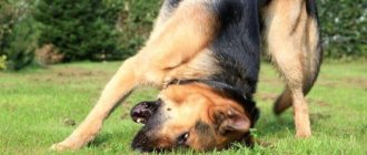
Ringworm (dermatophytosis) is a disease of dogs caused by fungal microorganisms. It is characterized by the appearance of hairless areas on the skin with a clearly defined contour.
With ringworm, a pet's coat looks dull, the hairs become dry, break easily and fall out. The spread of dermatophytosis is carried out not only by contagious, but also by household objects. The disease is classified as an anthropozoonosis, so human infection is possible.
Ringworm is completely cured in an average of 1 month, but in advanced cases, more time is required for treatment.
Routes of infection
The causative agents of ringworm in dogs are fungi from the Dermatophytes group, such as Trichophyton, Microsporam, Achoreon. Each species causes a separate disease, called trichophytosis, microsporia and favus. Animals affected by Microsporum canis are common. Very rarely, Trichophyton mentagrophytes and Microsporum gypseum parasitize dogs.
Ways of infection with dermatophytosis:
- Contact with a sick animal. The carriers of ringworm are stray dogs, cats, rodents and ticks. Dermatophytosis develops when a fungal spore gets on the damaged surface of the skin.
- Household method. Fungal spores are carried on grooming items (bowls, slicker brushes, brushes, beds) or equipment (leashes, collars, harnesses).
- Environment. The pathogen forms spores and is therefore resistant to changes in temperature and humidity, as well as to the effects of disinfectants. It is possible that fungal spores may enter the dog’s body from the ground or plants.
Ringworm often affects puppies, young and old dogs, as well as weak animals that have had illness or surgery.
There is an increased risk of contracting lichen in working dogs of hunting breeds due to their contact with wild animals.
Therapy
Treatment should be carried out systemically. In the article we will present several drugs that have proven themselves to be the best for the destruction of pathogenic fungi in dogs. Duration of treatment is at least six weeks. So here are the medications:
- Itraconazole This remedy has proven itself to be excellent among veterinarians all over the world.
- Antibiotics from the griseofulvin group. Old but extremely effective drugs. Their disadvantages include increased toxicity, so only your veterinarian should prescribe the medication.
- Terbinafine and fluconazole may be used. Quite effective drugs with an acceptable level of toxicity for the animal.
Symptoms and clinical picture
It takes a week, sometimes a month, from infection to the appearance of visible signs of the disease. There are cases when the minimum incubation period of ringworm lasted 2 days, and the maximum lasted 3 months.
The development of dermatophytosis in dogs occurs in two stages:
- Stage I. From a sick animal, the pathogen enters the epidermis of another dog. Next, the microorganism produces toxins and enzymes that loosen the stratum corneum of the epidermis and penetrates the hair follicle. The process is accompanied by itching and redness of the skin. Scratching the site of entry of the pathogen leads to the spread of the fungus throughout the body.
- Stage II . The pathogen destroys the hair root, resulting in malnutrition and hair loss. The skin looks dry and crusts form on its surface. Foci of dermatophytosis lesions become visible.
Symptoms of ringworm in dogs:
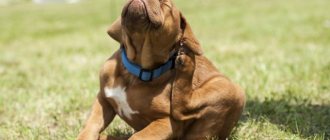
- The coat is dull;
- the pet itches often;
- hairs become brittle and fall out;
- redness, bumps on the surface of the skin;
- the skin is dry and flaky;
- areas of balding skin are round in shape with clear boundaries.
Signs of dermatophytosis
The incubation period for lichen ranges from 3-4 days to several weeks. The duration of the disease depends on the age of the dog and the state of its immune system.
Predisposed to lichen disease:
- Young dogs, especially under 1 year;
- Newborns;
- Decreased immunity;
- Parasites;
- Increased humidity and ambient temperature.
Ringworm is more common among some dog breeds. These include:
- Jack Russell Terrier;
- Yorkshire Terrier (possible carrier without signs of disease. Among other dog breeds, asymptomatic carrier is rare);
- Pekingese.
Not all animals will show signs of disease upon contact with the pathogen. If a dog has good immunity, then it is highly likely that it will not become infected.
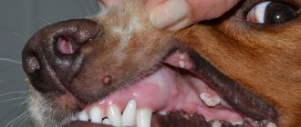
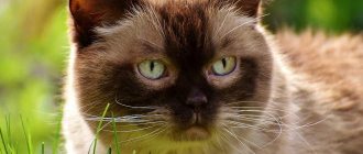
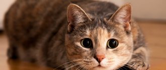
The most characteristic sign of the development of lichen on an animal’s body is the appearance of round bald spots (alopecia) of various sizes. In these places, the fur becomes very fragile and the skin peels off greatly. Sometimes weeping crusts appear, the skin underneath becomes pink. Their appearance on the body is often accompanied by itching. As dermatophytosis develops, the number of such bald spots will increase. The photo below shows a typical sign of lichen on the face:
A specific lesion in lichen is the kerion. This is the name given to a round, well-defined lesion. The skin in such places is thickened, pink, and may become wet. Most often, the kerion can be seen on the face or on the limbs.
What lichen may look like:
- Bald spots (alopecia). The most common symptom. Can be on any part of the body - muzzle, body, paws, tail;
- Wet crusts. The skin and coat may become wet when secondary microflora attaches;
- Dandruff, fragile hair;
- Complete loss of fur;
- Itching (rare).
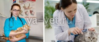
If you notice a small area of hairless hair on your dog’s body, it is best to take him to the clinic immediately. The doctor will conduct the necessary diagnostics, and based on the results, he will be able to say what caused the appearance of alopecia, and then prescribe the correct treatment. You should not treat such bald spots with any means in advance, as this may complicate further diagnosis. Self-treatment (for example, iodine) can only bring temporary improvement.
Establishing diagnosis
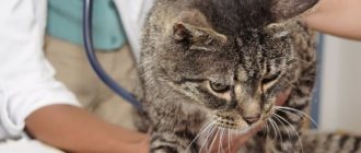
A veterinarian can make a preliminary diagnosis after examining the dog using a Wood's lamp. When you point the lamp at the affected area of the skin, the fungi glow green.
To make a final diagnosis, a deep skin scraping is taken from the animal and laboratory tests are performed. To determine the pathogen, biological material is inoculated on a nutrient medium.
For a complete clinical picture, blood and urine tests are taken.
Environment
It is very important to treat the environment correctly and thoroughly!
After all, spores spread with the animal’s fur throughout the apartment. For clothes, blankets, beds, it is recommended to wash them with boiling water or at 90 degrees, then carefully iron them or use a steam generator.
To treat premises, use Liverazol at a dilution of 1:100 or a 3-4% solution of Chlorhexidine. It is recommended to vacuum and wash the room daily. It is recommended to trim or shave the affected areas on your pet's skin. Animals with confirmed infection must be isolated from other pets. If there are a lot of animals in the room, it is recommended to use medicated shampoos or Liverazol for prevention.
Treatment of ringworm
The animal is isolated in a separate room. Bowls are washed with hot water. Bedding and equipment (collars, leashes) are washed or replaced with new ones. The premises are wet cleaned daily.
How to treat dermatophytosis in a dog ? The first step is to contact a veterinary clinic. There is no need to smear anything on hairless areas of the skin yourself, as the clinical picture will be distorted. It will be difficult for a doctor to establish an accurate diagnosis and therefore prescribe the correct treatment.
Drugs are prescribed depending on the type of dermatophytosis.
If a diagnosis of microsporia is made, which is confirmed by laboratory tests, the two-time monovalent Miccanis vaccine is used.
When the etiology of the disease is unknown, associated vaccines are used to treat ringworm in dogs: Vakderm, Vakderm-F, Microderm and Polivak-TM. Administration is carried out twice, in advanced cases three times, the interval between injections is 10-14 days.
When the fungus develops extensively on the animal’s body, antibiotics containing griseofulvin and amphotericin are used. The drugs are administered over a period of 3-5 weeks, depending on the stage of the disease.
Preparations for external treatment of damaged skin with ringworm:
- Salicylic acid solution (10%);
- lime sulfur solution (2%);
- tincture of iodine (5%);
- enilconazole solution (2%);
- salicylic ointment (5-10%).
Tincture of iodine and salicylic ointment are used in combination. First, apply iodine tincture, and after 1 hour salicylic ointment.
In addition to general medications, there are also special ointments against dermatophytosis, which stop the spread of the fungus, reduce inflammation and itching. These include: Miconazole (2%), Yam ointment, Zoomikol, Tiabendazole, Fungin, Ekalin.
Simultaneously with these drugs, the coat of the sick animal is treated once every 5 days with a 3-5% formaldehyde solution. Spray the drug all over the animal’s body, and then comb it first against the grain, and then along the grain.
If there is a large area of skin affected by dermatophytosis, the use of external fungicidal preparations is undesirable, since the substances included in their composition, in large dosages, have a toxic effect on the dog.
Throughout the treatment of ringworm, vitamin preparations (Complivit, Aevit, Gamavit) are used, which accelerate the restoration of hair follicles and hair growth.
Doctor, he has shingles!
Almost a third of the dermatologist's patients come with a suspicion of this diagnosis. Let's figure out whether this is really the case or whether there is something else hidden under these lesions.
This article will discuss inflammatory spontaneous focal alopecia (bald area of skin), since it is the main clinical sign of dermatophytosis (in common parlance - lichen).
Complete or partial hair loss is called alopecia. It can be primary, when the disease directly leads to inhibition of the hair growth cycle (for example, endocrinopathy) or secondary, as a result of injury and inflammation. In terms of prevalence, it can be diffuse, with a blurred exact boundary, or focal, local in a small area. According to the nature of hair loss, it can be spontaneous, the hair falls out entirely, or self-induced, the hair has broken ends as a result of injury.
The main differential diagnoses for spontaneous focal alopecia are dermatophytosis, demodicosis and bacterial folliculitis.
Let's look at these diseases in more detail. Let's start with dermatophytosis (lichen).
Dermatophytosis
- a fungal disease that affects the surface layers of the skin, coat and claws of cats and dogs. Ringworm in cats and ringworm in dogs are similar in their causes and progression. There are several types of dermatophytosis involved in the development of the disease:
- Microsporum canis - exists on the surface of dogs and cats, spores persist in the environment for up to 1.5 years;
- Microsporum gypseum - lives in the soil;
- Trichophyton mentagrophytes and Microsporum persicolor - carriers are rodents and wild animals.
Clinical signs
The lesions may have a “classic” appearance - rounded areas of baldness, redness with scales along the periphery of the area, often located asymmetrically on the head or paws. As a rule, the initial stage is characterized by focal hair loss and is often not accompanied by itching. In more advanced cases, the lesions can be extensive, with scabs and pustules. In case of infection with Trichophyton mentagrophytes, furunculosis is possible. Dermatophytosis can also be in the form of miliary dermatitis of cats - a papulocrustic rash (tubercles and crusts) over a large area of the body, but without hair loss.
Dogs very often, especially in the summer, can develop pododermatitis - inflammation of the paw pads. It can be itchy, the lesions are deep, fistulous. Dogs are characterized by the formation of a kerion - a clearly limited nodular formation with a scab (crusts) or erosion on the surface. This reaction is the result of an immune response to the penetration of dermatophytosis. Cats have their own kerion, it is called dermatophytic mycetoma - deep nodular lesions with or without ulceration, formed when dermatophytosis penetrates the dermis and subcutaneous tissue. Persian cats and longhaired cats are predisposed to mycetoma.
Diagnostic tests
- Luminescent diagnostics (50% of strains will glow; if there is no glow, this does not exclude dermatophytosis)
- Trichoscopy/scraping (examination of hair, for the presence of dermatophytosis spores)
- Cytology (examination under a microscope of stained smears from a suspected kerion or mycetoma. The method is used in combination with trichoscopy and/or scraping)
- Sowing on nutrient media (the most reliable research method. Determines the type of dermatophytosis, carriage in an asymptomatic carrier, allows you to find out whether to finish or continue treatment, because with clinical resolution, the animal is not always healthy)
- Biopsy (there are cases when this type of study is the only one to make a final diagnosis).
Treatment
It varies depending on the age of the animal, the severity of the disease, the presence of concomitant chronic pathologies, pregnancy during the period of the disease and a number of other factors.
It is worth noting that in healthy young animals the disease can go away WITHOUT treatment. But more often complex therapy is necessary:
- systemic drugs - itraconazole, ketoconazole, terbinafine, fluconazole;
- external treatments - enilconazole, shampoos with ketoconazole, miconazole, terbinafine for topical use, lime sulfur solution;
- environmental treatment - solutions with a high concentration of bleach, high temperature (washing clothes at 60°, treating furniture with a steam iron) or enilconazole in the form of sprays and checkers.
If several animals are kept in a house or nursery, an action plan is developed to identify infected and uninfected animals, separate them into isolated groups, therapy in each group, treatment of the premises and methods of monitoring treatment.
Ringworm vaccines are not effective in either preventing or treating dermatophytosis!
And since a number of animals recover even without treatment several months after the onset of the disease, this gives reason to evaluate the result of the vaccine as effective. The treatment period usually takes several months. To monitor treatment, examination and monthly cultures are necessary.
The pet is considered clinically healthy after two negative culture results with an interval of 1 month.
Forecasts depend on the general condition of the animal, the number of animals in the house and the type of dermatophytosis.
Author: Galiullina O.V.
Prevention of dermatophytosis
To prevent the disease, the same vaccines against dermatophytosis are used as for treatment. The injection is given twice. Vaccination provides the dog with immune protection throughout the year.
Try to avoid contact between your domestic dog and stray animals. At the first sign of ringworm, contact your veterinarian. In the early stages, dermatophytosis can be treated easily and quickly.
Remember!
- A person can become infected with ringworm from a sick dog.
- An accurate diagnosis can be established only through laboratory tests.
- The disease is treatable. The more advanced the ringworm, the longer it will take to recover.
- The main method of prevention is hygiene and timely vaccination of your pet.
Please rate the article. We tried our best:)
Reasons for appearance
The sources of fungal infection are sick mammals, with direct or indirect contact with which pathogens enter the skin and penetrate through microscopic lesions. Not every dog can get sick, but only those at risk.
Provoking factors are considered:
- poor nutrition without a sufficient amount of natural meat and other necessary components in the diet;
- weakened immunity, typical for puppies, elderly or sick animals;
- metabolic, hormonal or vitamin imbalance;
- antibiotic treatment;
- helminthic infestations, especially of a chronic nature;
- lack of proper conditions of detention according to sanitary, hygienic and temperature requirements.
In addition, the cause of dermatomycosis can be constant trauma to the skin in pets sitting on a leash or leading a sedentary lifestyle in small enclosures.
Important! The incubation period of the disease ranges from 1 week to 1 month, but can last up to 3 months. Throughout this period, the dog is a carrier of the infection and poses a danger to humans.
Ringworm in dogs (dermatophytosis)
Your dog has shingles. These words can stupefy almost any person. This disease is shrouded in a lot of myths:
- lichen cannot be treated, and sick animals must be euthanized
- you can only get lichen from animals
- The diagnosis of lichen can be established by shining a special lamp
- lichen can be cured by sprinkling gunpowder/sulfur on the wound/applying Yam ointment/injecting a vaccine/performing a shamanic dance with a tambourine
Which of this is fiction, and which has some basis? How to understand that it is lichen? What to do if your dog has shingles? How to get out of the situation with a minimum of losses? How to prevent its occurrence? Let's find out!
How to diagnose lichen in a dog?
The diagnosis of lichen in a dog (dermatophytosis) cannot be made only on the basis of examination! For diagnosis, a combination of 2-3 research methods will be required.
There are several methods for diagnosing lichen:
- That same “magic” lamp (Wood’s lamp) – research using ultraviolet light of a certain spectrum. Simple, cheap, but not very cheerful. Since more than 50% (!) of fungal colonies (dermatophytes) do not produce a characteristic green glow. Therefore, if a doctor shines a lamp on a dog, but nothing “shines,” this does not exclude the possibility that the puppy or adult dog is actually sick. And some artifacts (for example, talc from medical gloves) can give a “false” glow. The method is used solely as an auxiliary method. It is from the location of the detected glow that the doctor will take the next analysis.
- Trichoscopy (skin scraping) - examination of hair from the lesion site under a microscope. It is a more accurate diagnostic method. Detection of spores in a scraping will in most cases be a sufficient basis for making a diagnosis. The absence of spores in the scraping does not exclude the disease/carriage (with a probability of about 30%).
- Sowing on a special nutrient medium is the most accurate method for diagnosing lichen (dermatophytosis). Using culture, you can not only confirm/exclude the disease, but also identify carriers of dermatophyte spores, determine the exact type of pathogen, and also monitor treatment.
- Cytology or skin histology - usually used when kerion is suspected. It is rarely used, as it is expensive and not very accessible in our country.
That is, if you have picked up a puppy and want to check it for lichen, the doctor will conduct a general examination, and then:
- Conduct a Wood's lamp examination
- There is no glow and no lesions - he will suggest doing a culture to exclude carrier status.
- There are lesions, no glow - scrapings from the lesions and culture will be taken.
- There are lesions, there is a glow - he will take scrapings from those areas where there is a glow and suggest sowing.
- The scraping result is positive - treatment will be prescribed.
- The result of the scraping is negative - they will suggest culture and/or PCR diagnostics.