What is patella
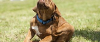
Patella (also called “knee dislocation”) is a disease characterized by displacement of the knee cap relative to its usual position in the femoral ligament of the bone. As a result, the dog is not able to move normally and lameness appears, which in some cases is only slightly pronounced, and in others is characterized by almost complete tightening of the affected limb due to pain in it. If the disease affects several limbs at once and actively develops, it is likely that the dog will soon be unable to walk at all.
There are several reasons for the development of patella:
- hereditary factor (the first signs of the disease begin to appear at about 4 months, less often from 2-3 weeks after birth, as soon as the puppies try to run);
- injuries to a pet, received during an unsuccessful jump, falling from a height, fights with relatives;
- age-related changes in muscle ligaments and individual muscles, which leads to their relaxation and, as a result, displacement of the cup in the knee.
Important! If the disease is of a genetic nature, such dogs are not allowed to be bred further, especially since females suffer more from the disease.
Why is a luxated kneecap dangerous?
With Patella Luxation, the biomechanics of the knee joint and the function of the pelvic limb as a whole are disrupted. The animal cannot fully lean on its paw, which manifests itself as lameness. Curvature of the hind limbs is often observed. Constant slipping of the patella leads to trauma to the cartilage and ligamentous structures of the knee joint and ultimately to arthritis. Constant pain and inflammation leads to destruction of the joint, the animal experiences discomfort, is depressed, inactive, appetite decreases and the body’s general resistance to resist infections.
Types of disease
The type of joint displacement allows us to divide the patella in dogs into 2 main types: lateral luxation with a shift to the outer part of the thigh and medial dislocation, in which it shifts closer to the inside of the hind limb. The latter option is typical for 75-80% of situations, with approximately 15-20% of them involving dislocations of both hind legs.
Lateral luxation
This variant of the disease is more often diagnosed in large breed dogs and is clearly visible visually (the hind legs are curved outward). If dislocations affect both hind legs of an animal at once, they will have an “X-shaped” arrangement, which is often important for hip dysplasia. In some situations, you can notice something is wrong after just six months of the puppy’s life.
Medial dislocation
The medial variety is more relevant for small animals and often manifests itself in the proximal displacement of the tibia to the inner part. Certain disorders in the hip joint cannot be ruled out: for example, the development of osteochondritis of the femoral head or a change in the angle of placement of the hip bone. The pathology often appears on both legs at once and is a genetic disease for many dwarf dog breeds.
Risk factors depending on age and breed
There are a number of factors predisposing to dislocation:
- hip dysplasia;
- deviation of their angle of inclination;
- congenital bone growth disorders;
- weak muscles or ligaments.
In small breed dogs, an insufficiently developed groove between the condyles of the knee joint most often provokes a dislocation of the patella. This feature is inherited genetically.
This pathology also occurs in other dog breeds: large St. Bernards, retrievers, bull terriers, and German shepherds. Other possible causes for joint deformation include:
- large body weight and excessive loads;
- limb injury and ligament rupture;
- wear of the condyles;
- weak ligaments with x-shaped position of the hind legs. In this case, lateral dislocation occurs more often.
How the disease manifests itself at different stages
Signs of the disease may vary depending on the specific stage of the disease, which is also sometimes considered as a criterion for dividing patella into types. Each stage has its own characteristics.
Did you know? If you don't wash your dog's paws well after a walk, they will start to smell like corn chips.
First
At the very beginning of the development of the patella, the patella moves from its groove in a state of complete rest, for example, when the animal lies down and does not load its paws in any way. As soon as the position of the body changes and the pet stands up, at first it may limp until everything falls into place. Sometimes the pet holds the limb suspended for a long time, and then leans on it again. In an extended position of the paw, in the medial direction, the owner himself can pull the knee pad out of the joint, after which it will return to its usual place in the knee block. There should be no crunching during such manipulations. In some situations, this problem is associated with age-related weakness of the ligaments and may disappear on its own as the puppy grows, without requiring surgical intervention.
Second
In this case, displacement of the cup is observed much more often, and the dog does not rely on the limb from time to time, even during prolonged movement. The movement of the animal may be accompanied by crunching of the limbs, and the toes of the affected paw are usually turned inward. If both limbs are damaged, the dog leans on them, but tries not to straighten them too much. At the second stage of the disease, the bone is already rotated by more than 30 degrees, and the head of the hip is slightly deflected in the joint. The patella shifts in the medial direction, and the joint of the tibia gradually turns outward, making it difficult to fully rest on the affected paw. Constant friction of a dislocated joint can cause erosion and severe changes that will require surgery in the future.
We advise you to learn how to cure canine elbow bursitis.
Third
Patella at this stage is characterized by a regular shift of the patella, with the dog's hip bone everted by almost 50 degrees. The pet can only step lightly on the affected limb, allowing it to rest more and more time. The hock joint is no longer in line with the knee joint, shifting when the animal moves to the right or left. The groove where the kneecap should be placed narrows and acquires a flatter surface. With a bilateral violation, the dog moves on half-bent legs, in small steps. It is almost impossible to correct the medial dislocation on your own at this stage of the patella, and if this is possible, it will happen again very soon. This cannot be done without surgical intervention.
Fourth
Patella stage four is the most complex form of the disease, in which the pelvic bone is rotated 50–90 degrees, the dog constantly limps and this is clearly visible visually. When feeling the kneecap, it can be felt just above the hip joint on the inside of the thigh. The groove fills with bone tissue and begins to bulge, or it bulges but is empty. Due to pain and discomfort, the animal has to move erratically on bent limbs. Of course, there can no longer be any question of any reduction here, and the only way to restore mobility to the pet will be the fastest possible surgical intervention.
Important! The most effective surgery on the kneecap will be only in animals up to a year old, while the bone tissue is still capable of regeneration. In the future, the percentage of successful surgical intervention decreases and it is only possible to slow down further tissue destruction.
Symptoms of the disease and its severity
Medial luxation of the patella has the main early sign - intermittently occurring and disappearing lameness in the dog.
With patella dislocation and joint damage in dogs, their symptoms increase and progress as the disease progresses. In veterinary medicine, there is a special classification that evaluates the degree of displacement and deformation of the hind limbs.
Depending on the severity, the following stages of the disease are distinguished:
- The first stage of the disease does not yet bring the dog noticeable discomfort and pain. The bone moves from its correct location when the animal is in a relaxed state, for example, during sleep. When the dog tries to stand up, the patella does not immediately get into the desired position. The dog can straighten the joint on its own by extending its paw. The short-term lameness that occurs, followed by a normal gait, is characteristic of the first stage of the disease. At this stage of the disease, spontaneous displacement of the patella during movement practically does not occur.
- The second stage is characterized by frequent displacement of the patella. The patella can no longer return to its place on its own. At this stage, the cartilage tissue is already worn away, and inflammation occurs in the joint.
- At the third stage of the disease, the patella is constantly in a displaced state, the hip bone is turned out almost 50 degrees. When the bone is set, it stays slightly in the anatomically correct place. The injured limb may be half-bent; the dog almost does not step on it.
- In the fourth, most severe degree of the disease, the kneecap can no longer be reduced. The hip bone is turned 90 degrees, the dog cannot step on the affected leg. If both hind legs are affected, the dog loses the ability to move.
As each stage of the disease progresses, more and more damage occurs to the knee cartilage. Its elasticity decreases and there is a risk of osteoarthritis. The inflammatory process progresses, accompanied by pain.
Diagnostics
To confirm the diagnosis of patella and assess the severity of the disease, a specialist needs to perform several diagnostic procedures:
- During a visual examination by a doctor, the incorrect position of the hind limbs is noted, even in cases where there is no obvious lameness, and the animal’s movement is also assessed after a change in body position (for example, when a dog gets up after a long period of lying or sitting). The owner may be asked to simply walk the dog around the room straight or in a circle.
- During the examination, palpation of the affected paw is also allowed, in order to determine the position of the joint, the exit of the patella from the groove and the strength of the swelling.
- It would also be appropriate to conduct several orthopedic tests: for example, a compression test, a test for pain and the stage of dislocation.
X-ray data, always in two main projections: lateral and direct, help the doctor to verify his assumptions and obtain more accurate data in each individual case. In addition to the knee joint, the hip joint is also subject to such examination. The finished photographs will show the extent of their damage, the degree of rotation of the bones and the condition of the groove in the knee. An additional macroscopic examination makes it possible to determine the level of wear of the cartilage tissue, the presence of fibrosis and contracture, stretching of the joint capsule, and a microscopic examination makes it possible to see the fibrosis of the articular cartilage and synovitis. Joint swelling and lameness do not always indicate the presence of patella in dogs, so it is recommended to undergo a full examination.
Surgical correction
The reasons for the development of the disease, the age of the dog, and the state of its health determine exactly how surgical care will be provided.
However, favorable prognosis can be expected only if the operation was performed at the second or third stage of the disease. The fourth stage cannot be corrected.
Methods of surgical correction depending on age and cause of dislocation:
- If lameness appears in a young growing dog, then surgery is necessary to avoid severe skeletal deformation. In the first year of life, the surfaces of the articular bones are still capable of regeneration; success from surgery in this case is more possible. At this age, it is advisable to perform chondroplasty. In this case, areas of cartilage that are least subject to physical stress are used as implants.
- If the notch in the knee joint is not deep enough and the articular bone is poorly fixed, trochleoplasty is performed. This is the deepening of the groove of the femur where the patella is located. To do this, a section of cancellous bone is cut out in the form of a wedge.
- To limit the rotation of the knee joint, it is stabilized by drilling channels in the tibia, applying and tightening a non-absorbable thread.
- If the joint capsule is overstretched, it is reduced by cutting out a flap.
- When ligaments are torn, they are sutured.
- In case of severe deformity and the impossibility of joint plastic surgery, an artificial fracture of the limb is performed - osteotomy. The fragments are fixed in an anatomically correct position.
The post-operative recovery period is long and difficult for the dog. She should not be allowed to put any weight on her operated paw for several weeks or even months.
After surgical treatment, cases of recurrence of joint dislocations are quite possible. To minimize risks, keep the joint mobile, and avoid muscle atrophy, the dog needs to be provided with proper nutrition, good care, and physiotherapy.
Treatment of patella in dogs
Based on the stage of development of the disease, adequate treatment is selected to eliminate all the unpleasant consequences of a dislocated knee joint in dogs. In the first and second cases, it is quite possible to get by with medications, but in more advanced situations, the only way out will be surgical intervention. Each stage of treatment must be carried out in accordance with generally accepted requirements, taking into account the individual characteristics of the existing case.
Important! Use medications containing methylsulfanylmethane with caution. It relieves pain well, but the dog does not control the force of the load, which leads to rapid wear of the damaged joint.
Drugs
Traditional treatment for diagnosed first degree and some types of second degree consists of the use of chondroprotective, anti-inflammatory and analgesic agents. The best option for the latter drugs would be formulations based on chondroitin and glucosamine, which are best given to the pet along with drugs that stimulate collagen production in the animal’s body.
Modern vitamin complexes containing the aforementioned chondroitin, glucosamine and collagen hydrolysate will also be useful for your pet. You can achieve optimal effectiveness of their use by alternately using several variants of such remedies at once, giving them to the dog in courses. In this way, it is possible to avoid the addictive effect, effectively relieving inflammation and restoring joint tissue in the affected limb.
Surgical intervention
Certain types of patella of the second degree, as well as diseases of the third and fourth, are treated only surgically, by trochleoplasty or trochlear chondroplasty. The essence of the first operation lies in deepening the groove, that is, the place where the kneecap is usually located, for which a section of cancellous bone has to be removed. Subsequently, the fibrocartilage grows again and lines the block of the femur.
Did you know? Famous dog actors sign their contracts themselves with a paw print. For example, such prints were taken from the German shepherd Rin Tin Tin, whose activity in cinema occurred in 1922–1932.
Trochlear chondroplasty is prescribed mainly for young animals, up to six months of age. During surgical procedures, part of the cartilage tissue is peeled off, the subchondral portion of the bone is deepened inside, and then the cartilage tissue is grafted so that it covers the formed groove. The most favorable prognosis will be after surgery at the second and third stages, but repeated dislocations occur in approximately 48% of operated dogs, so the owner will have to provide his pet with the highest quality postoperative care (with intensive physical therapy) and gentle conditions for further life.
Drug treatment
The disease is treated depending on its severity, as well as the dog’s age and health condition.
If a dog's kneecap dislocation has not worsened beyond the first stage, then treatment can be carried out with medications.
They act like:
- painkillers;
- anti-inflammatory;
- chondroprotective.
Glycosaminoglycan drugs are medicines for humans, but they also help animals well. They contain components that are found in cartilage. If there is damage to the joints, then these medications:
- deliver the necessary substances to the damaged area;
- strengthen cartilage;
- relieve inflammation;
- restore elasticity.
Methods such as manual therapy and acupuncture have proven themselves quite well.
If a dog has a predisposition to joint dislocations, then all such procedures should begin from puppyhood. This way it will be possible to minimize the manifestations of the disease.
Which dogs are at risk?
Patella is considered a hereditary disease characteristic of ornamental animals.
During breeding, representatives of the following breeds are tested for this disease:
- Pomeranian Spitz;
- cocker spaniel (almost all varieties);
- Yorkshire Terrier;
- Japanese Chin;
- chow-chow;
- Shar Pei;
- beagle;
- Tibetan Terrier;
- Labrador Retriever;
- Parson Russell Terrier.
Of the large dogs, Newfoundlands, Mastiffs, and Mountain Dogs suffer more often than others from dislocation of the knee joint. Small Chihuahuas and Toy Terriers can injure their paws even when jumping from chairs or sofas, which can later lead to complications such as patella.
How to recognize the degree of injury
In severe stages of the disease, lameness becomes chronic.
This classification includes four degrees. Each of them manifests itself differently and determines future treatment:
- 1st degree
: after an injury, the kneecap returns to its place; - 2nd degree
: when bent, it acquires an unnatural position, in rare cases it falls into place; - 3rd degree
: dislocation occurs during movement, when the dog bends and extends the paw; - 4th degree
: the cup cannot take its natural position and is constantly in a state of dislocation.
In severe stages, lameness becomes chronic, while in the initial stages it appears periodically and may suddenly disappear.
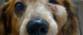
Symptoms of the disease
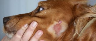
This is what a luxated kneecap looks like in a dog.
- In the first stage, the knee cannot bend if the cup is not in place. In such cases, dogs rarely experience any complications. As a rule, treatment is carried out without surgery, since this condition can very rarely lead to degenerative changes.
- The second stage often leads to osteoarthritis. When walking, the cartilage is severely damaged due to the constant sliding of the cup outside the articular block. This condition often leads to degenerative changes, so in these cases surgery is indicated.
- The third stage is characterized by persistent dislocation, so it is impossible to bring the kneecap to its natural state manually. Even if the dislocation is eliminated without the use of surgical manipulation, the dog will soon experience a second displacement.
- The fourth stage, like the previous one, is also characterized by the stability of the dislocation. In such cases, even surgery does not promise a favorable outcome. Surgery is warranted in dogs under one year of age.
Osteoarthritis of the knee joint in a dog.
Clinical symptoms and their manifestation do not always correspond to a certain stage.
But, despite the fact that this classification is general, it makes it possible to monitor how the disease develops in young dogs. It is also useful in cases where surgery is planned.
Often only during surgery can the doctor determine the necessary treatment methods, but the clinical stage is also very useful. For example, if at the initial stage there are no symptoms of the disease, then conservative therapy is carried out. In addition, special clamps are used to help keep the kneecap in the correct position.
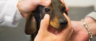
Diagnostic methods
The type of displacement can be determined by palpation.
To prescribe effective treatment, it is necessary to establish the exact cause of the injury, as well as determine its extent.
Basic, basic diagnostic methods are carried out by palpation. To confirm suspicions, doctors take an x-ray of the injured limb.
Using palpation, the specialist determines the type of displacement of the kneecap (inward or outward) and determines whether it is possible to move it back or not. Dog X-rays are taken in two positions - straight and lateral. The procedure makes it possible to determine the degree of dislocation, as well as find the cause of the unnatural position of the kneecap. In some cases, a ligament rupture can lead to a dislocation. This reason must be confirmed or excluded.
Doctors are required to take x-rays
, which will help determine whether the dog has. This disease occurs as a result of disturbances in the blood supply to the head of the femur.
Establishing diagnosis
Dog owners should take into account the fact that when making a diagnosis, the doctor should not be guided solely by the results of a general examination. Thus, in many cases, animals may experience lameness due to various orthopedic diseases. Only a comprehensive examination, including X-ray examination, will help confirm suspicions or exclude them. The procedure should help assess the condition of not only the knee, but also the hip joint.
An x-ray will help confirm or rule out suspicions.
How is the patella test performed?
The test for patella in dogs of small breeds is carried out at the age of 6-8 months, and in representatives of large breeds - no earlier than 12-14 months after the birth of the puppy. The procedure itself is absolutely painless for the pet and does not require the use of anesthesia, because the doctor simply feels the knee joint, determines the position of the cup and enters the data of his observation into a special international certificate (Patella examination). During the examination, the dog’s gait is also assessed, and if the veterinarian has any doubts about the animal’s health, he may also prescribe an x-ray. For testing, the breeder needs to provide the specialist with the animal itself, its pedigree card, puppy card, and veterinary passport.
Find out why your dog has poor coordination and what needs to be done.
Prevention
Considering the huge percentage of the genetic nature of the disease, first of all, it is necessary to exclude from breeding all dogs with a confirmed diagnosis. For this purpose, before the planned mating, mandatory testing for patella luxation of all animals with a breed predisposition is provided. Even if the owner is not a breeder, but the dog’s breed is at risk, testing can be carried out as early as six months of age.
Owners of dogs without any congenital knee defects need to monitor the animal's weight and carefully avoid any potential traumatic situations.
source
Causes of pathology
The disease is provoked by genetic, hereditary pathologies, the consequence of which is abnormal bone development. Injuries and age-related changes lead to dislocation much less frequently.
Heredity
In this case, the dislocation is associated with congenital anomalies. This may also be a pathological development of the skeleton during the growth period.
Predisposition to the disease appears by 4 months. Dislocations begin to appear frequently.
Limb injuries
Unsuccessful jumps and falls are rare causes of cup dislocation. Any adult and large dog can get a knee injury.
Age-related changes
Over the years, the dog's bones become brittle and the ligaments and muscles become weak. Any strain can cause injury. It is important not to overload pets over 8 years old.