Perthes disease is a fairly common orthopedic problem in small breeds of dogs, which leads to severe lameness and subsequently impaired ability to support the affected limb. Perthes disease most often affects such dwarf dog breeds as the Yorkshire Terrier, Toy Terrier, West Highland White Terrier, Cairn Terrier, Miniature Pinscher, Chihuahua, Spitz, Miniature Poodle and their mixed breeds. There is no gender predisposition to the disease in dogs; both females and males are affected. Perthes disease can affect both hip joints at once or develop in one. In veterinary practice, this disease should be given great attention, since in a large number of literary sources the disease is described as a genetic pathology (inherited). Therefore, this disease should be monitored very carefully by dog breeders, and affected dogs should not be allowed to be bred.
Causes of Perthes disease in dogs
The causes of the development of Perthes disease have not been reliably proven. Some theories include a genetic predisposition of dogs to the disease; according to other literary sources, the disease can develop as a result of a strong and long-term load on the hip joint due to weakness of the muscular system in the joint area, in particular on the femoral head; according to other literary sources, it has been proven that hormonal violations affect the development of the disease.
Another name for the disease is Perthes's, which is aseptic necrosis of the femoral head. The mechanism of development of this disease lies in a decrease in vascularization, that is, a deterioration in blood flow to the head of the femur, since the blood flow decreases, the nutrition of the bone substance and marrow is correspondingly disrupted, then aseptic necrosis occurs and subsequently it is destroyed.
Histological examination of the affected femoral head and neck in Perthes disease revealed that after necrosis, trabecular collapse occurs, which subsequently leads to instability and rupture of the subchondral cartilage and disruption of the congruence of the articular surfaces of the hip joint. Further, all this leads to the development of secondary osteoarthritis, after which the avascular bone is replaced by granulation tissue.
What to do if there are no apparent reasons
If a dog is limping on its front leg without visible damage, what should you do? If there is a slight disturbance in gait, you should not burden the dog with walks, but give it the opportunity to rest for several days. If this is not associated with illness, then the animal’s well-being will improve on its own.
If the condition worsens, the dog should be transported to a veterinarian and treated based on the established diagnosis.
Regular examination by a specialist helps to avoid serious problems.
Note! Only a doctor can make a correct diagnosis and identify the cause of the problem.
Symptoms of Perthes disease in dogs
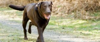
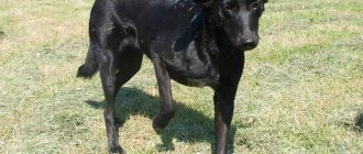
Perthes disease develops gradually. Typically, the first clinical signs of the disease appear in puppies at 5-6 months. Sometimes, especially after active games, a dog begins to take care of its paw; it can be almost unnoticeable, especially for owners, to limp or not actively load the sore limb. In Perthes disease, as a rule, clinical signs develop gradually. When the disease reaches a later stage of development and the femoral head is greatly altered, atrophy of the thigh muscles actively progresses in many small dogs, this can be easily noticed by comparing the diseased paw with a healthy one.
With bilateral damage to the hip joints in Perthes disease, intermittent claudication is observed; the dog may limp on one leg or the other. With severe pain in both hip joints, the dog may try not to walk at all or try to move, as if bouncing, in order to avoid stress on the sore joints.
Atrophy of the thigh muscles in a toy terrier
Very pronounced clinical signs of Perthes disease can appear in dogs as a result of a pathological fracture of the femoral neck or simultaneous destruction of the femoral head. Typically, such pronounced clinical signs manifest themselves in a complete lack of support on the affected limb, or the dog slightly steps on its toes during a slow walk, and when the step increases, it stops using its paw and runs on three feet.
Causes of the condition
To determine why your dog suddenly stopped running and walking normally, you need to know the main causes of lameness. After all, the inability to fully walk and run causes serious discomfort to the dog.
Injuries of varying severity
A dog can become lame as a result of mechanical injury, which often occurs during normal walking.
An insect bite can cause illness
Common types of injuries in dogs:
- puncture wounds;
- cuts;
- splinters;
- dislocation;
- fracture;
- joint bruises;
- sprain.
Note! Snow that has accumulated between your pet's toes can also cause injury to a limb.
Diseases
Not only people, but also dogs suffer from diseases of the joints and bones. Malaise can occur not only due to age-related changes, but also in puppies. Most often, this is caused by injuries acquired long ago, increased physical activity, and an unbalanced diet.
Common musculoskeletal diseases in dogs:
- arthritis;
- osteoarthritis;
- dysplasia.
Interdigital cyst
The reason for the development of cysts in dogs has not yet been fully studied. According to veterinarians, the appearance of nodules between the fingers can be caused by violations of the integrity of the subcutaneous tissue, hair follicles or sebaceous glands.
The cause is sometimes mechanical, chemical or thermal effects, as well as parasitic and infectious damage.
Chemical or thermal burns
A dog can damage its paw when exposed to high or low temperatures. Burns occur upon contact with aggressive agents such as alkali, fuel oil, acid, gasoline. A pet can burn a limb with boiling water or jump onto a hot metal sheet.
On a note! Lameness also appears after walking in winter, since during this period the paths are sprinkled with special reagents that irritate the skin.
Diagnosis of Perthes disease in dogs
Diagnosis of Perthes disease at a late stage of the disease does not present much difficulty for veterinary specialists.
On general examination, all dogs with Perthes disease will have pain in the hip joint, especially when abducted backward. Dogs with constant pain may not eat well and will be depressed. All dogs will have lameness of varying degrees, up to a complete lack of support on the affected paw.
Perthes disease, radiographic image
Yorkshire Terrier, Perthes disease of both hip joints, destruction of the femoral head on the right, arthroplasty of the hip joint was performed on the left.
Clinical examination of dogs at a late stage of the disease usually reveals atrophy of the muscles of the thigh (which is very noticeable with unilateral damage to the hip joint) or both hips, crepitus in the joint area due to a violation of the congruence of the articular surfaces or due to destruction of the femoral head. In some dogs, as a result of atrophy, the greater trochanter of the femur will protrude greatly; this is important for your veterinarian to differentiate from a dislocation.
In most cases, an X-ray examination of the hip joint in the ventrodorsal projection is sufficient to confirm the diagnosis. If the dog has very severe pain, then sedation is possible in order not to unnecessarily injure the already sore joint. In the early stages of the disease, radiographic images of the femoral head and neck will show increased bone density, changes in the shape of the femoral head due to ischemic necrosis, and changes in the intra-articular space. In the later stages of Perthes disease, the radiographic image will show signs of osteoarthritis, osteophytes as a result of destruction of the femoral head in the acetabulum, and in some cases the pathological fracture line of the femoral neck is clearly visible.
Of great clinical significance in the early stages of the development of Perthes disease, when there is minor pain and no atrophy of the hip muscles, is CT diagnostics of the hip joint. A computed tomography scan of the head and neck of the femur will show the initial stages of pathological restructuring of bone tissue, which cannot be recognized on an x-ray examination. The purpose of CT scanning for Perthes disease is, first of all, early diagnosis of the disease and, accordingly, early implementation of therapeutic measures, which is important for the subsequent quality of life of the dog.
Fracture
Fractures of limb bones can be open or closed. An open fracture is the most dangerous, as soft tissues, joints, and muscles are damaged, and severe bleeding occurs. Fragments of bone tissue can be observed through the open wound.
It is difficult to diagnose a closed fracture at home, but it is characterized by the following symptoms:
- The injured limb swells;
- The dog flexes its affected leg, moves on three legs, or stops walking altogether;
- The injured limb may be unnaturally deployed;
- The pet experiences severe pain upon palpation.
Treatment of fractures in a veterinary clinic includes the application of an immobilizing bandage and splint, as well as further restriction of mobility for a period of 30 days (depending on the age of the animal). The dog's movement is restricted using a crate.
For some complex fractures, it is impossible to do without surgical intervention and surgery.
Treatment of Perthes disease in dogs
Treatment for Perthes disease can be of two types: therapeutic and surgical. When choosing a therapeutic treatment for Perthes disease, one must be guided by the type of pathological changes in the joint, that is, with a pathological fracture of the femoral neck, therapy will clearly not give good results. Therapeutic treatment is advisable when the congruence of the articular surfaces is not greatly changed, there is no large number of osteophytes, severe muscle atrophy and there is no pathological fracture.
Therapy includes the use of non-steroidal anti-inflammatory drugs that eliminate pain and, accordingly, relieve inflammation, such as Loxicom, Previcox, Meloxicam, etc. All drugs must be used strictly in the exact dosage and after meals so as not to irritate the gastric mucosa. Also important in the conservative treatment of Perthes disease is maintaining a rest regime for 1-2 months; the dog should avoid excessive stress on the femoral head so as not to lead to its pathological fracture. The use of physiotherapy for Perthes disease also has its place; for example, electrophoresis with anti-inflammatory drugs can be used to relieve local inflammation.
Surgical treatment of Perthes disease is used as the main type of treatment for complete or partial destruction of the femoral head, pathological fracture, severe muscle atrophy and severe osteoarthritis of the hip joint. This type of treatment involves removing the cause of the pain, that is, removing the femoral head. This type of operation is called resection arthroplasty of the hip joint, which is used in our clinic. During this operation, to avoid rubbing the resected surface of the thigh on the acetabulum and causing pain, the gluteal muscle is inserted. After the operation, the dog is prescribed antibiotics and painkillers for 7 days, the sutures are removed on the 10th day, and then rehabilitation measures begin.
After surgery, proper rehabilitation of the dog is extremely important, which should include: massage, passive movements of the joint, swimming, the use of weights on the affected limb and other activities that help restore the motor function of the joint. Properly performed rehabilitation measures are 80% of the success of postoperative treatment.
With an illiterate approach to rehabilitation, the dog may not begin to use the operated paw at all; as a rule, this happens due to the owners’ reluctance to work with the dog or because of pity and fear of hurting the dog. If it is impossible to carry out rehabilitation measures, it is better to seek help from a veterinary rehabilitation specialist.
Symptoms and types
Lameness can affect one or more limbs and ranges in severity from a minor problem to complete loss of the ability to bear body weight on the affected leg.
If only one thoracic limb is affected, then when the body weight is transferred to it, the dog’s head and neck move upward, and when resting on a healthy paw, the front part of the dog’s body lowers.
If only one hind limb is affected, then when leaning on it, the pelvis sinks, and when leaning on the healthy paw, it rises.
If there is a double pelvic lameness, the front legs are placed lower to shift the weight forward.
There may also be two variants of lameness intensification - when it becomes more noticeable after a period of high activity, and vice versa, when the dog limps immediately after a long sleep, and then “paces.”
Other signs accompanying lameness:
- Soreness and associated decrease in activity.
- Limitation of the range of movement, the dog may refuse to do things that were easy to do before - jumping, climbing stairs, jumping into a car, onto a sofa, and so on.
- Decrease in muscle volume (atrophy).
- Changing the position in which the dog stands or lies, including while sleeping. If the dog begins to take relaxed positions on its back much less often, this may indirectly indicate a pain syndrome.
- Change in gait.
- Neurological manifestations - the dog places its paws incorrectly and drags them.
- Changes in the size and shape of bones and/or joints.
- Sounds (crepitus) when moving the joints.
Forecast
The prognosis for recovery from Perthes disease is usually good. The prognosis largely depends on the type of treatment and the degree of change in the femoral head. With a therapeutic type of treatment, the prognosis for restoration of normal joint function is relative; according to literature data, out of 89 dogs, only 19 had normal joint function.
When choosing surgical treatment and performing all rehabilitation measures, the prognosis for restoration of normal joint function reaches up to 90% of the total number of operated dogs.
Author of the article: veterinary surgeon, specialist in traumatology, orthopedics and neurology Ekaterina Sergeevna Maslova
Diagnostics
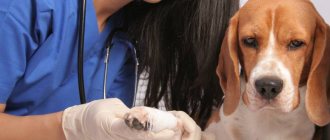
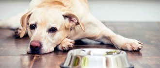
Joint pain can last from a few days to several months and can range from mild to severe. The veterinarian will examine your dog to rule out underlying conditions other than bone inflammation. X-rays and blood tests will be used to find the cause and make a proper diagnosis.
In most cases, inflammation will be the cause of pain and can be reduced with medication.
Injuries
These causes of lameness in dogs, along with bruises and sprains, are quite common. Wounds (most often) are bitten and lacerated, which is bad. Such injuries take a long time to heal and cause many “side effects,” with lameness being perhaps the most harmless. Brief description of these injuries:
- They are more often found on the hind limbs (this is where dogs are bitten by their relatives during fights).
- Wounds are often accompanied by crushing and tissue ruptures, including ligaments.
- In addition to bites, broken nails, insect bites, and grass stubble embedded in the paw pads can cause lameness. In these cases, the animal begins to fall on its hind paw, but the front limb, as a rule, is supported by weight.